. 2007 Mar;117(3):627-35.
doi: 10.1172/JCI28253. Epub 2007 Feb 22.

Abnormal thyroid hormone metabolism in mice lacking the monocarboxylate transporter 8

Affiliations

Abnormal thyroid hormone metabolism in mice lacking the monocarboxylate transporter 8

Marija Trajkovic et al. J Clin Invest. 2007 Mar.

Abstract

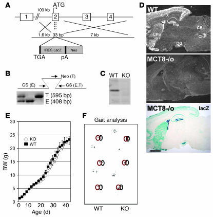
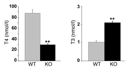
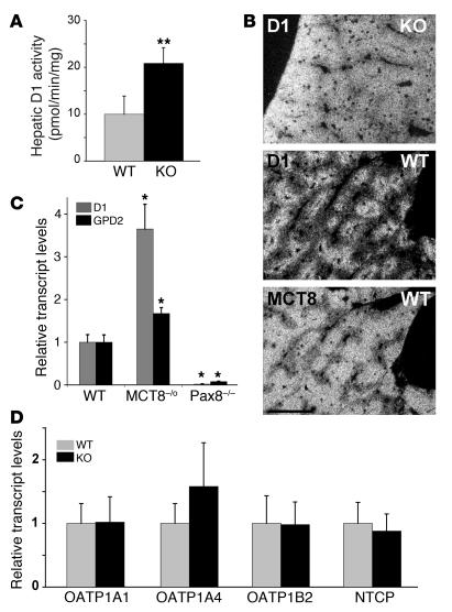
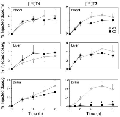
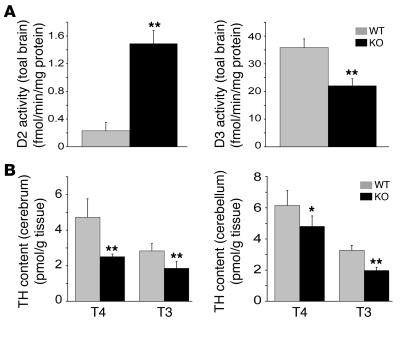
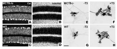
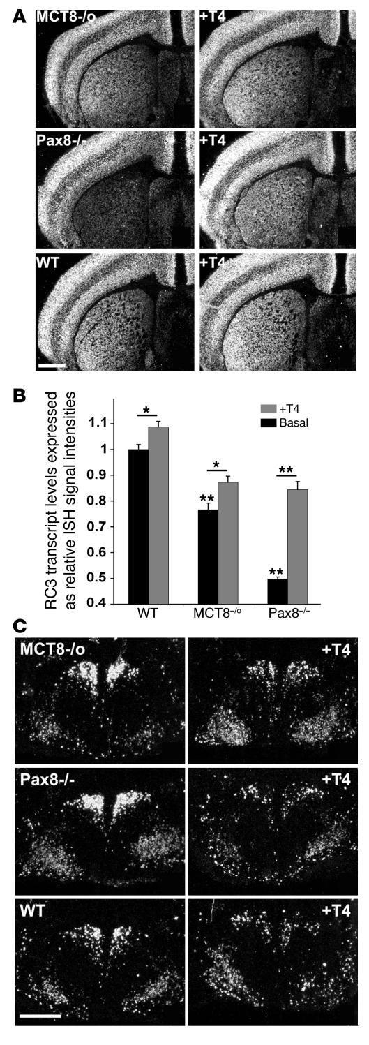
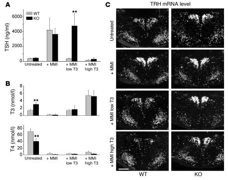
In humans, inactivating mutations in the gene of the thyroid hormone transporter monocarboxylate transporter 8 (MCT8; SLC16A2) lead to severe forms of psychomotor retardation combined with imbalanced thyroid hormone serum levels. The MCT8-null mice described here, however, developed without overt deficits but also exhibited distorted 3,5,3'-triiodothyronine (T3) and thyroxine (T4) serum levels, resulting in increased hepatic activity of type 1 deiodinase (D1). In the mutants' brains, entry of T4 was not affected, but uptake of T3 was diminished. Moreover, the T4 and T3 content in the brain of MCT8-null mice was decreased, the activity of D2 was increased, and D3 activity was decreased, indicating the hypothyroid state of this tissue. In the CNS, analysis of T3 target genes revealed that in the mutants, the neuronal T3 uptake was impaired in an area-specific manner, with strongly elevated thyrotropin-releasing hormone transcript levels in the hypothalamic paraventricular nucleus and slightly decreased RC3 mRNA expression in striatal neurons; however, cerebellar Purkinje cells appeared unaffected, since they did not exhibit dendritic outgrowth defects and responded normally to T3 treatment in vitro. In conclusion, the circulating thyroid hormone levels of MCT8-null mice closely resemble those of humans with MCT8 mutations, yet in the mice, CNS development is only partially affected.


Figures

Figure 1
Figure 1. Generation and analysis of MCT8-null mice.
(A) Targeting strategy for the MCT8-knockout mouse. As described in Methods, 33 bp of exon 2 were replaced by a lacZ/neomycin reporter cassette. pA, polyadenylation signal; TGA, stop codon. (B) Homologous recombination was confirmed by PCR analysis using 3 primers recognizing the endogenous (E) and the targeted (T) allele as indicated and described in Methods. GS, gene-specific primer; Neo, neomycin cassette–specific primer. (C) Absence of MCT8 protein in MCT8-deficient animals was demonstrated by Western blot analysis of liver homogenates. (D) Deletion of the MCT8 gene was further confirmed by radioactive ISH demonstrating that MCT8 mRNA expression in sagittal brain sections of wild-type animals is completely diminished in sections derived from MCT8-null mice. Deletion of the MCT8 gene was also demonstrated by the expression of lacZ in MCT8–/o animals. CBL, cerebellum; ChP, choroid plexus; CPu, caudate-putamen; CTX, cerebral cortex; DG, dentate gyrus; Hip, hippocampus. Scale bar in D: 1.5 mm. (E) MCT8-null mice develop normally, as indicated by the growth curve. (F) Gait analysis did not reveal any signs of ataxia.
Figure 2
Figure 2. Serum thyroid hormone levels in 21-day-old male MCT8-null mice and wild-type littermates.
Per group, 12 animals were analyzed. Bars represent the mean ± SEM of values obtained in each group. **P < 0.0001.
Figure 3
Figure 3. Analysis of D1 expression in the liver of MCT8-null and wild-type animals.
(A) D1 enzymatic activity was determined in liver and kidney preparations from 21-day-old MCT8-null mice and wild-type littermates. (B) ISH analysis of hepatic MCT8 and D1 expression in wild-type mice and D1 expression in MCT8-null animals. In MCT8-null mice, expression of D1 in liver was highly upregulated compared with the expression of D1 in liver of control animals. Expression of MCT8 in wild-type animals is shown in the lower panel. Scale bar: 450 μm. (C) Hepatic D1 and GPD2 mRNA levels were determined in 21-day-old MCT8-null mice, wild-type littermates, and athyroid Pax8–/– mice by real-time PCR. Bars in A and C represent the mean ± SEM of values obtained in each group. *P < 0.05; **P < 0.0001. (D) Transcript levels of putative hepatic thyroid hormone transporters were examined in the liver of MCT8-null mice and controls by real-time PCR. This analysis revealed no statistical differences in the expression levels for the organic anion transporting polypeptides OATP1A1, OATP1A4, and OATP1B2 and the Na+/taurocholate-cotransporting polypeptide NTCP between the different genotypes.
Figure 4
Figure 4. Entry of 125I-labeled iodothyronines into the brain and liver of MCT8-null mice and wild-type littermates.
Adult animals (4 per experiment) were s.c. injected with 2 × 10⁶ cpm [125I]T4 or [125I]T3. At indicated time points, blood samples were taken and the radioactivity was determined. Subsequently, the animals were perfused with saline. Liver and brain were then removed and weighed, and the radioactivity was determined. Results are presented as the percentage of injected dose normalized to the amount of tissue (in ml or g). *P < 0.05.
Figure 5
Figure 5. Analysis of the thyroid state in the brain of MCT8-null mice.
(A) Compared with those in wild-type littermates, brain D2 activities are increased in MCT8-null mice, whereas D3 activities are decreased. Bars represent the mean ± SEM of values obtained in each group. (B) Determination of the tissue thyroid hormone content revealed decreased T4 and T3 levels in the cerebrum and cerebellum of MCT8-null mice, respectively. Bars represent the mean ± SD of values obtained in each group. *P < 0.05; **P < 0.005.
Figure 6
Figure 6. Cerebellar analysis of MCT8-null mice.
Calbindin (Cal) (A and C) and Hoechst (B and D) staining of cerebellar vibratome sections from 12-day-old MCT8-null mice (A and B) and wild-type controls (C and D) did not reveal any differences with regard to the development of Purkinje cells (A and C) or the maturation of the external granule cell layer (B and D). When cultured without T3 as described in Methods, Purkinje cells from MCT8-null mice (E) and wild-type controls (G) showed severely impaired development. In the presence of 1 nM T3, the dendritic development of cultured Purkinje cells derived from MCT8 mutants (F) was stimulated just as strongly as that of Purkinje cells from wild-type controls (H). Scale bar in C (for AD): 100 μm; in G (for EH): 50 μm.
Figure 7
Figure 7. Expression of the T3-regulated genes RC3 and TRH in the striatum and in the PVN.
(A) Compared with that in wild-type controls, RC3 mRNA expression in the striatum was modestly decreased in MCT8-null mice and strongly in athyroid Pax8–/– mice. After treatment with T4 (200 ng/g BW for 3 consecutive days), striatal RC3 mRNA levels increased in MCT8 mutants and in Pax8–/– mice as well as in control animals. Scale bar: 1.5 mm. (B) The evaluation was validated by NIH image analysis of the hybridization signals. *P < 0.05; **P < 0.0001. (C) In hypothalamic PVN neurons, ISH analysis revealed highly upregulated TRH mRNA levels in MCT8-null mice and athyroid Pax8–/– mice compared with control animals. After treatment with T4 (200 ng/g BW for 3 consecutive days), TRH mRNA expression was strongly reduced in wild-type animals as well as in MCT8 mutants and Pax8–/– mice. Scale bar: 450 μm.
Figure 8
Figure 8. Effect of the thyroid state on serum TSH, T3, and T4 levels and hypothalamic TRH mRNA levels in MCT8-null mice and wild-type littermates.
Adult MCT8-null mice and wild-type littermates were rendered hypothyroid by MMI/perchlorate treatment for 12–14 weeks as described in Methods. As a consequence, serum TSH levels (A) and ISH signal intensities reflecting TRH transcript levels (C) were markedly increased in animals of both genotypes, whereas serum T3 and T4 levels (B) were close to the detection limit. In wild-type animals, s.c. injection of a low dose of T3 (5 ng/g BW for 3 consecutive days) was sufficient to normalize not only serum T3 and TSH levels but also to restore to normal TRH mRNA levels in the PVN, whereas in MCT8-null animals, neither TSH nor TRH expression was found to be suppressed in the presence of normal serum T3 concentrations. Only rendering the MCT8-null mice hyperthyroid by injecting a high dose of T3 (25 ng/g BW for 3 days) resulted in a downregulation of serum TSH levels, whereas TRH expression in the PVN was only slightly reduced. Scale bar: 330 μm. **P < 0.0001.

Similar articles

Cited by

References

    1. Porterfield S.P., Hendrich C.E. The role of thyroid hormones in prenatal and neonatal neurological development — current perspectives. Endocr. Rev. 1993;14:94–106. - PubMed
    1. Oppenheimer J.H., Schwartz H.L. Molecular basis of thyroid hormone-dependent brain development. Endocr. Rev. 1997;18:462–475. - PubMed
    1. de Escobar G.M., Obregon M.J., del Rey F.E. Maternal thyroid hormones early in pregnancy and fetal brain development. Best Pract. Res. Clin. Endocrinol. Metabol. 2004;18:225–248. - PubMed
    1. Jones S.A., Thoemke K.R., Anderson G.W. The role of thyroid hormone in fetal and neonatal brain development. Current Opinion in Endocrinology and Diabetes. 2005;12:10–16.
    1. Bernal J. Thyroid hormones and brain development. Vitam. Horm. 2005;71:95–122. - PubMed

Publication types

MeSH terms