

Acute modulation of cortical oscillatory activities during short trains of high-frequency repetitive transcranial magnetic stimulation of the human motor cortex: a combined EEG and TMS study
- PMID: 17318833
- PMCID: PMC6870897
- DOI: 10.1002/hbm.20371
Acute modulation of cortical oscillatory activities during short trains of high-frequency repetitive transcranial magnetic stimulation of the human motor cortex: a combined EEG and TMS study
Abstract
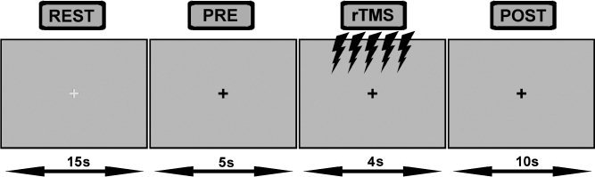
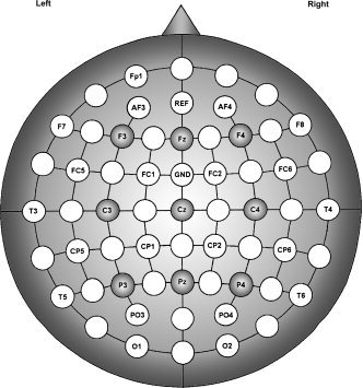
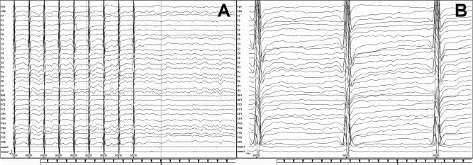
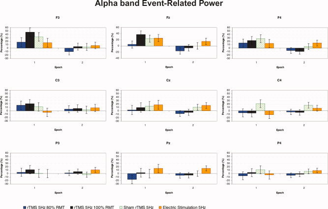
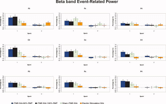
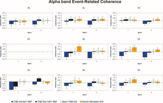
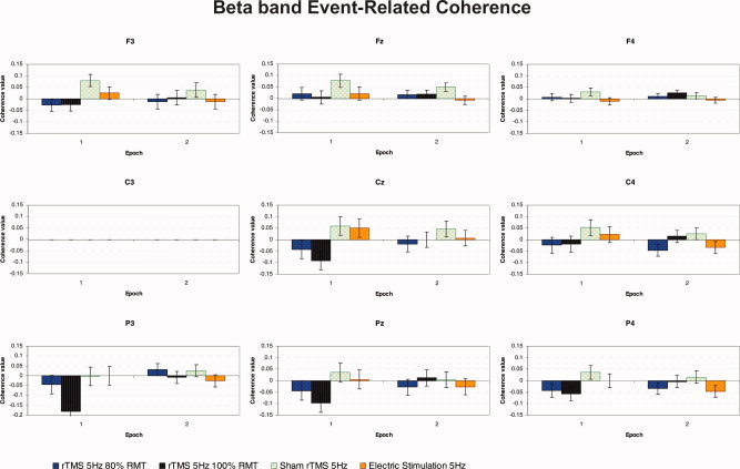
In this study, a combined repetitive transcranial magnetic stimulation/electroencephalography (rTMS/EEG) method was used to explore the acute changes of cortical oscillatory activity induced by intermittent short trains of high-frequency (5-Hz) rTMS delivered over the left primary motor cortex (M1). We evaluated the electrophysiological reaction to magnetic stimulation during and 2-4 s after 20 trains of 20-pulses rTMS, using event-related power (ERPow) that reflects the regional oscillatory activity of neural assemblies, and event-related coherence (ERCoh) that reflects the interregional functional connectivity of oscillatory neural activity. These event-related transformations were for the upper alpha (10-12 Hz) and beta (18-22 Hz) frequency ranges, respectively. For the alpha band, threshold rTMS and subthreshold rTMS induced an ERPow increase during the trains of stimulation mainly in frontal and central regions ipsilateral to stimulation. For the beta band, a similar synchronization of cortical oscillations for both rTMS intensities was seen. Moreover, subthreshold rTMS affected alpha-band activity more than threshold rTMS, inducing a specific ERCoh decrease over the posterior regions during the trains of stimulation. For beta band, the decrease in functional coupling was observed mainly during threshold rTMS. These findings provide a better understanding of the cortical effects of high-frequency rTMS, whereby the induction of oscillations reflects the capacity of electromagnetic pulses to alter regional and interregional synaptic transmissions of neural populations.
Figures
Similar articles
-
Human cortical theta reactivity to high-frequency repetitive transcranial magnetic stimulation.Hum Brain Mapp. 2012 Sep;33(9):2224-37. doi: 10.1002/hbm.21355. Epub 2011 Aug 5. Hum Brain Mapp. 2012. PMID: 21823206 Free PMC article.
-
Modulation of cortical oscillatory activities induced by varying single-pulse transcranial magnetic stimulation intensity over the left primary motor area: a combined EEG and TMS study.Neuroimage. 2005 Oct 1;27(4):896-908. doi: 10.1016/j.neuroimage.2005.05.013. Neuroimage. 2005. PMID: 16054397 Clinical Trial.
-
The neural response to transcranial magnetic stimulation of the human motor cortex. I. Intracortical and cortico-cortical contributions.Exp Brain Res. 2006 Nov;175(2):231-45. doi: 10.1007/s00221-006-0551-2. Epub 2006 Jun 17. Exp Brain Res. 2006. PMID: 16783559
-
Study and modulation of human cortical excitability with transcranial magnetic stimulation.J Clin Neurophysiol. 1998 Jul;15(4):333-43. doi: 10.1097/00004691-199807000-00005. J Clin Neurophysiol. 1998. PMID: 9736467 Review.
-
Measuring Brain Stimulation Induced Changes in Cortical Properties Using TMS-EEG.Brain Stimul. 2015 Nov-Dec;8(6):1010-20. doi: 10.1016/j.brs.2015.07.029. Epub 2015 Jul 17. Brain Stimul. 2015. PMID: 26275346 Review.
Cited by
-
Modulation of EEG functional connectivity networks in subjects undergoing repetitive transcranial magnetic stimulation.Brain Topogr. 2014 Jan;27(1):172-91. doi: 10.1007/s10548-013-0277-y. Epub 2013 Mar 8. Brain Topogr. 2014. PMID: 23471637 Free PMC article.
-
The relationship between brain oscillatory activity and therapeutic effectiveness of transcranial magnetic stimulation in the treatment of major depressive disorder.Front Hum Neurosci. 2013 Feb 26;7:37. doi: 10.3389/fnhum.2013.00037. eCollection 2013. Front Hum Neurosci. 2013. PMID: 23550274 Free PMC article.
-
Deep brain stimulation in persistent vegetative States: ethical issues governing decision making.Behav Neurol. 2014;2014:641213. doi: 10.1155/2014/641213. Epub 2014 Mar 16. Behav Neurol. 2014. PMID: 24803730 Free PMC article. Review.
-
Evaluating frontal and parietal contributions to spatial working memory with repetitive transcranial magnetic stimulation.Brain Res. 2008 Sep 16;1230:202-10. doi: 10.1016/j.brainres.2008.07.008. Epub 2008 Jul 11. Brain Res. 2008. PMID: 18662678 Free PMC article.
-
Interhemispheric and Intrahemispheric Connectivity From the Left Pars Opercularis Within the Language Network Is Modulated by Transcranial Stimulation in Healthy Subjects.Front Hum Neurosci. 2020 Mar 17;14:63. doi: 10.3389/fnhum.2020.00063. eCollection 2020. Front Hum Neurosci. 2020. PMID: 32256324 Free PMC article.
References
-
- Bestmann S, Baudewig J, Siebner HR, Rothwell JC, Frahm J ( 2003): Subthreshold high‐frequency TMS of human primary motor cortex modulates interconnected frontal motor areas as detected by interleaved fMRI‐TMS. Neuroimage 20: 1685–1696. - PubMed
-
- Bestmann S, Baudewig J, Siebner HR, Rothwell JC, Frahm J ( 2004): Functional MRI of the immediate impact of transcranial magnetic stimulation on cortical and subcortical motor circuits. Eur J Neurosci 19: 1950–1962. - PubMed
-
- Brasil‐Neto JP, Cohen LG, Panizza M, Nilsson J, Roth BJ, Hallett M ( 1992): Optimal focal transcranial magnetic activation of the human motor cortex: Effects of coil orientation, shape of the induced current pulse, and stimulus intensity. J Clin Neurophysiol 9: 132–136. - PubMed
-
- Di Lazzaro V, Oliviero A, Berardelli A, Mazzone P, Insola A, Pilato F, Saturno E, Dileone M, Tonali PA, Rothwell JC ( 2002a): Direct demonstration of the effects of repetitive transcranial magnetic stimulation on the excitability of the human motor cortex. Exp Brain Res 144: 549–553. - PubMed
-
- Di Lazzaro V, Oliviero A, Mazzone P, Pilato F, Saturno E, Dileone M, Insola A, Tonali PA, Rothwell JC ( 2002b): Short‐term reduction of intracortical inhibition in the human motor cortex induced by repetitive transcranial magnetic stimulation. Exp Brain Res 147: 108–113. - PubMed
MeSH terms
LinkOut - more resources
Full Text Sources
