. 2007 Feb 23:2:5.
doi: 10.1186/1749-8104-2-5.

Antagonism between Notch and bone morphogenetic protein receptor signaling regulates neurogenesis in the cerebellar rhombic lip

Affiliations

Antagonism between Notch and bone morphogenetic protein receptor signaling regulates neurogenesis in the cerebellar rhombic lip

Robert P Machold et al. Neural Dev. .

Abstract

Background: During the embryonic development of the cerebellum, neurons are produced from progenitor cells located along a ventricular zone within dorsal rhombomere 1 that extends caudally to the roof plate of the fourth ventricle. The apposition of the caudal neuroepithelium and roof plate results in a unique inductive region termed the cerebellar rhombic lip, which gives rise to granule cell precursors and other glutamatergic neuronal lineages. Recently, we and others have shown that, at early embryonic stages prior to the emergence of granule cell precursors (E12), waves of neurogenesis in the cerebellar rhombic lip produce specific hindbrain nuclei followed by deep cerebellar neurons. How the induction of rhombic lip-derived neurons from cerebellar progenitors is regulated during this phase of cerebellar development to produce these temporally discrete neuronal populations while maintaining a progenitor pool for subsequent neurogenesis is not known.

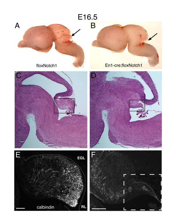
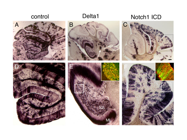
Results: Employing both gain- and loss-of-function methods, we find that Notch1 signaling in the cerebellar primordium regulates the responsiveness of progenitor cells to bone morphogenetic proteins (BMPs) secreted from the roof plate that stimulate the production of rhombic lip-derived neurons. In the absence of Notch1, cerebellar progenitors are depleted during the early production of hindbrain neurons, resulting in a severe decrease in the deep cerebellar nuclei that are normally born subsequently. Mechanistically, we demonstrate that Notch1 activity prevents the induction of Math1 by antagonizing the BMP receptor-signaling pathway at the level of Msx2 expression.

Conclusion: Our results provide a mechanism by which a balance between neural induction and maintenance of neural progenitors is achieved in the rhombic lip throughout embryonic development.


Figures

Figure 1
Figure 1
Notch and BMP signaling components in the embryonic cerebellar primordium. Sagittal cryosections of E12.5 wild-type brains were stained by in situ hybridization with antisense probes for Notch1, Mash1, Hes5, Msx2, Math1 and Delta1. Differentiating neural precursors in the ventricular zone (VZ) and rhombic lip (RL) are delineated by Mash1 and Math1, respectively. Msx2 expression is indicative of BMP receptor signaling, and overlaps with the Math1+ territory at this stage. Hes5 and Delta1 are expressed throughout the cerebellar progenitor population, and are generally indicative of high and low Notch1 activity, respectively. The field shown in these panels corresponds to the box in the schematic (inset). Scale bar represents 300 μm.
Figure 2
Figure 2
Conditional loss of Notch1 in the early embryonic cerebellum results in precocious differentiation at the expense of the progenitor pool. (a) Whole mount β-galactosidase staining of an E9.5 En1cre; Rosa-stopLacZ reveals the extent of recombination across the mid-hindbrain region. (b-g) In situ hybridization with antisense probes for (b, c)Notch1, (d, e)Mash1, and (f, g) Math1 on sagittal cryosections of the cerebellar primordia from E10.5 floxNotch1 (b, d, f) and En1cre; floxNotch1 (c, e, g) embryos. (h, i) Immunohistochemistry for Math1 on E10.5 floxNotch1 (h) and En1cre; floxNotch1 (i) tissue cryosections. (j-o) In situ hybridization with antisense probes for (j, k)Notch1, (l, m) Mash1, and (n, o) Math1 on sagittal cryosections of the embryonic cerebella from E12.5 floxNotch1 (j, l, n) and Engrailed1-cre; floxNotch1 (k, m, o) embryos. The r1/r2 boundary is indicated by the arrow in (k). Brackets in (m, n) indicate the extent of Math1 induction in the VZ. (p, q) Immunohistochemistry for Math1 (white arrows) on E12.5 floxNotch1 (p) and En1cre; floxNotch1 (q) tissue cryosections. Scale bars in (g, o) represent 300 μm.
Figure 3
Figure 3
Conditional loss of Notch1 in the cerebellar primordium results in severe hypoplasia and loss of Purkinje cell precursors. (a, b) Sagittal whole mount view of E16.5 floxNotch1 (a) and En1-cre; floxNotch1 (b) brains. The arrows in (a, b) indicate the position of the cerebellum. (c-f) Sagittal cryosections of floxNotch1 (c, e) and En1-cre; floxNotch1 (d, f) cerebella stained by hematoxylin and eosin histochemistry (c, d) or by immunohistochemistry for calbindin (e, f). Boxes in (c, d) indicate the approximate photographic fields in (e, f). Dashed box in (f) delineates the residual cerebellum in the mutant embryo. Scale bars in (e, f) represent 300 μm.
Figure 4
Figure 4
Short term fate map of Math1-lacZ cells generated after conditional ablation of Notch1 in the cerebellar primordium. (a-d) Coronal sections from E14.5 floxNotch1 (a, c) and En1-cre; floxNotch1 (b, d) embryos stained for β-galactosidase activity. Long arrows indicate the approximate position of the MPT and LPB neurons, and short arrows indicate the position of the DCN. (e, f) Immunohistochemical staining for calbindin on adjacent sections to (a, b) shows an increase in the LPB neuron hindbrain population (white arrows). The asterisk in (e) indicates calbindin positive afferents that are absent in the mutants. Quantification of β-gal+ cells in the control and mutant brains across rostral to caudal tissue sections shows the decrease in DCN in the mutant. Scale bar in (f) represents 300 μm. CB, cerebellum; MB, midbrain; RL, rhombic lip.
Figure 5
Figure 5
Notch1 activity regulates cell fate decisions in the embryonic cerebellum. Pseudotyped bicistronic retroviruses expressing PLAP and full length Delta1 or the Notch1 ICD were injected into the ventricle of E9.5 embryos in utero using ultrasound backscatter microscopy, and the pups sacrificed at P21. Brains of infected animals were cryosectioned sagittally and stained for alkaline phosphatase using NBT/BCIP histochemistry. (a, d) Control experiment with retrovirus expressing alkaline phosphatase alone. (b, e) Retroviral expression of Delta1 in E9.5 cerebellar progenitors yields granule cells at P21; the inset in (e) shows sections stained by immunohistochemistry for PLAP (red) and Zic2 (green). (c, f) Retroviral expression of the Notch1 ICD in E9.5 cerebellar progenitors results in Bergmann glia at P21; inset in (f) shows sections stained for PLAP (green) and BLBP (red). Scale bars in (c, f) represent 300 μm. ICD, intracellular domain; IGL, internal granule layer; ML, molecular layer.
Figure 6
Figure 6
Interaction between Notch and BMP receptor signaling in the cerebellar primordium. (a-f) Stage 10 to 12 chick embryos were electroporated in ovo with a GFP reporter plasmid and the expression plasmids described below. After two days of incubation, embryos were processed for cryosectioning, and the sections processed by in situ hybridization for Cath1. Adjacent sections were photographed for GFP and Cath1 in each experiment. (a, b) GFP alone. (c, d) GFP and caBMPR1b. (e, f) GFP, Notch1 ICD, and caBMPR1b. In the case of caBMPR1b and Notch1 ICD/caBMPR1b electroporations, additional sections were stained by immunohistochemistry for GFP (green) and phosphorylated (g, i, j) Smad1 and (h, k, l) Msx1/2 expression (red; green and red channels are shown separately to the left of each overlay). The rostral cerebellar primordium is outlined with a dashed oval. In the caBMPR and Notch1 ICD/caBMPR Msx1/2 stainings shown in (k, l), GFP/Msx1/2+ and GFP/Msx1/2- cells were counted across sections from three electroporated cerebella each and represented as columns. Scale bar in (f) represents 100 μm.

Similar articles

Cited by

References

    1. Miale IL, Sidman RL. An autographic analysis of histogenesis in the mouse cerebellum. Exp Neurol. 1961;4:277–296. doi: 10.1016/0014-4886(61)90055-3. - DOI - PubMed
    1. Alder J, Cho NK, Hatten ME. Embryonic precursor cells from the rhombic lip are specified to a cerebellar granule neuron identity. Neuron. 1996;17:389–399. doi: 10.1016/S0896-6273(00)80172-5. - DOI - PubMed
    1. Wingate RJ. The rhombic lip and early cerebellar development. Curr Opin Neurobiol. 2001;11:82–88. doi: 10.1016/S0959-4388(00)00177-X. - DOI - PubMed
    1. Wang VY, Zoghbi HY. Genetic regulation of cerebellar development. Nat Rev Neurosci. 2001;2:484–491. doi: 10.1038/35081558. - DOI - PubMed
    1. Akazawa C, Ishibashi M, Shimizu C, Nakanishi S, Kageyama R. A mammalian helix-loop-helix factor structurally related to the product of Drosophila proneural gene atonal is a positive transcriptional regulator expressed in the developing nervous system. J Biol Chem. 1995;270:8730–8738. doi: 10.1074/jbc.270.3.1342. - DOI - PubMed

Publication types

MeSH terms