Review
. 2007 May 22;581(11):2150-5.
doi: 10.1016/j.febslet.2007.01.093. Epub 2007 Feb 16.

Virus membrane fusion

Affiliations
Review

Virus membrane fusion

Winfried Weissenhorn et al. FEBS Lett. .

Abstract

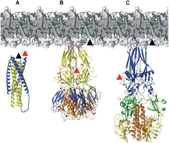
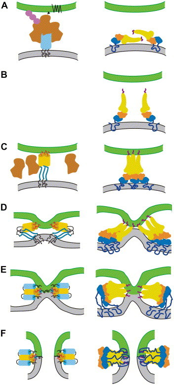
Membrane fusion of enveloped viruses with cellular membranes is mediated by viral glycoproteins (GP). Interaction of GP with cellular receptors alone or coupled to exposure to the acidic environment of endosomes induces extensive conformational changes in the fusion protein which pull two membranes into close enough proximity to trigger bilayer fusion. The refolding process provides the energy for fusion and repositions both membrane anchors, the transmembrane and the fusion peptide regions, at the same end of an elongated hairpin structure in all fusion protein structures known to date. The fusion process follows several lipidic intermediate states, which are generated by the refolding process. Although the major principles of viral fusion are understood, the structures of fusion protein intermediates and their mode of lipid bilayer interaction, the structures and functions of the membrane anchors and the number of fusion proteins required for fusion, necessitate further investigations.


Figures

Fig. 1
Fig. 1
Structural motifs of viral fusion proteins. Ribbon diagrams of representative structures of class I, II and III fusion proteins in their proposed post-fusion conformations positioned with respect to the lipid bilayer. The positions of both membrane anchors at the tip of the elongated structures are indicated by black (fusion peptide, fp) and red (transmembrane, TM) arrows. (A) HIV-1 gp41 core structure; (B) Flavivirus fusion protein E and (C) VSV glycoprotein G. Structural elements, which undergo change from pre-fusion to post-fusion are show in different colors (B and C). The structural changes of gp41 from a pre-fusion to a post-fusion conformation are still unknown.
Fig. 2
Fig. 2
Similar fusion models evolved for class I (left panel) and class II (right panel) fusion proteins. (A) Receptor binding alone (e.g., HIV-1, CD4 and CXCR4 or CCR5) or coupled to endozytosis (e.g., influenza virus HA, TBE E) leads to conformational changes outlined in panels B to F. (B) A transition in oligomeric state is accompanied by fusion peptide target membrane interaction in case of class II. Intermediate monomeric structures have to be also postulated for Rhabdovirus G and paramyxovirus F. Whether they play a role in other class I mediated fusion reactions (influenza virus HA, HIV-gp41) remains to be determined and if so it might be very short lived. (C) Transient intermediate states where the fusion peptide is anchored in the membrane might induce initial curvature. This might involve several fusion proteins, which might cluster via fusion peptide interactions. (D) Initial refolding of the C-terminal region leads to further apposition of the bilayers. Although this step may keep strict trimeric symmetry at the N-terminus, its C-terminal region must be flexible. (E) Final zipping up of the outer layers might induce hemifusion controlled by both membrane anchors. (F) The membrane anchors also play a critical role in fusion pore opening and possibly expansion.

Similar articles

Cited by

References

    1. Skehel J.J., Wiley D.C. Receptor binding and membrane fusion in virus entry: the influenza hemagglutinin. Annu. Rev. Biochem. 2000;69:531–569. - PubMed
    1. Harrison S.C. Mechanism of membrane fusion by viral envelope proteins. Adv. Virus Res. 2005;64:231–261. - PMC - PubMed
    1. Kielian M., Rey F.A. Virus membrane-fusion proteins: more than one way to make a hairpin. Nat. Rev. Microbiol. 2006;4:67–76. - PMC - PubMed
    1. Lamb R.A., Paterson R.G., Jardetzky T.S. Paramyxovirus membrane fusion: lessons from the F and HN atomic structures. Virology. 2006;344:30–37. - PMC - PubMed
    1. Stiasny K., Heinz F.X. Flavivirus membrane fusion. J. Gen. Virol. 2006;87:2755–2766. - PubMed

Publication types