. 2007 Mar;47(7):946-54.
doi: 10.1016/j.visres.2006.12.015. Epub 2007 Feb 22.

Human and macaque pupil responses driven by melanopsin-containing retinal ganglion cells

Affiliations

Human and macaque pupil responses driven by melanopsin-containing retinal ganglion cells

Paul D R Gamlin et al. Vision Res. 2007 Mar.

Abstract

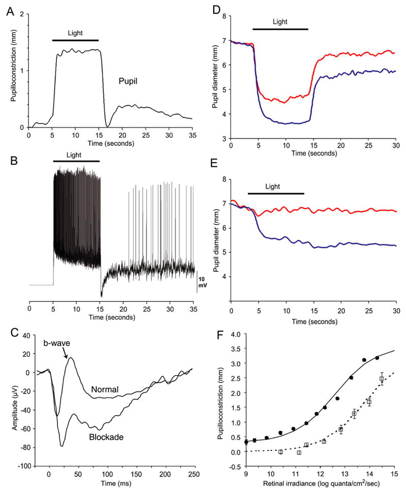
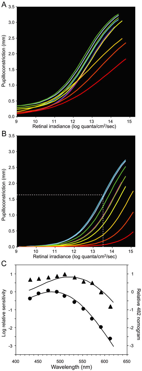
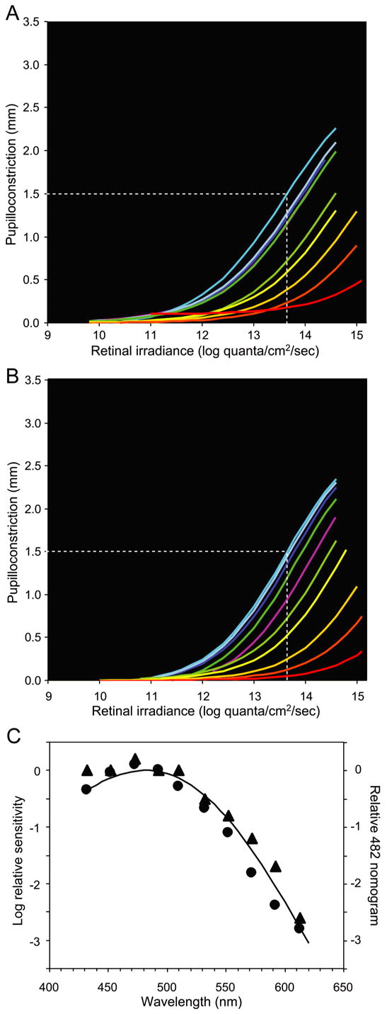
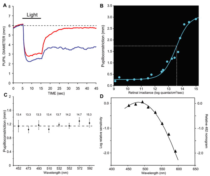
Melanopsin, a novel photopigment, has recently been localized to a population of retinal ganglion cells that display inherent photosensitivity. During continuous light and following light offset, primates are known to exhibit sustained pupilloconstriction responses that resemble closely the photoresponses of intrinsically-photoreceptive ganglion cells. We report that, in the behaving macaque, following pharmacological blockade of conventional photoreceptor signals, significant pupillary responses persist during continuous light and following light offset. These pupil responses display the unique spectral tuning, slow kinetics, and irradiance coding of the sustained, melanopsin-derived ganglion cell photoresponses. We extended our observations to humans by using the sustained pupil response following light offset to document the contribution of these novel ganglion cells to human pupillary responses. Our results indicate that the intrinsic photoresponses of intrinsically-photoreceptive retinal ganglion cells play an important role in the pupillary light reflex and are primarily responsible for the sustained pupilloconstriction that occurs following light offset.


Figures

Figure 1
Figure 1
Pupillary and ganglion cell responses in macaques. (A) Averaged pupillary responses (n=5) showing sustained pupilloconstriction to a 10-second pulse of light (493 nm, 13.3 log quanta/cm2/second). At light OFF, there is a transient pupil dilation followed by a sustained pupilloconstriction. The transient pupil dilation is not always evident, since it depends on prior stimulus conditions and the magntitude of the sustained, post-stimulus pupil constriction. (B) In vitro, intracellular recording from an intrinsically-photoreceptive retinal ganglion cell. For further details see Dacey et al. (2005). A 10-second pulse of light (470 nm, 13.5 log quanta/cm2/second) was presented. Note the similarity between the retinal ganglion cell response and the pupillary response. (C) In vivo, flash electroretinogram under normal conditions (upper trace) and following intravitreal injection of L-AP4/CNQX/D-AP5 (lower trace). The b wave is virtually eliminated following this pharmacological blockade. (D) Normal condition. Averaged responses (n=3) of the pupil to 493 nm light of 14.0 log quanta/cm2/second irradiance (blue trace), and 613 nm light of 14.1 log quanta/cm2/second irradiance (red trace). Following light extinction, there is a pronounced sustained pupilloconstriction that, in this case, masks the transient pupil dilation often seen at light OFF. (E) Pharmacological blockade condition. Averaged responses (n=3) of the pupil to 493 nm light of 14.0 log quanta/cm2/second irradiance (blue trace), and 613 nm light of 14.1 log quanta/cm2/second irradiance (red trace). Note the robust and sustained pupillary responses elicited by light of 493 nm but not of 613 nm. (F) Retinal irradiance-pupillary response plots for 532 nm irradiance for the normal condition (●) and during pharmacological blockade (□) (SEM error bars).
Figure 2
Figure 2
Pupillary responses of macaques during a light stimulus under normal conditions and during pharmacological blockade. (A) Retinal irradiance-pupillary response plots under normal conditions. (B) Retinal irradiance-pupillary response plots after pharmacological blockade. The white dotted line indicates the retinal irradiance at 470 nm required to produce half-maximal pupilloconstriction. In panels A and B, line color represents an approximation of stimulus wavelength. (C) Spectral sensitivity data derived from panels A and B. The data in normal condition (▲) is poorly fit (R2 = 0.77) by a best-fit, Vitamin A1 pigment nomogram (peak sensitivity at 522 nm). The data obtained during pharmacological blockade (●) is well fit (R2 = 0.99) by a Vitamin A1 pigment nomogram with peak sensitivity at 482 nm.
Figure 3
Figure 3
Post-stimulus, sustained pupillary responses of macaques under normal conditions and during pharmacological blockade. (A) Retinal irradiance-pupillary response plots under normal conditions. (B) Retinal irradiance-pupillary response plots after pharmacological blockade. In both A and B, the white dotted line indicates the retinal irradiance at 470 nm required to produce half-maximal pupilloconstriction, line color represents an approximation of stimulus wavelength, and Pmax = 3.0 mm was used for fitting the Hill equations. (C) Spectral sensitivity data derived from panels A and B. The solid curve, a Vitamin A1 pigment nomogram with peak sensitivity at 482 nm, closely matches the data obtained both under normal conditions (▲, R2 = 0.98) (Best fit λmax 483 nm) and during pharmacological blockade (●, R2 = 0.97) (Best fit: λmax 476 nm).
Figure 4
Figure 4
Post-stimulus, sustained pupillary responses in humans. (A) Averaged responses (n=3) of the pupil to 493 nm light of 14.1 log quanta/cm2/second irradiance (blue trace), and 613 nm light of 14.1 log quanta/cm2/second irradiance (red trace). (B) Retinal irradiance-pupillary response plot for 493 nm irradiance. The white dotted line indicates the retinal irradiance required to produce half-maximal pupilloconstriction. (C) Criterion pupil response. Magnitude of the sustained post-stimulus pupillary response after light OFF at eight different wavelengths (SEM error bars). The retinal irradiance used at each wavelength is indicated above the data point. Each stimulus irradiance was calculated to produce the same sustained pupil constriction if these responses were mediated solely by a Vitamin A1 pigment nomogram with a peak sensitivity at 482 nm. (D) Spectral sensitivity data derived from the results shown in C. For each wavelength, the difference between the measured and criterion pupilloconstriction in C was converted into an irradiance difference value based on the irradiance-pupilloconstriction response curve in B. This difference value was then combined with the stimulus irradiance value to determine the retinal irradiance required to produce the criterion pupil response at that wavelength. The solid curve, a Vitamin A1 pigment nomogram with peak sensitivity at 482 nm, closely matches the data (▲, R2 = 0.99).

Similar articles

Cited by

References

    1. Alpern M, Campbell FW. The behaviour of the pupil during dark-adaptation. J Physiol Lond. 1962;165:5–7p.
    1. Alpern M, Ohba N. The effect of bleaching and backgrounds on pupil size. Vision Res. 1972;12:943–951. - PubMed
    1. Baylor DA, Nunn BJ, Schnapf JL. The photocurrent, noise and spectral sensitivity of rods of the monkey Macaca fascicularis. J Physiol. 1984;357:575–607. - PMC - PubMed
    1. Baylor DA, Nunn BJ, Schnapf JL. Spectral sensitivity of cones of the monkey Macaca fascicularis. J Physiol. 1987;390:145–160. - PMC - PubMed
    1. Berson DM, Dunn FA, Takao M. Phototransduction by retinal ganglion cells that set the circadian clock. Science. 2002;295:1070–1073. - PubMed

Publication types

MeSH terms