

doi: 10.1016/j.visres.2007.01.007.
Epub 2007 Feb 22.
The length of Henle fibers in the human retina and a model of ganglion receptive field density in the visual field
Affiliations
- PMID: 17320143
- PMCID: PMC2077907
- DOI: 10.1016/j.visres.2007.01.007
Item in Clipboard
The length of Henle fibers in the human retina and a model of ganglion receptive field density in the visual field
Vision Res.
2007 Oct.
Abstract
An experimental study of lateral displacement of ganglion cells (GCs) from foveal cones in six human retinas is reported. At 406-675 microm in length, as measured in radially oriented cross-sections, Henle fibers are substantially longer than previously reported. However, a new theoretical model indicates that the discrepancies in these reports are mainly due to meridional differences. The model takes into account the effects of optical degradation and peripheral ON/OFF asymmetry and predicts a central GC:cone ratio of 2.24:1. It provides estimates of cumulative counts and GC receptive field density at 0 degrees -30 degrees along the principal meridians of the visual field.
Figures

Histological section, showing the receptoral and post-receptoral components of GC displacement. Glycol methacrylate section, stained methylene blue - azure II, includes the fovea and nasal parafovea from an 82 yr old male (second donor listed in Table 3). A. Low magnification view created by photomontage of multiple higher magnification images. Sites illustrated in panels B,D and C,E are indicated. Bar, located at fovea, 200 μm. B. Site at 1.87 mm eccentricity in the layer of inner segments. Henle fiber length (as traced through zone delimited by red arrowheads) is 0.21 mm. Post-receptoral replacement (as traced through zone delimited by green arrowheads) is zero. The two ends of the displacements were projected onto the external limiting membrane (yellow lines), and lengths were measured along this membrane between the yellow arrows. Bar, 100 μm. C. Site at 0.52 mm eccentricity in the layer of inner segments. Henle fiber is 0.52 mm. Post-receptoral replacement is 0.073 mm, for a total of 0.595 mm. Bar, 100 μm. D, E. Detail of sites shown in B, C, respectively. Contrast in Henle fiber layer is selectively enhanced for illustrative clarity. ONL, outer nuclear layer; HF, Henle fibers; INL, inner nuclear layer; GCL, ganglion cell layer. Inner segment layer is just barely visible at top of panel E. Bars, 20 μm. F. Schematic of Henle fiber layer, viewed from vitreal aspect. The appearance of Henle fibers at section levels pf (parafovea) and f (foveal center) are shown. Fibers at f are mostly long, whereas in parafoveal sections, fibers are shorter. Cross-sectional profiles of those directly superior (or inferior) to the foveal center are circular.

Lateral displacement of GC along the horizontal meridian, including receptoral (fibers of Henle, FH), post-receptoral (bipolar-GC processes, BP-GC) components, and total displacement (Total). Displacements measured for this 82 yr old male donor (second listed in Table 4) were closest to the curve fit to total displacement for all 6 retinas (Fig. 3). The location of a GC nasal or temporal to the foveal center is shown as eccentricity in the GC layer. The distance to a cone inner segment projected along the external limiting membrane (displacement) is shown as a positive value along the y-axis. The bar denotes the approximate position of the optic disk. Lengths are corrected for shrinkage.

Total displacement along the horizontal meridian pooled from 6 human retinas. The coefficients of the best fitting cubic splines (solid lines) for nasal (r2 = 0.89) and temporal (r2 = 0.86) horizontal meridian are given in Table 4. Confidence intervals are shown as dashed lines.

Visual acuity and neural acuity in central vision, according to the model, averaged for principal meridians of the visual field. Experimental studies show inter-individual variation but confirm that the two curves do not differ significantly beyond 5° eccentricity (Green, 1970).
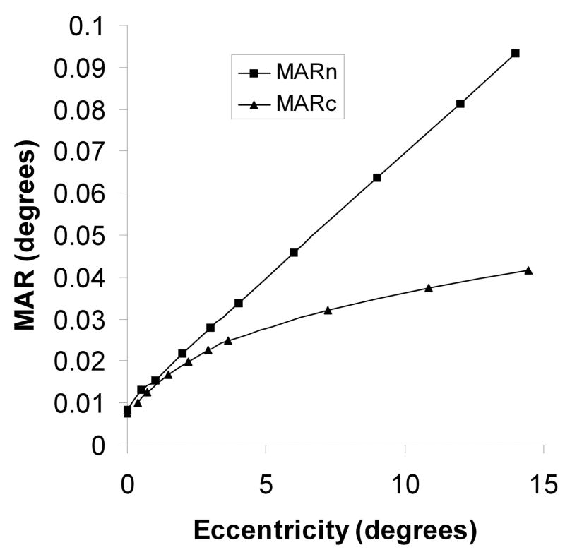
The neural minimum angle of resolution (MARn) in degrees of the eye according to the model. This is assumed to coincide with the MAR of the OFF-midget ganglion cell mosaic. Also shown is the MAR of the cone mosaic (MARc), based on the data of Curcio, Sloan, Kalina and Hendrickson (1990). Data are averages of principal meridians.

The computed mean displacement of ganglion cells from the position of their cone inner segments averaged from principal meridians, according to the model.

The computed mean displacement of ganglion cells from the position of their cone inner segments averaged from principal meridians, as a function of GC soma position.
References
-
- Ahmad KM, Klug K, Herr S, Sterling P, Schein S. Cell density ratios in a foveal patch in macaque retina. Visual Neuroscience. 2003;20:189–209. - PubMed
-
- Azzopardi P, Cowey A. Models of ganglion cell topography in the retina of macaque monkeys and their application to sensory cortical scaling. Neuroscience. 1996;72(3):617–625. - PubMed
-
- Charman WN. Limits on visual performance set by the eye’s optics and the cone mosaic. Ch 17 in Limits of vision. In: Kulikowski JJ, Walsh V, Murray IJ, Cronley Dillon J, editors. Vision and visual dysfunction. Vol. 5. MacMillan; London: 1991. pp. 81–96.
-
- Conradi N, Sjöstrand J. A morphometric and stereologic analysis of ganglion cells of the central human retina. Graefes Archive for Clinical and Experimental Ophthalmology. 1993;231:169–174. - PubMed
Publication types
MeSH terms
Grants and funding
LinkOut - more resources
Full Text Sources
Miscellaneous
