Review
. 2007 Jun;1771(6):746-60.
doi: 10.1016/j.bbalip.2007.01.011. Epub 2007 Jan 24.

Glycolipid transfer proteins

Affiliations
Review

Glycolipid transfer proteins

Rhoderick E Brown et al. Biochim Biophys Acta. 2007 Jun.

Abstract

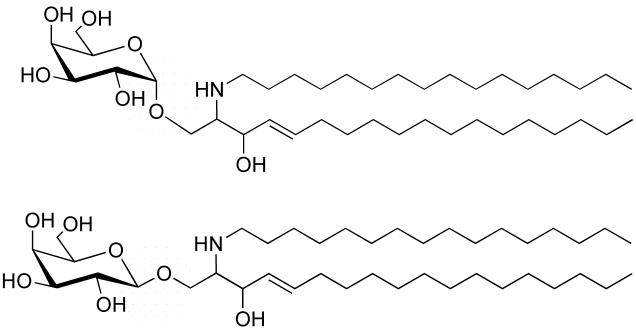
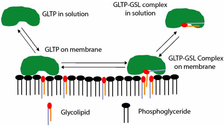
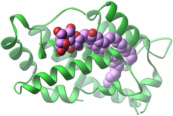
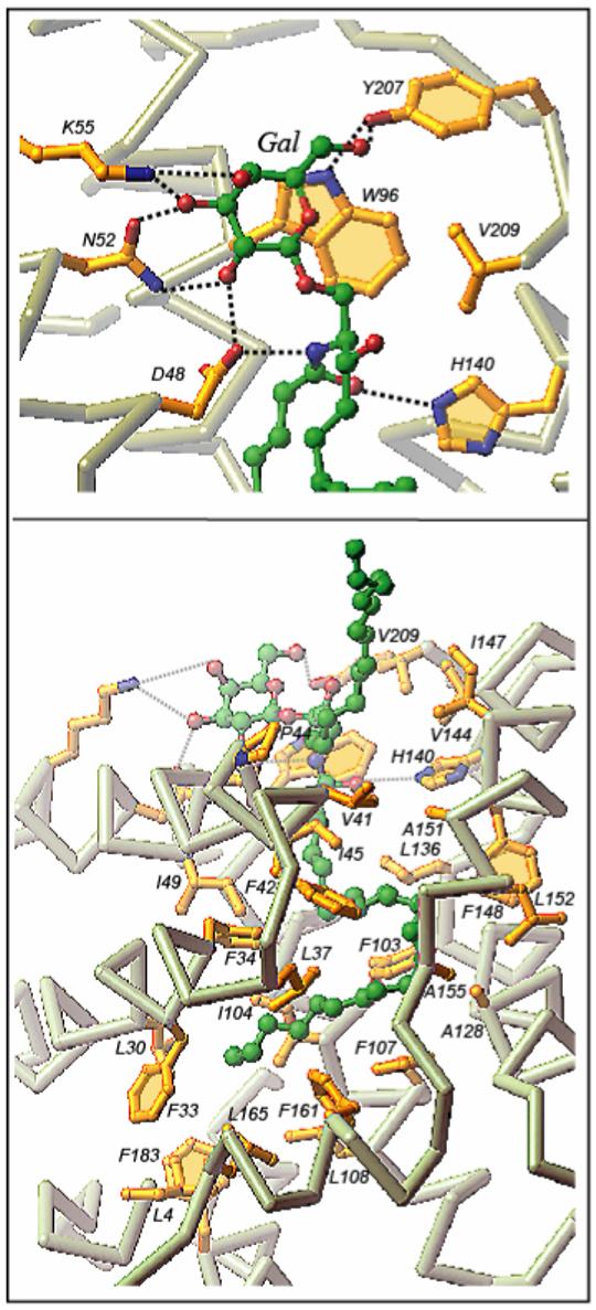
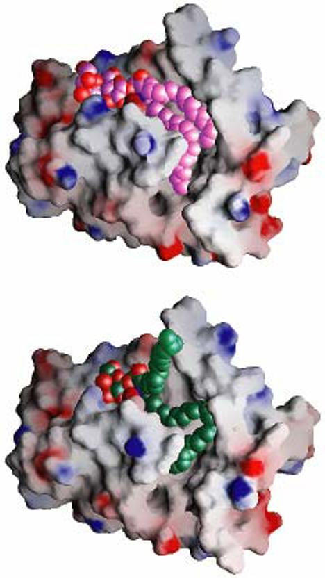
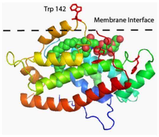
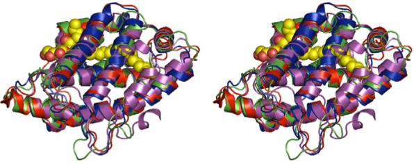
Glycolipid transfer proteins (GLTPs) are small (24 kDa), soluble, ubiquitous proteins characterized by their ability to accelerate the intermembrane transfer of glycolipids in vitro. GLTP specificity encompasses both sphingoid- and glycerol-based glycolipids, but with a strict requirement that the initial sugar residue be beta-linked to the hydrophobic lipid backbone. The 3D architecture of GLTP reveals liganded structures with unique lipid-binding modes. The biochemical properties of GLTP action at the membrane surface have been studied rather comprehensively, but the biological role of GLTP remains enigmatic. What is clear is that GLTP differs distinctly from other known glycolipid-binding proteins, such as nonspecific lipid transfer proteins, lysosomal sphingolipid activator proteins, lectins, lung surfactant proteins as well as other lipid-binding/transfer proteins. Based on the unique conformational architecture that targets GLTP to membranes and enables glycolipid binding, GLTP is now considered the prototypical and founding member of a new protein superfamily in eukaryotes.


Figures

Figure 1
Figure 1
N-Palmitoyl-galactosylceramide showing galactose linked in either alpha (upper) or beta (lower) configuration to the ceramide backbone. While the beta-linked form of GalCer (and GlcCer) are normal GSL constituents of mammals, the alpha-linked forms are not. The alpha-linked form of GalCer is found in Agelas maurifianus, a marine sponge. This glycolipid and its synthetic analog, KRN7000, have been used as specific activators of a subset of Natural Killer Cells known to be involved in the regulation of certain autoimmune diseases [118].
Figure 2
Figure 2
Typical resonance energy transfer assay for monitoring real-time kinetics of GLTP-mediated intervesicular transfer of glycolipid. (Upper panel). In this case, the fluorophore pairs undergoing energy transfer are anthrylvinyl-galactosylceramide (energy donor) and DiO-C16 (energy acceptor) in ethanol (Normalized spectras). (a) Excitation of anthrylvinyl-GalCer (λem=440 nm). (b) Emission of anthrylvinyl-GalCer at λex=370 nm. (c) Excitation of DiO-C16 (λem=580 nm). (d) Emission of DiO-C16 at λex=440 nm. (Lower Panel). Schematic shows the observed signal response during the movement of fluorescently labeled lipid between donor and acceptor bilayer vesicles. Donor vesicles contain a low molar ratio of a fluorescently labeled lipid substrate (i.e. anthrylvinyl-GalCer or BODIPY-glucosylceramide) and a non-transferable energy acceptor or quencher (i.e. perylenoyl-triglyceride or DiO-C16) in a suitable membrane matrix. Acceptor vesicles are usually added in 10-fold excess or more, and the system is allowed to equilibrate. Next, GLTP (1 μg) is added and the change in fluorescence intensity at the RET energy donor fluorescence maxima is recorded. The transfer reaction rate can be calculated from the obtained transfer curve by using the value for total fluorescence intensity obtained after Triton X-100 addition. The detergent causes a complete disruption of the vesicle system.
Figure 3
Figure 3
Mechanism of GLTP-mediated transfer of GSL between membranes in vitro. Glycolipid transfer kinetics, GLTP-membrane partitioning, and GLTP/GSL complex structural measurements support a mechanism in which GLTP acts as a GSL carrier that shuttles between membranes. Because GLTP in a GSL-free state displays a relatively weak protein/membrane-binding propensity [57], GLTP can be expected to readily partition on and off the membrane surface. The rapid lateral diffusions rates of lipids in fluid-phase membranes and the confinement of the glycolipid ligand to a membrane surface will likely enhance the capacity of GLTP to associate with a glycolipid molecule among other membrane lipids. The sugar moiety of the glycolipid acts as a primary specificity determinant, while the ceramide amide functional group orients the entry of the hydrocarbon chain(s) through the cleft-like gate. There is an increased interaction propensity centered about the cleft-like GLTP gate, that is surrounded by aromatic surface residues (e.g. W142, W96, Y153, Y157, Y207, Y81) and a half dozen Lys residues, which are known to interact favorably with membrane interfaces [91,92], thereby potentially facilitating the opening of the gate in the membrane-associated state and entry of the acyl chain into the hydrophobic channel of the GLTP. The accommodation limits of the hydrophobic tunnel, shown by the crystal structures of the GSL-GLTP complexes, strongly suggest that sphingosine is the last lipid part to enter GLTP and, most likely, the first to depart GLTP upon interaction with a membrane. GLTP is shown in green, phospholipids in black, and GSL with a red polar headgroup and the ceramide region made up of a lavender (longer) acyl chain and orange (shorter) sphingoid chain. Note that the depictions detailing the acquisition and liganding of glycolipid by GLTP (right-side of this figure) summarize structural data findings depicted in greater detail in Figures 4, 5, and 6.
Figure 4
Figure 4
Crystal structure of the 18:1 LacCer-GLTP complex. The GLTP is shown in a green ribbon representation. The carbon atoms of the LacCer are shown in a lavender-colored space-filling representation. The red- and blue-colored atoms in LacCer represent oxygen and nitrogen, respectively. Adapted from Figure S1 (panel C) of Malinina et al. [79].
Figure 5
Figure 5
GSL-GLTP interactions in the 24:1 GalCer-GLTP complex. (Upper panel) 24:1 GalCer headgroup (sugar and amide) interactions with GLTP recognition center residues. Hydrogen bonds are shown by black dashed lines. The bound GSL atoms colored green, red and blue represent carbon, oxygen and nitrogen atoms, respectively. The GLTP Cα-backbone is colored light grey, the side chains are shown in gold, and oxygen and nitrogen are in red and blue, respectively. (Lower panel) 24:1 GalCer ceramide chain interactions with the GLTP hydrophobic tunnel residues. The longer acyl chain occupies the tunnel while the shorter sphingosine chain is directed outwards. Reproduced from Figure 2 of Malinina et al. [79].
Figure 6
Figure 6
‘Gate-removed’ electrostatic surface views of the GLTP hydrophobic tunnel accommodating GSLs. The GLTP is shown in an electrostatic surface representation (blue, positive; red, negative; grey, neutral), with gate residues 33 and 35 to 45 removed to make the tunnel and its contents visible. (Upper panel) Structure of the 18:1 LacCer-GLTP complex exhibiting the sphingosine-in mode. The carbon atoms of the 18:1 LacCer are shown in lavender color and space filling representation. Both lipid chains optimally fit into the available space of the hydrophobic tunnel. (Lower panel) Structure of the 24:1 GalCer-GLTP complex exhibiting the sphingosine-out mode. The carbon atoms of the 24:1 GalCer are shown in a green space filling representation. The long acyl chain, bent in a serpentine fashion, occupies the available space of the hydrophobic tunnel, resulting in an outward positioning of the sphingosine chain. Reproduced from Figure 3 of Malinina et al. [79].
Figure 7
Figure 7
Putative membrane interaction region of GLTP, showing the location of the three tryptophan residues (red color). LacCer is shown in space-filling mode with carbon, oxygen, and nitrogen atoms colored green, red, and blue, respectively. The membrane interface is represented by the black dashed line with Trp142 penetrating the membrane surface. The figure was adapted from the Orientations of Proteins in Membranes (OPM) Database website, (http://opm.phar.umich.edu/families.php?family=117), which uses a computational approach to optimize the spatial arrangement of protein structures in lipid bilayers [96,97].
Figure 8
Figure 8
Stereo view of the X-ray structure (1.36 Å) of bovine GLTP (shown in red) with bound ganglioside GM3 (in yellow), superimposed are the structurally modeled HET-C2 homologue (in blue) and the Arabidopsis thaliana GLTP-like protein ACD11 (in green). The crystal structure (PDB 2I3F) of the GLTP-like protein from the algae Galdieria sulphuraria is shown in lavender.

Similar articles

Cited by

References

    1. Metz RJ, Radin NS. Glucosylceramide uptake from spleen cytosol. J. Biol. Chem. 1980;255:4463–4467. - PubMed
    1. Metz RJ, Radin NS. Purification and properties of a cerebroside transfer protein. J. Biol. Chem. 1982;257:12901–12907. - PubMed
    1. Abe A, Yamada K, Sasaki T. A protein purified from pig brain accelerates the intermembranous translocation of mono- and dihexosylceramides, but not the translocation of phospholipids. Biochem. Biophys. Res. Commun. 1982;104:1386–1393. - PubMed
    1. Wong M, Brown RE, Barenholz Y, Thompson TE. Glycolipid transfer protein from bovine brain. Biochemistry. 1984;23:6498–6505. - PubMed
    1. Abe A, Sasaki T. Purification and some properties of the glycolipid transfer protein from pig brain. J. Biol. Chem. 1985;260:11231–11239. - PubMed

Publication types

LinkOut - more resources