

Characterization of circulating osteoblast lineage cells in humans
- PMID: 17320497
- PMCID: PMC1920541
- DOI: 10.1016/j.bone.2006.12.064
Characterization of circulating osteoblast lineage cells in humans
Erratum in
- Bone. 2007 Oct;41(4):741
Abstract
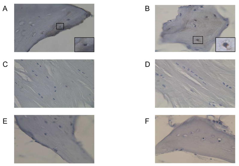
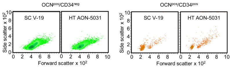
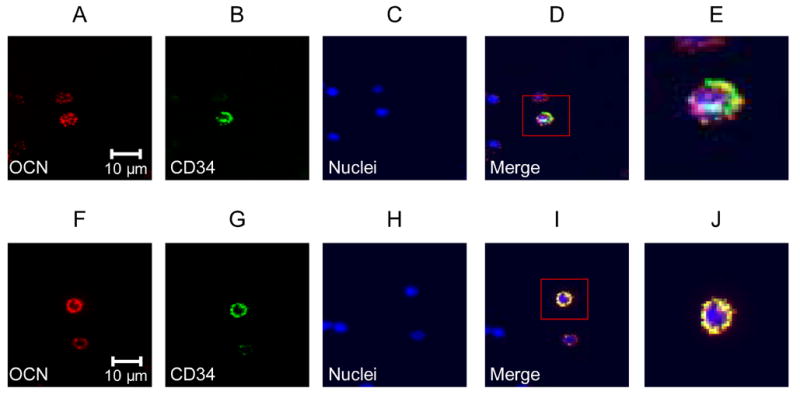
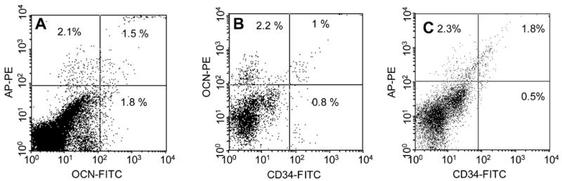
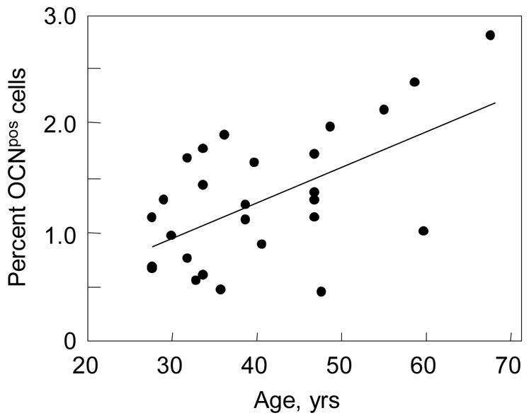
We recently identified circulating osteoblastic cells using antibodies to osteocalcin (OCN) or alkaline phosphatase (AP). We now provide a more detailed characterization of these cells. Specifically, we demonstrate that 46% of OCN positive (OCN(pos)) cells express AP, and 37% also express the hematopoietic/endothelial marker CD34. Using two different anti-OCN antibodies and forward/side light scatter characteristics by flow cytometry, we find that OCN(pos) cells consist of two distinct populations: one population exhibits low forward/side scatter, consistent with a small cell phenotype with low granularity, and a second population has higher forward/side scatter (larger and more granular cell). The smaller, low granularity population also co-expresses CD34, whereas the larger, more granular cells are CD34 negative. Using samples from 26 male subjects aged 28 to 68 years, we demonstrate that the concentration of circulating OCN(pos) cells increases as a function of age (R=0.59, P=0.002). By contrast, CD34(pos) cells tend to decrease with age (R=-0.31, P=0.18); as a consequence, the ratio of OCN(pos):CD34(pos) cells also increase significantly with age (R=0.54, P=0.022). These findings suggest significant overlap between circulating cells expressing OCN and those expressing the hematopoietic/endothelial marker CD34. Further studies are needed to define the precise role of circulating OCN(pos) cells not only in bone remodeling but rather also potentially in the response to vascular injury.
Figures


Similar articles
-
Identification of circulating murine CD34+OCN+ cells.Cytotherapy. 2018 Nov;20(11):1371-1380. doi: 10.1016/j.jcyt.2018.07.004. Epub 2018 Oct 16. Cytotherapy. 2018. PMID: 30340982
-
Circulating immature osteoprogenitor cells and arterial stiffening in postmenopausal osteoporosis.Nutr Metab Cardiovasc Dis. 2011 Sep;21(9):636-42. doi: 10.1016/j.numecd.2010.01.015. Epub 2010 May 31. Nutr Metab Cardiovasc Dis. 2011. PMID: 20554181
-
Human umbilical cord blood-derived CD34-positive endothelial progenitor cells stimulate osteoblastic differentiation of cultured human periosteal-derived osteoblasts.Tissue Eng Part A. 2014 Mar;20(5-6):940-53. doi: 10.1089/ten.TEA.2013.0329. Epub 2013 Dec 3. Tissue Eng Part A. 2014. PMID: 24168264
-
Monoclonal antibodies as tools for studying the osteoblast lineage.Microsc Res Tech. 1996 Feb 1;33(2):128-40. doi: 10.1002/(SICI)1097-0029(19960201)33:2<128::AID-JEMT4>3.0.CO;2-P. Microsc Res Tech. 1996. PMID: 8845513 Review.
-
GPR37 and its neuroprotective mechanisms: bridging osteocalcin signaling and brain function.Front Cell Dev Biol. 2024 Nov 20;12:1510666. doi: 10.3389/fcell.2024.1510666. eCollection 2024. Front Cell Dev Biol. 2024. PMID: 39633709 Free PMC article. Review.
Cited by
-
Circulating osteogenic precursor cells in type 2 diabetes mellitus.J Clin Endocrinol Metab. 2012 Sep;97(9):3240-50. doi: 10.1210/jc.2012-1546. Epub 2012 Jun 27. J Clin Endocrinol Metab. 2012. PMID: 22740707 Free PMC article.
-
Circulating osteogenic precursor cells.Crit Rev Eukaryot Gene Expr. 2010;20(2):171-80. doi: 10.1615/critreveukargeneexpr.v20.i2.70. Crit Rev Eukaryot Gene Expr. 2010. PMID: 21133846 Free PMC article. Review.
-
Journey into Bone Models: A Review.Genes (Basel). 2018 May 10;9(5):247. doi: 10.3390/genes9050247. Genes (Basel). 2018. PMID: 29748516 Free PMC article. Review.
-
Assessment of bone turnover and bone quality in type 2 diabetic bone disease: current concepts and future directions.Bone Res. 2016 Mar 22;4:16001. doi: 10.1038/boneres.2016.1. eCollection 2016. Bone Res. 2016. PMID: 27019762 Free PMC article. Review.
-
Cell replication in craniofacial periosteum: appositional vs. resorptive sites.J Anat. 2011 Mar;218(3):285-97. doi: 10.1111/j.1469-7580.2010.01336.x. Epub 2011 Jan 12. J Anat. 2011. PMID: 21223257 Free PMC article.
References
-
- Friedenstein AJ, Petrakova KV, Kurolesova AI, Frolova GP. Heterotopic of bone marrow. Analysis of precursor cells for osteogenic and hematopoietic tissues. Transplantation. 1968;6:230–247. - PubMed
-
- Price PA, Williamson MK, Lothringer JW. Origin of the vitamin K-dependent bone protein found in plasma and its clearance by kidney and bone. J Biol Chem. 1981;256:12760–12766. - PubMed
Publication types
MeSH terms
Substances
Grants and funding
LinkOut - more resources
Full Text Sources
Other Literature Sources
