. 2007 May 11:1145:28-40.
doi: 10.1016/j.brainres.2007.01.103. Epub 2007 Feb 1.

ATP-sensitive potassium currents reduce the PGE2-mediated enhancement of excitability in adult rat sensory neurons

Affiliations

ATP-sensitive potassium currents reduce the PGE2-mediated enhancement of excitability in adult rat sensory neurons

Xian Xuan Chi et al. Brain Res. .

Abstract

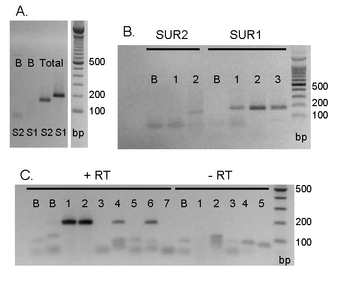
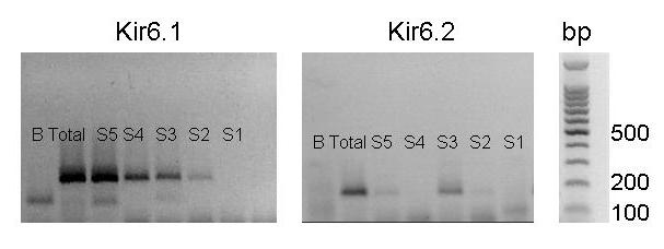
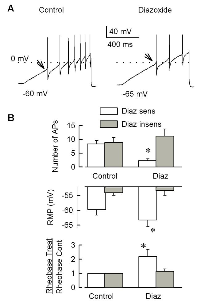
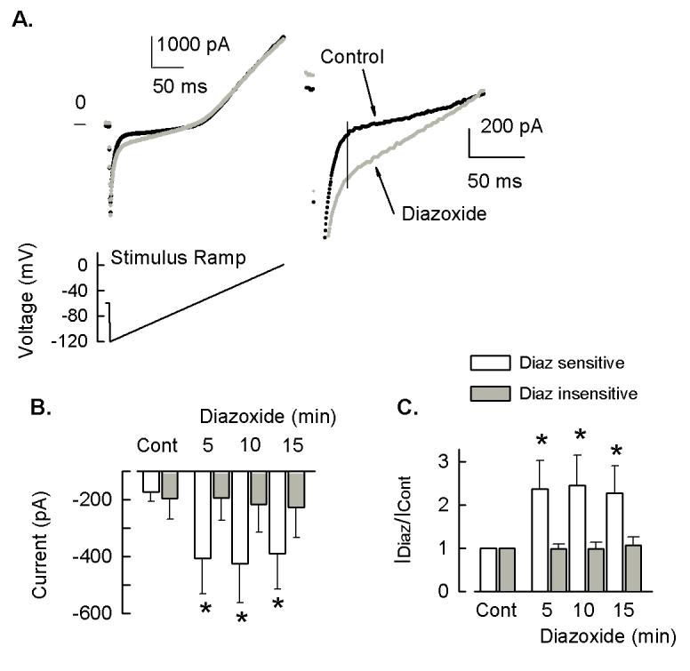
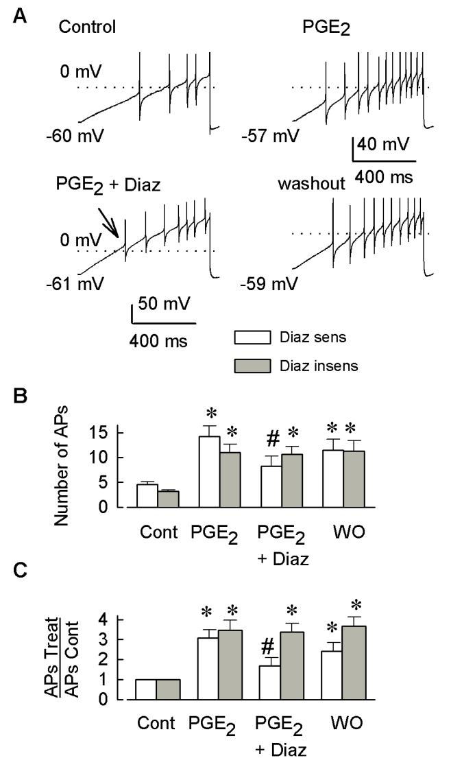
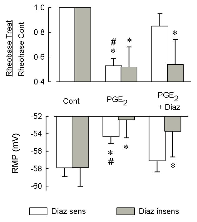
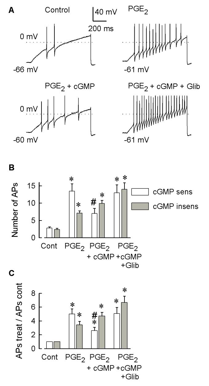
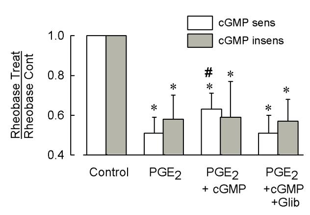
Behavioral studies have shown that the hyperalgesia arising from inflammatory agents, such as prostaglandin E(2) (PGE(2)), can be antagonized by activators of the ATP-sensitive potassium current (K(ATP)). This observation raises questions as to whether this suppression results from a direct action on sensory neurons and what are the cellular mechanisms giving rise to this inhibition. We found that small to medium diameter sensory neurons isolated from the L4-6 DRGs expressed the mRNAs for Kir6.1, Kir6.2, and SUR1. In perforated-patch clamp recordings from acutely dissociated sensory neurons from the young adult rat, exposure to 300 microM diazoxide, a K(ATP) channel agonist, significantly hyperpolarized the resting membrane potential, reduced the number of action potentials evoked by a ramp of depolarizing current, and increased the amplitude of inward K(ATP) currents evoked by the voltage ramp. Similar results were obtained with the protonophore FCCP, which is known to reduce the levels of intracellular ATP and lead to the activation of K(ATP). Only a subpopulation of sensory neurons was sensitive to diazoxide whereas other neurons were unaffected. Treatment with 1 microM PGE(2) significantly enhanced the excitability of these small to medium diameter capsaicin-sensitive sensory neurons; this enhancement was reversed by subsequent exposure to diazoxide in a subpopulation of neurons. Similar to diazoxide, exposure to 8-Br-cyclic GMP antagonized the PGE(2)-induced increase in excitability. The effects of 8-Br-cyclic GMP could be reversed by exposure to glibenclamide, an antagonist of K(ATP) channels. As with diazoxide, only a subpopulation of sensory neurons were affected by 8-Br-cyclic GMP. These results demonstrate that activation of K(ATP) can reverse the sensitization produced by PGE(2) and may be an important means to modulate the enhanced excitability that results from inflammatory or injury conditions.


Figures

Figure 1
Figure 1
Sensory neurons express the mRNA for the SUR subtypes SUR1 and SUR2. Panel A illustrates that both SUR1 (S1) and SUR2 (S2) were detected in a sample from an intact ganglia preparation (labelled total) whereas no PCR products were detected in the blanks (B). Panel B shows that the mRNA for SUR1 was detected in two different samples obtained from two groups of five small to medium diameter sensory neurons (SUR1 lanes 1 and 3). SUR1 was also detected in a sample (lane 2) obtained from a different intact ganglia preparation than shown in panel A. The primer pair for SUR2 did not detect SUR2 in a sample from five small to medium diameter sensory neurons (SUR2 lane 1, same cDNA as used for SUR1 lane 3). However, SUR2 was detected (SUR2 lane 2) in the sample from the same intact ganglia that exhibited SUR1 (SUR1 lane 2). Samples that did not contain any cDNA are shown as the blanks (lanes labelled B). Panel C demonstrates that in the presence of reverse transcriptase (+RT), PCR products for SUR1 were detected in total cDNA from whole ganglia (lanes 1 and 2) and from two (lanes 4 and 6) single cell RT-PCR samples but not in three others (lanes 3, 5, and 7). In the absence of reverse transcriptase (−RT), no products were detected for either total cDNA (lanes 1 and 2) or for single cells (lanes 3-5). No products were detected in the blank samples (lanes labelled B). For all panels, the base pair ladder (labelled bp) is shown on the right.
Figure 2
Figure 2
Sensory neurons express the mRNA for the KATP channel subtypes Kir6.1 and Kir6.2. The left panel shows the mRNA for Kir6.1 in individual single cells (labeled S1, S2, S3, S4 and S5) and an intact ganglia preparation (labeled total). The middle panel shows the mRNA for Kir6.2 in individual single cells (labeled S1, S2, S3, S4 and S5) and in the same intact ganglia preparation as shown in the left panel (labeled total). For all panels, the base pair ladder (labeled bp) is shown on the right. No PCR products were detected in the no template control (lanes labeled B).
Figure 3
Figure 3
Diazoxide, a KATP channel opener, reduces the excitability of small to medium diameter capsaicin-sensitive sensory neurons. Panel A illustrates a current clamp recording from a representative neuron wherein the ramp of depolarizing current elicits 7 APs whereas after a 5 min exposure to 300 μM diazoxide the number is reduced to 3. Note that the resting membrane potential hyperpolarizes by 5 mV after diazoxide. The dotted line represents the level of 0 mV. The panels shown in B summarize the results obtained for those neurons that were either sensitive (n=8) or insensitive (n=9) to diazoxide. The top panel represents the effects of a 5 min exposure to 300 μM diazoxide on the number of APs evoked by the ramp. After treatment 3 of 8, diazoxide-sensitive neurons did not fire any APs (see Results). The middle panel demonstrates that diazoxide hyperpolarizes the resting membrane potential in those sensitive neurons. The bottom panel shows the effects of diazoxide on the normalized rheobase where the rheobase after diazoxide is divided by their respective control values. The asterisks indicate a significant difference from the control values (p<0.05).
Figure 4
Figure 4
Diazoxide increases the inward KATP current in small to medium diameter capsaicin-sensitive sensory neurons. Top left panel in A shows an example of the inward current evoked by the voltage ramp (left bottom panel in A) from a representative neuron for the control and after a 5 min exposure to 300 μM diazoxide. This enhancement of the current by diazoxide is shown at greater resolution in the right panel of A . Panel B summarizes the amplitude of the currents obtained for those neurons that were either sensitive (n=9) or insensitive (n=6) to diazoxide measured at −110 mV. Panel C illustrates the results obtained with diazoxide for these same neurons when the current values have been normalized to their respective control values measured at −110 mV. The asterisks indicate a significant difference from the control values (p<0.05).
Figure 5
Figure 5
Diazoxide reverses the sensitization produced by PGE2. The top left panel of A shows a current-clamp recording from a representative small to medium diameter capsaicin-sensitive sensory neuron. A ramp of depolarizing current (1 s duration, 5000 pA final amplitude) was used to evoke APs under control conditions. The top right panel demonstrates the increase in the number of evoked APs after a 5 min treatment with 1 μM PGE2. The bottom left panel shows the reduction in the number of APs after a 5 min exposure to 300 μM diazoxide (Diaz) in the presence of PGE2. The bottom right panel demonstrates that a 5 min washout with normal Ringers can reverse the reduction in APs produced by diazoxide. The numbers to the left of each trace represent the resting membrane potential; the dotted lines indicate the 0 mV level; the calibration scales in the middle is for the top and bottom right traces whereas the scale to the left is for the bottom left panel only. The effects of these different treatments on the number of evoked APs are summarized in panel B. Panel C illustrates the effects of these treatment conditions after the number of APs have been normalized to that obtained for each neuron under their respective control conditions. The asterisks represent a significant difference between the values obtained under control conditions and after a particular treatment (p<0.05, RM ANOVA). The # represents a significant difference between the PGE2 and PGE2 plus diazoxide treatments (p<0.05 RM ANOVA).
Figure 6
Figure 6
Diazoxide reverses the PGE2-induced increase in excitability. The top panel shows that diazoxide decreases the rheobase after diazoxide when normalized to their respective control values. Sensory neurons were sequentially exposed to 1 μM PGE2 for 5 min, then 1 μM PGE2 plus 300 μM diazoxide. The bottom panel shows that diazoxide reverses the PGE2-induced depolarization of the resting membrane potential (RMP). The asterisks represent a significant difference between the values obtained under control conditions and after a particular treatment (p<0.05, RM ANOVA). The # represents a significant difference between the PGE2 and PGE2 plus diazoxide treatments (p<0.05 RM ANOVA).
Figure 7
Figure 7
In a subpopulation of sensory neurons, 8-Br-cGMP reverses the sensitization produced by PGE2; subsequent treatment with glibenclamide can reverse the suppressive effects of 8-Br-cGMP. Panel A illustrates, in a representative neuron, that under control conditions the depolarizing ramp evoked 2 APs (top left); a 5 min exposure to 1 μM PGE2 produced sensitization as indicated by the increased number of APs (top right). Treatment with 100 μM 8-Br-cGMP (5 min) reduced the increase in the number of APs produced by PGE2 (bottom left), but only in a subpopulation of sensory neurons. Subsequent treatment with 20 μM glibenclamide for 5 min reverses the suppressive effects of 8-BrcGMP (bottom right panel). APs were evoked by a ramp of current that was 1 sec in duration with a final amplitude of 5000 pA. Each dotted line indicates 0 mV. The values at the beginning of each trace represent the resting membrane potential. Panel B summarizes the effects of sequential treatment of sensory neurons with PGE2, PGE2 + 8-Br-cGMP, and finally PGE2 + 8-Br-cGMP + glibenclamide on the number of APs evoked by the ramp. Panel C represents the effects for the different treatment conditions after they have been normalized to their respective control responses. Asterisks represent a significant difference between values obtained under control and the different treatment conditions (p<0.05, RM ANOVA). The # represents a significant difference between the values obtained for the PGE2 + 8-Br-cGMP condition vs. PGE2 alone or the PGE2 + 8-Br-cGMP + glibenclamide (p<0.05, RM ANOVA).
Figure 8
Figure 8
8-Br-cGMP reverses the PGE2-induced decrease in rheobase of neurons. These results were obtained from those same neurons represented in Fig. 7. Asterisks represent a significant difference between values obtained under control and the different treatment conditions (p<0.05, RM ANOVA). The # represents a significant difference between the values obtained for the PGE2 + 8-Br-cGMP condition vs. PGE2 alone or the PGE2 + 8-Br-cGMP + glibenclamide (p<0.05, RM ANOVA).

Similar articles

Cited by

References

    1. Aguilar-Bryan L, Clement JP, 4th, Gonzalez G, Kunjilwar K, Babenko A, Bryan J. Toward understanding the assembly and structure of KATP channels. Physiol. Rev. 1998;78:227–245. - PubMed
    1. Aley KO, McCarter G, Levine JD. Nitric oxide signaling in pain and nociceptor sensitization in the rat. J. Neurosci. 1998;18:7008–7014. - PMC - PubMed
    1. Allen TGJ, Brown DA. Modulation of the excitability of cholinergic basal forebrain neurones by KATP channels. J. Physiol. 2004;554:353–370. - PMC - PubMed
    1. Alves D, Duarte I. Involvement of ATP-sensitive K+ channels in the peripheral antinociceptive effect induced by dipyrone. Eur. J. Pharmacol. 2002;444:47–52. - PubMed
    1. Ammala C, Moorhouse A, Ashcroft FM. The sulphonylurea receptor confers diazoxide sensitivity on the inwardly rectifying K+ channel Kir6.1 expressed in human embryonic kidney cells. J. Physiol. 1996;494:709–714. - PMC - PubMed

Publication types

MeSH terms