. 2007 Apr 15;304(2):722-34.
doi: 10.1016/j.ydbio.2007.01.022. Epub 2007 Feb 7.

Hedgehog signaling regulates the amount of hypaxial muscle development during Xenopus myogenesis

Affiliations

Hedgehog signaling regulates the amount of hypaxial muscle development during Xenopus myogenesis

Benjamin L Martin et al. Dev Biol. .

Abstract

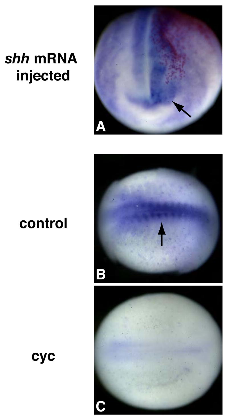
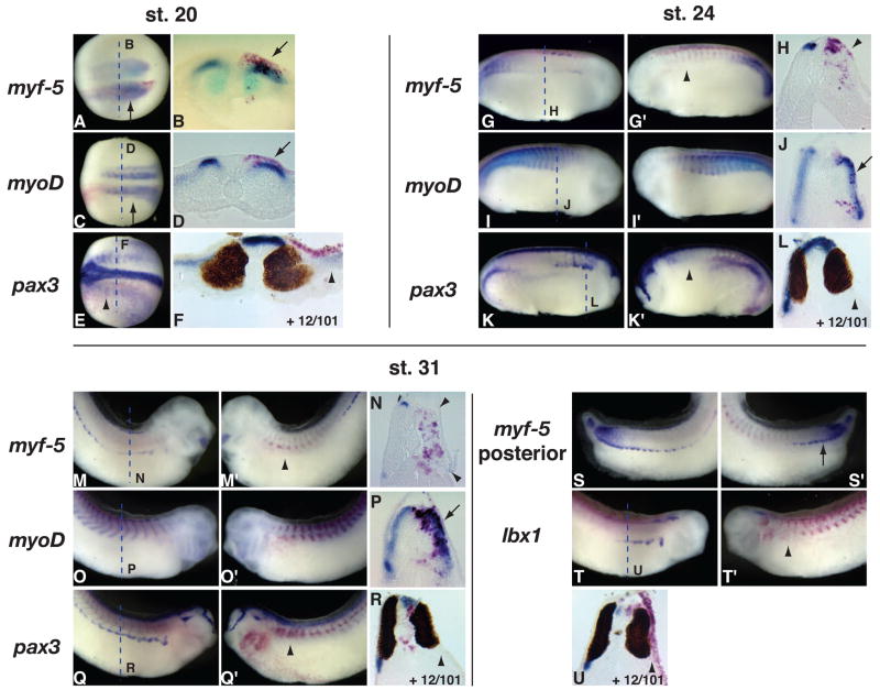
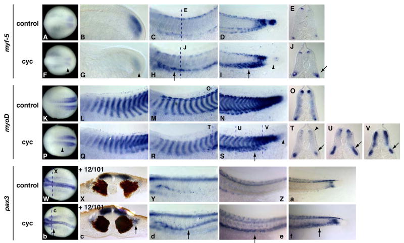
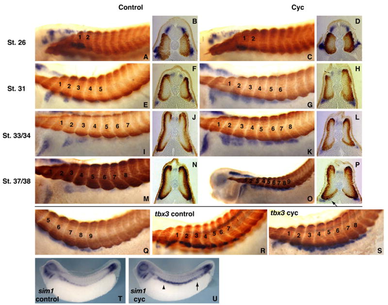
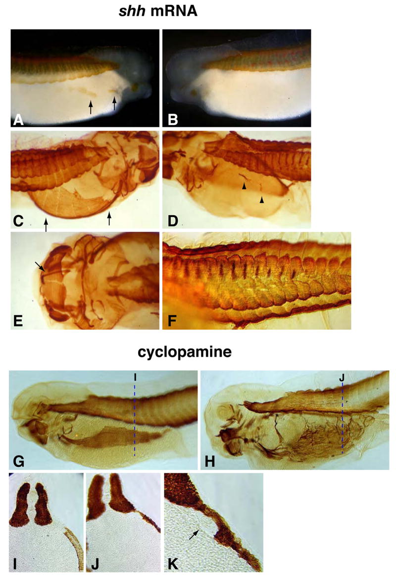
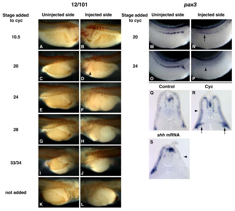
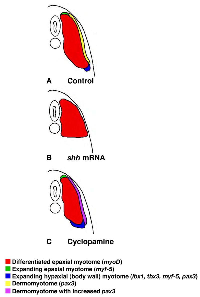
Hedgehog (Hh) signaling is proposed to have different roles on differentiation of hypaxial myoblasts of amniotes. Within the somitic environment, Hh signals restrict hypaxial development and promote epaxial muscle formation. On the other hand, in the limb bud, Hh signaling represses hypaxial myoblast differentiation. This poses the question of whether differences in response to Hh signaling are due to variations in local environment or are intrinsic differences between pre- and post-migratory hypaxial myoblasts. We have approached this question by examining the role of Hh signaling on myoblast development in Xenopus laevis, which, due to its unique mode of hypaxial muscle development, allows us to examine myoblast development in vivo in the absence of the limb environment. Cyclopamine and sonic hedgehog (shh) mRNA overexpression were used to inhibit or activate the Hh pathway, respectively. We find that hypaxial myoblasts respond similarly to Hh manipulations regardless of their location, and that this response is the same for epaxial myoblasts. Overexpression of shh mRNA causes a premature differentiation of the dermomyotome, subsequently inhibiting all further growth of the epaxial and hypaxial myotome. Cyclopamine treatment has the opposite effect, causing an increase in dermomyotome and a shift in myoblast fate from epaxial to hypaxial, eventually leading to an excess of hypaxial body wall muscle. Cyclopamine treatment before stage 20 can rescue the effects of shh overexpression, indicating that early Hh signaling plays an essential role in maintaining the balance between epaxial and hypaxial muscle mass. After stage 20, the premature differentiation of the dermomyotome caused by shh overexpression cannot be rescued by cyclopamine, and no further embryonic muscle growth occurs.


Figures

Figure 1
Figure 1. Schematic illustrating early myotome growth in anterior-most somites
At stage 20, anterior-most somites that will contribute to the hypaxial body wall muscle have already undergone an initial phase of myf-5 expression and have differentiated to form the primary epaxial myotome (A). Subsequent expansion of the epaxial myotome occurs in the dorsal and ventral domains of the somites (B). At later stages, the ventral region contributes myoblasts to the hypaxial body wall muscles (C).
Figure 2
Figure 2. Hh manipulations affect patched expression
A stage 20 embryo that was injected in one cell at the 2-cell stage with shh mRNA exhibits increased ptc-2 expression on the injected side (A, anterior view, arrow). A stage 20 control treated embryo shows normal ptc-2 expression in the somites (B, arrow), which is lost in a cyclopamine treated sibling (C).
Figure 3
Figure 3. Shh over-expression promotes initial epaxial muscle differentiation but inhibits markers of epaxial myotome expansion and hypaxial body wall myoblasts
Shh mRNA was injected into one cell at the 2-cell stage. Embryos are pictured as dorsal views with anterior to the left and the injected side down (A, C, E), transverse sections with the injected side on the right (B, D, F, H, J, L, N, P, R, U), or lateral views with the uninjected side facing right and injected side facing left (all other panels). Dashed lines indicate the approximate positions of the transverse sections. Red staining represents lineage tracing of co-injected beta-galactosidase mRNA. At stage 20, there is an increase of myf-5 (A, B, arrows, A shown from a posterior vantage point) and myoD (C, D, arrows) and a decrease of pax3 (E, F, arrowheads) expression on the injected side of the embryo. At stage 24 and 31, an increase of myoD expression on the injected side is still observed (I–J, O–P, arrows). The expression of myf-5 in the expanding dorsal and ventral region of the myotome is lost on the injected side (G–H, M–N, arrowheads), but expression in posterior somites where initial myogenesis is occurring is normal (S, S’, arrow). pax3 and lbx1 expression are also lost on the injected side (K–L, Q–R, T–U, arrowheads). Transverse sections of pax3 and lbx1 expression are co-labeled with the 12/101 antibody. At stage 20, there is no significant difference in differentiated muscle on the injected side of the embryo (F). At stages 24 and 31, a wider, shorter amount of differentiated muscle is observed on the injected side (L, R, U).
Figure 4
Figure 4. – Cyclopamine treatment elicits the opposite effect of shh overexpression
Stage 20, 24, 31, and 33/34 cyclopamine treated embryos were compared to control embryos for myf-5, myoD, and pax3 expression. At stage 20, myf-5 (A, F) and myoD (K, P) expression are reduced in cyclopamine treated embryos (F, P, arrowheads), while pax3 (W, X, b, c) is upregulated (b, c, arrows). This is especially evident in transverse sections, where pax3 expression is stronger along the lateral surface of the differentiated myotome in cyclopamine treated embryos (c, arrow) than in controls (X). At stage 24, myf-5 (B, G) is significantly reduced in the posterior of a cyclopamine treated embryo (G, arrowhead) when compared to a control (B). At this stage, myoD (L, Q) is reduced in the anterior-posterior extent of expression in anterior somites in cyclopamine treated embryos (Q). Also at stage 24, pax3 expression (Y, d) is strongly upregulated in the ventral region of somites in cyclopamine treated embryos (e, arrow). By stage 31, myf-5 expression (C–E, H–J) is strongly upregulated in the ventral domain of somites in cyclopamine treated embryos (H–J, arrows), while initial expression in forming somites is still lost (I, arrowhead). The expression of myoD is similarly upregulated in the ventral region of somites in cyclopamine treated embryos (R–V, arrows), particularly in posterior somites (S, arrow), and lost in the newly formed somites of the tailbud (S, arrowhead). At stage 33/34, the expression of pax3 (Z-a, e–f) remains upregulated in the ventral regions of somites in cyclopamine treated embryos, in both the anterior (e compared to Z, arrow) and posterior (f compared to a, arrow).
Figure 5
Figure 5. Cyclopamine treatment expands hypaxial specific markers medially and posteriorly
The expression of lbx1 (A–Q), tbx3 (R, S), and sim1 (T, U) were examined in control (A, B, E, F, I, J, M, N, R, T) and cyclopamine treated (C, D, G, H, K, L, O, P, Q, S, U) embryos. Trunk somites in lbx1 and tbx3 probed embryos are labeled with numbers, with the most anterior trunk somite labeled 1. At stage 26 there are no significant differences in lbx1 expression between control (A, B) and cyclopamine treated embryos (C, D). At stages 31 (E–H), 33/34 (I–L), and 37/38 (M–Q), lbx1 expression is expanded one somite posterior compared to controls, and is expanded medially towards the notochord (P, arrow, compare to N). The image in Q is a higher power image of the embryo in O. The expression of tbx3 is also expanded one somite posteriorly in cyclopamine treated embryos (S compared to R). The expansion of the hypaxial dermomyotome marker sim1 is also observed in stage 32 cyclopamine treated embryos (U) in both the anterior somites where it is normally expressed (arrowhead) and in the posterior somites (arrow), as compared to a control embryo (T).
Figure 6
Figure 6. Hh effects on differentiated muscle are consistent with earlier effects on gene markers
Embryos were injected in one cell at the 2-cell stage with shh mRNA (A–F) or treated with cyclopamine (G–K) and stained with 12/101. At stage 37 (A, B), hypaxial muscles are present on the uninjected side of the embryo (A, arrows), but are completely absent on the injected side (B). At stage 42, the hypaxial muscles have grown considerably on the uninjected side (C, arrows), but are still absent on the injected side except for very small muscle remnants (D, arrowheads). A ventral view of a stage 42 head shows the presence of the somite derived geniohyoideus muscle on the uninjected side (E, arrow), which is absent on the injected side (E). Tail epaxial myotomes of a stage 42 embryo are considerably shorter on the injected side (F, foreground) than on the uninjected side (F, background). Cyclopamine treatment causes and excess of hypaxial muscle at stage 39 (H) relative to a control embryo (G). Dashed lines indicate the position of transverse sections shown in I and J. Transverse sections of the embryos pictured in H and G indicate that the hypaxial and epaxial myotomes are continuous in cyclopamine treated embryos (J), whereas they are not in control embryos (I). Differentiated muscle lies adjacent to the pronephric duct in cyclopamine treated embryos (K, arrow).
Figure 7
Figure 7. Dermomyotome and hypaxial muscles cannot be rescued after premature differentiation of myoblasts
Embryos were injected in one cell at the 2-cell stage with shh mRNA and subsequently added to cyclopamine at stages 10.5 (A, B), 20 (C, D, M, N), 24 (E, F, O, P), 28 (G, H), 33/34 (I, J), never added to cyclopamine (K, L, S), added to cyclopamine but not injected with shh (R), or not treated (Q). Embryos were fixed at stage 42 (A–L), 31 (M–P), and 33/34 (Q–S). Embryos fixed at stage 42 were examined with the 12/101 antibody, while the rest were probed for pax3 expression. When injected embryos are added to cyclopamine at stage 10.5, there is no difference in hypaxial muscle between the injected and uninjected side (B compared to A). When added at stage 20, hypaxial muscles are normal except for in the most anterior, where they are missing (D, arrowhead). When added to cyclopamine at stage 24 and beyond, or not added at all, hypaxial muscles are missing on the injected side (F compared to E, H to G, J to I, L to K). When injected embryos are added to cyclopamine at stage 20 and fixed at stage 31, there is some recovery of pax3 expression on the injected side (N compared to M, arrow), while those added at stage 24 do not exhibit any pax3 recovery (P compared to O, arrowhead). A transverse section through a stage 33/34 embryo that has only been treated with cyclopamine and stained for pax3 expression (R), exhibits a medial expansion of pax3 at the ventral edge of the somite (arrow) and an increase in pax3 expression along the lateral edge of the somite (arrowhead) as compared to a control embryo (Q). A transverse section of a stage 33/34 embryo that has been injected with shh mRNA in one cell at the 2-cell stage shows a complete loss of pax3 expression in the somite on the injected side (S, arrowhead).
Figure 8
Figure 8

Similar articles

Cited by

References

    1. Amthor H, Christ B, Patel K. A molecular mechanism enabling continuous embryonic muscle growth - a balance between proliferation and differentiation. Development. 1999;126:1041–53. - PubMed
    1. Amthor H, Christ B, Weil M, Patel K. The importance of timing differentiation during limb muscle development. Curr Biol. 1998;8:642–52. - PubMed
    1. Bober E, Franz T, Arnold HH, Gruss P, Tremblay P. Pax-3 is required for the development of limb muscles: a possible role for the migration of dermomyotomal muscle progenitor cells. Development. 1994;120:603–12. - PubMed
    1. Borycki AG, Brunk B, Tajbakhsh S, Buckingham M, Chiang C, Emerson CP., Jr Sonic hedgehog controls epaxial muscle determination through Myf5 activation. Development. 1999;126:4053–63. - PubMed
    1. Brand-Saberi B, Christ B. Evolution and development of distinct cell lineages derived from somites. Curr Top Dev Biol. 2000;48:1–42. - PubMed

Publication types

LinkOut - more resources