

Vascular dementia in leukoaraiosis may be a consequence of capillary loss not only in the lesions, but in normal-appearing white matter and cortex as well
- PMID: 17320909
- PMCID: PMC1989116
- DOI: 10.1016/j.jns.2007.01.015
Vascular dementia in leukoaraiosis may be a consequence of capillary loss not only in the lesions, but in normal-appearing white matter and cortex as well
Abstract
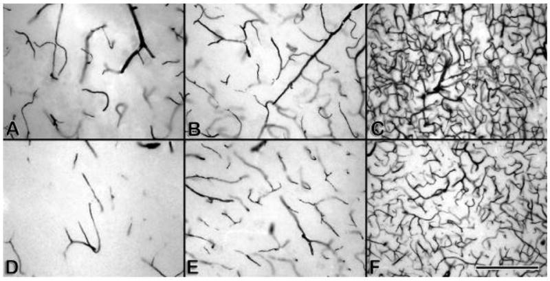
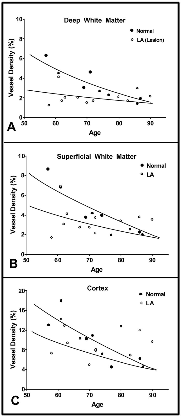
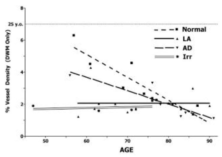
We investigated capillary density in 12 subjects with leukoaraiosis (LA), in 9 age-matched normal subjects, in 7 cases of Alzheimer's disease (AD), and 4 after whole-brain irradiation for brain tumors. In the LA study (which as been published), autopsy brains were evaluated by MRI. The presence of LA was indicated by confluent or patchy areas of hyperintensity in the deep white matter. We employed a stereology method using computerized image processing and analysis to determine microvascular density. Afferent vessels (arterioles and capillaries, but not veins or venules) were stained for alkaline phosphatase in 100 microm thick celloidin sections. Microvascular density in LA lesions in the deep white matter (2.56%) was significantly lower than in the corresponding deep white matter of normal subjects (3.20%, p=0.0180). LA subjects demonstrated decreased vascular density at early ages (55-65 years) when compared to normal subjects. Our findings indicate that LA affects the brain globally, with capillary loss, although the parenchymal damage is found primarily in the deep white matter. In ongoing studies of the deep white matter in AD brains, we found a pattern of decreased vascular density compared to normal, as well as an age-related decline. In the four irradiated brains, we found very low vessel densities, similar to those found in LA, without an additional age-related decline.
Figures
Similar articles
-
Microvascular changes in the white mater in dementia.J Neurol Sci. 2009 Aug 15;283(1-2):28-31. doi: 10.1016/j.jns.2009.02.328. Epub 2009 Mar 5. J Neurol Sci. 2009. PMID: 19268311 Free PMC article.
-
Quantification of afferent vessels shows reduced brain vascular density in subjects with leukoaraiosis.Radiology. 2004 Dec;233(3):883-90. doi: 10.1148/radiol.2333020981. Radiology. 2004. PMID: 15564412
-
Review: cerebral microvascular pathology in ageing and neurodegeneration.Neuropathol Appl Neurobiol. 2011 Feb;37(1):56-74. doi: 10.1111/j.1365-2990.2010.01139.x. Neuropathol Appl Neurobiol. 2011. PMID: 20946471 Free PMC article. Review.
-
Morphometric analysis of arteriolar tortuosity in human cerebral white matter of preterm, young, and aged subjects.J Neuropathol Exp Neurol. 2007 May;66(5):337-45. doi: 10.1097/nen.0b013e3180537147. J Neuropathol Exp Neurol. 2007. PMID: 17483690
-
Sorting out the clinical consequences of ischemic lesions in brain aging: a clinicopathological approach.J Neurol Sci. 2007 Jun 15;257(1-2):17-22. doi: 10.1016/j.jns.2007.01.020. Epub 2007 Feb 23. J Neurol Sci. 2007. PMID: 17321551 Review.
Cited by
-
Imaging of Angiogenesis in White Matter Hyperintensities.J Am Heart Assoc. 2023 Nov 7;12(21):e028569. doi: 10.1161/JAHA.122.028569. Epub 2023 Oct 27. J Am Heart Assoc. 2023. PMID: 37889177 Free PMC article.
-
Development of capillary dysfunction in Alzheimer's disease.Front Aging Neurosci. 2024 Aug 29;16:1458455. doi: 10.3389/fnagi.2024.1458455. eCollection 2024. Front Aging Neurosci. 2024. PMID: 39267722 Free PMC article. Review.
-
Assessment of microvascular rarefaction in human brain disorders using physiological magnetic resonance imaging.J Cereb Blood Flow Metab. 2022 May;42(5):718-737. doi: 10.1177/0271678X221076557. Epub 2022 Jan 26. J Cereb Blood Flow Metab. 2022. PMID: 35078344 Free PMC article.
-
The effect of exercise on the cerebral vasculature of healthy aged subjects as visualized by MR angiography.AJNR Am J Neuroradiol. 2009 Nov;30(10):1857-63. doi: 10.3174/ajnr.A1695. Epub 2009 Jul 9. AJNR Am J Neuroradiol. 2009. PMID: 19589885 Free PMC article.
-
The science of stroke: mechanisms in search of treatments.Neuron. 2010 Jul 29;67(2):181-98. doi: 10.1016/j.neuron.2010.07.002. Neuron. 2010. PMID: 20670828 Free PMC article. Review.
References
-
- Hachinski VC, Potter P, Merskey H. Leuko-araiosis. Arch Neurol. 1987;44:21–3. - PubMed
-
- Munoz DG, Hastak SM, Harper B, Lee D, Hachinski VC. Pathologic correlates of increased signals of the centrum ovale on magnetic resonance imaging. Arch Neurol. 1993;50:492–7. - PubMed
-
- Pantoni L, Garcia JH. The significance of cerebral white matter abnormalities 100 years after Binswanger’s report. A review Stroke. 1995;26:1293–301. - PubMed
-
- Pantoni L, Garcia JH. Pathogenesis of leukoaraiosis: a review. Stroke. 1997;28:652–9. - PubMed
Publication types
MeSH terms
Substances
Grants and funding
LinkOut - more resources
Full Text Sources
