Review
. 2007 Jun 15;462(2):176-88.
doi: 10.1016/j.abb.2007.01.020. Epub 2007 Feb 8.

Cell shrinkage and monovalent cation fluxes: role in apoptosis

Affiliations
Review

Cell shrinkage and monovalent cation fluxes: role in apoptosis

Carl D Bortner et al. Arch Biochem Biophys. .

Abstract

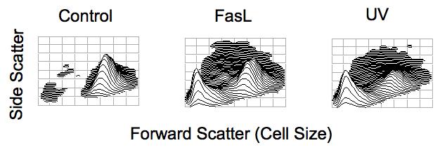
The loss of cell volume or cell shrinkage has been a morphological hallmark of the programmed cell death process known as apoptosis. This isotonic loss of cell volume has recently been term apoptotic volume decrease or AVD to distinguish it from inherent volume regulatory responses that occurs in cells under anisotonic conditions. Recent studies examining the intracellular signaling pathways that result in this unique cellular characteristic have determined that a fundamental movement of ions, particularly monovalent ions, underlie the AVD process and plays an important role on controlling the cell death process. An efflux of intracellular potassium was shown to be a critical aspect of the AVD process, as preventing this ion loss could protect cells from apoptosis. However, potassium plays a complex role as a loss of intracellular potassium has also been shown to be beneficial to the health of the cell. Additionally, the mechanisms that a cell employs to achieve this loss of intracellular potassium vary depending on the cell type and stimulus used to induce apoptosis, suggesting multiple ways exist to accomplish the same goal of AVD. Additionally, sodium and chloride have been shown to play a vital role during cell death in both the signaling and control of AVD in various apoptotic model systems. This review examines the relationship between this morphological change and intracellular monovalent ions during apoptosis.


Figures

Fig. 1
Fig. 1
Apoptotic volume decrease (AVD) as observed by changes in the light scattering properties of a cell during apoptosis. Jurkat T-cells were treated with 25 ng/ml Fas Ligand or 30 mJ/cm2 UV for 4 hours. Cells were examined for changes in their light scattering properties by flow cytometry. Forward scattered light is a direct measure of cell size, with cells having a high forward scatter value being of a larger cell size, while cells having a lower forward scatter value are of a smaller cell size. Side scatter is measure of cellular granularity or complexity, with cells having a higher side scatter value being more granular. Upon apoptotic stimulation, a decrease in the cells ability to scatter light in the forward direction, along with a transient increase in the cells ability to scatter light at a side angle is observed indicating a loss of cell volume characteristic of apoptosis.
Fig. 2
Fig. 2
Ionic regulation of apoptosis. Cells use numerous ionic transport mechanisms including channels, co-transporters, and exchangers to maintain a constant cell volume. Under anisotonic conditions, these ionic transport mechanisms are activated to combat an increase or decrease in cell size through inherent RVD and RVI responses respectively. However upon apoptotic stimulation, a change in intracellular ions results from the activation or inhibition of stimulus or cell type specific ionic transport mechanisms including ion channels and ionic transport proteins. Under normal conditions of high extracellular sodium the apoptotic-induced change in intracellular ions, specifically the loss of intracellular potassium, results in a loss of cell volume or AVD permitting the activation of apoptotic enzymes. Interestingly, in the absence of extracellular sodium, this change in intracellular ions does not induce cell shrinkage or AVD, but results in cell swelling. However the change in intracellular ions still permits the activation of the apoptotic machinery. Thus, the presence or absence of extracellular sodium (red arrows) has been shown to control whether an apoptotic cell physically shrinks or swells, respectively during cell death. In contrast and independent of the presence or absence of extracellular sodium, high extracellular potassium (green arrows) can inhibit AVD along with preventing the activation of the apoptotic machinery to protect cells from apoptosis.

References

    1. Kerr JF, Wyllie AH, Currie AR. Br. J. Cancer. 1972;26:239–257. - PMC - PubMed
    1. Wyllie AH. Nature. 1980;284:555–556. - PubMed
    1. Thomas N, Bell PA. Mol. Cell. Endocrinol. 1981;22:71–84. - PubMed
    1. Benson RSP, Heer S, Dive C, Watson JM. Am. J. Physiol. Cell Physiol. 1996;270:C1190–C1203. - PubMed
    1. Klassen NV, Walker PR, Ross CK, Cygler J, Lach B. Int. J. Radiat. Biol. 1993;64:571–581. - PubMed