. 2007 Apr;34(4):662-78.
doi: 10.1016/j.mcn.2007.01.007. Epub 2007 Jan 25.

The atypical cadherin flamingo regulates synaptogenesis and helps prevent axonal and synaptic degeneration in Drosophila

Affiliations

The atypical cadherin flamingo regulates synaptogenesis and helps prevent axonal and synaptic degeneration in Drosophila

Hong Bao et al. Mol Cell Neurosci. 2007 Apr.

Abstract

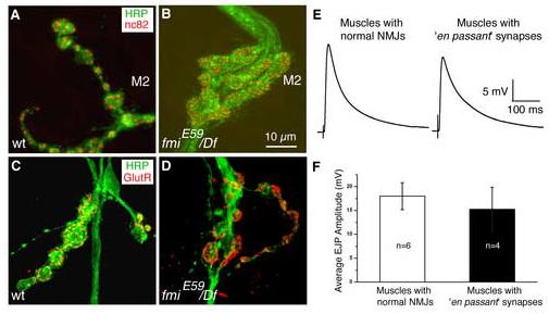
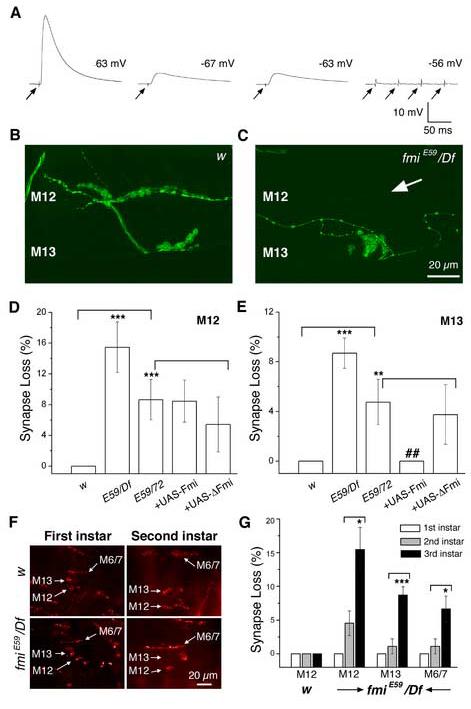
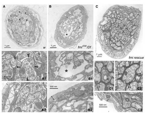
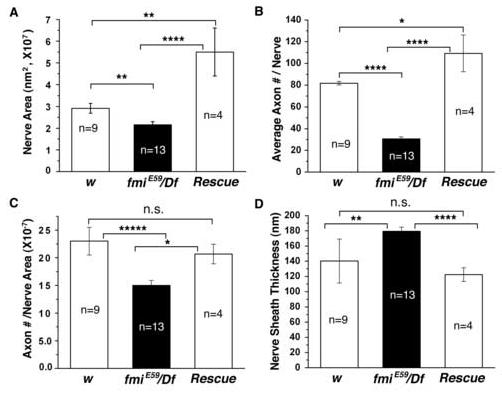
The formation of synaptic connections with target cells and maintenance of axons are highly regulated and crucial for neuronal function. The atypical cadherin and G-protein-coupled receptor Flamingo and its orthologs in amphibians and mammals have been shown to regulate cell polarity, dendritic and axonal growth, and neural tube closure. However, the role of Flamingo in synapse formation and function and in axonal health remains poorly understood. Here we show that fmi mutations cause a significant increase in the number of ectopic synapses on muscles and result in the formation of novel en passant synapses along axons, and unique presynaptic varicosities, including active zones, within axons. The fmi mutations also cause defective synaptic responses in a small subset of muscles, an age-dependent loss of muscle innervation and a drastic degeneration of axons in 3rd instar larvae without an apparent loss of neurons. Neuronal expression of Flamingo rescues all of these synaptic and axonal defects and larval lethality. Based on these observations, we propose that Flamingo is required in neurons for synaptic target selection, synaptogenesis, the survival of axons and synapses, and adult viability. These findings shed new light on a possible role for Flamingo in progressive neurodegenerative diseases.


Figures

Figure 1
Figure 1
Flamingo is expressed in motor axons and presynaptic terminals (A & A1). A confocal image of segmental nerve projection and presynaptic terminals on the bodywall muscle in a wild type embryo. The boxed area in panel A is enlarged below (A1). The nerve roots exiting the ventral nerve cord (VNC) of the central nervous system (CNS) are clearly immunoreactive to a Flamingo mAB (Fmi, green). The intersegmental nerve (ISN), segmental nerves SNa and SNb (indicated by arrows) are also stained for Flamingo. At this view level, presynaptic terminals at the NMJ are marked by a polyclonal Ab to the synaptic vesicle synaptotagmin I (Syt I, red). At NMJs, synaptotagmin I is enriched at the tip of axons. Further, synaptotagmin I immunoreactivity partially overlaps with Flamingo staining. The inset in panel A (left corner) shows Flamingo expression in both CNS and sensory cells in the peripheral nervous system (PNS). All embryos used in this and the following panels were approximately 18 hrs old. (B & C). In dissected embryonic bodywall muscles, Flamingo is detected weakly in muscles but at slightly higher levels in axons and presynaptic terminals. (D). The immunoreactivity for HRP (green, a neuronal membrane marker) and Flamingo (red) is found on the same CNS and axonal projections
Figure 2
Figure 2
Western blot of Flamingo in the larval CNS and the growth and locomotion of fmi mutant larvae (A). A Western blot of 3rd instar larval CNS (brain + VNC) or VNC in wild type (w), fmi mutant (fmiE59/Df), and rescued fmi mutant (fmiE59, Elav Gal4/fmi72, UAS-Fmi) is probed with a Flamingo antibody (upper panel). In a duplicated gel, an antibody to tubulin is used to examine the protein loading level. Note the presence of Flamingo in both CNS and VNC in the wild type and the rescued mutant and its absence in the fmi mutant. (B & C). Photographs of 3rd instar larvae in the wild type and the fmi mutant 7 days after hatching from the egg case. Note that the mutant larva is smaller in body size compared to the wild type. (D). Within additional 4-7 days, the mutant larva continues to forage in the food and grows to mature wandering 3rd instar larva, whose body size is larger than that of its wild type counterpart. (E). Still photographs illustrate the locomotion of the wild type larva and the mutant larva from panel D. The wild type larva quickly wanders off the penny, whereas the mutant larva crawls slowly.
Figure 3
Figure 3
Mutations in fmi cause a significant increase in the number of ectopic type I boutons (A-C). Representative immunocytochemical staining of the NMJs on muscles 12 and 13 with antibodies to the synaptic vesicle protein synaptotagmin I (Syt I, red) and the microtubule-associated protein Futsch (22C10, green) in the control (w, A), the fmiE59/ fmi72 (B), and rescued fmiE59/ fmi72 (C) larvae. Ectopic type I synapses on muscle 12 are indicated by the arrows in panel (B). The while lines mark the boundary between muscles 12 and 13. (D & E). Histograms show that fmi mutations significantly increase the fraction of muscles (muscle 12, D and muscle 13, E) receiving ectopic type I synapses compared to that in the w control larvae (* p<0.05). This defect is effectively rescued by neuronal expression of the wild type Flamingo in the fmiE59/ fmi72 mutant background (# p<0.05). (F). A representative image showing ectopic type II innervations on muscle 4 in fmi mutants labeled with the postsynaptic marker Dlg (red) and presynaptic marker Syt I (green). Type II synaptic boutons are marked by arrows. (G). Histogram plots show that the percentage of muscle 4 receiving ectopic type II synaptic input is significantly increased in fmiE59/Df and fmiE59/ fmi72 mutant larvae (***P<0.001). This defect can be effectively rescued to near wild type levels by neuronal expression of the wild type Flamingo in the fmiE59/ fmi72 mutant background, but only partially rescued by expression of the truncated Flamingo (### p<0.001; # P<0.05).
Figure 4
Figure 4
En passant synapses are found on muscles 2 and 3 in the fmi mutant (A & A1). Representative images of axons (HRP, green) and synapses (Syt I, red) on muscles 1, 2 and 3 in the control larvae (A). The NMJ indicated by the arrow is shown at higher magnification in panel (A1). Note the usual ‘beads-on-a-string’ pattern of synaptic boutons extended on the muscle surface. (B-B2). Representative images of axons (HRP, green) and synapses (Syt I, red) on muscles 2 and 3 in the fmiE59/Df larvae (B). The NMJs indicated by the arrows are shown at higher magnification below in panels (B1) and (B2). Compared to the NMJ in the control larva, these axons arrive on muscles 2 and 3 as one bundle, then defasciculate into individual axons and form boutons along the axon on muscles 2 and 3, and finally converge back to one nerve bundle and proceed to the next muscle. Synaptic vesicle proteins represented by Syt I are well retained within each en passant synapse. Note the irregular sized, often enlarged, synaptic boutons. (C). The formation of these en passant synapses can be partially suppressed by neuronal expression of the wild type Flamingo in the mutant background.
Figure 5
Figure 5
The en passant synapse contains presynaptic active zones and postsynaptic glutamate receptors and functions normally (A, B). Representative images of NMJs on muscle 2 stained for neuronal membranes (HRP, green) and active zones (nc82, red) in the wildtype larvae (panel A) and fmiE59/Df mutant larvae (panel B). (C, D). Representative images of NMJs on muscle 2 stained for neuronal membranes (HRP, green) and glutamate receptor III (red) in the wildtype larva (panel C) and fmiE59/Df mutant larva (panel D). (E, F). Representative EJPs from muscles with normal NMJs (panel E, left) and from muscles with ‘en passant’ synapses (panel E, right) in fmiE59/Df mutant larvae. The average amplitude of EJPs is slightly reduced in muscles with the ‘en passant’ synapse (panel F); however, this reduction is not statistically significant.
Figure 6
Figure 6
Axonal varicosities are found within the segmental nerve of the fmi mutant (A, B). Representative images of colocalization of the microtubule-associated protein Futsch (22C10, green) and the endocytotic protein DAP160 (red) on segmental nerves of control larvae (A) and fmiE59/Df mutant larvae (B), respectively. Note that DAP160 is present at low levels uniformly along the control segmental nerve. In contrast, some segmental nerves similar to the one shown in panels (B) have a string of bouton-like varicosities clustered on axons. Insets show one of the axonal varicosities at higher magnification. (C-F). Representative images showing colocalization of various synaptic (Hiw, nc82) and synaptic vesicle proteins (CSP, n-Syb, Syt I, VGluT) along segmental nerves of the fmi mutant larvae. Panels (C) show that the synaptic vesicle protein cysteine-string protein (CSP, green) colocalizes with the synaptic vesicle protein neuronal synaptobrevin (n-Syb, red) within the string of bouton-like varicosities along the segmental nerve. Panels (D) show that the peri-active zone protein Highwire (Hiw, green) colocalizes with the synaptic vesicle protein synaptotagmin I (Syt I, red) within the bouton-like varicosities. Panels (E) show that the active zone marker nc82 is clustered in bouton-like shapes along the segmental nerve (marked by HRP). Panels (F) show the localization of the Drosophila vesicular glutamate transporter (VGluT) along the segmental nerve (marked by HRP). (G & H). The axonal varicosities found in the fmiE9/fmi72 mutant (G) can be rescued by neuronal expression of the wild type Flamingo in the mutant background (F). Note the absence of Syt I-positive varicosities along the segmental nerve in the rescued fmi mutant. Arrowheads point to synaptic boutons normally found on bodywall muscles. However, this rescue is statistically significant but not a full rescue (see Fig. S3E).
Figure 7
Figure 7
The fmi mutant loses synaptic potentials and displays an age-dependent loss of NMJs (A). Examples of muscles in 3rd instar fmi mutant larvae that display normal (the first example of EJP), dramatically reduced (the 2nd and 3rd examples of EJPs) or no synaptic potentials (the last example, with four arrows) evoked by stimulating the segmental nerve. The resting potential of each muscle is shown. These recordings were conducted under identical experimental conditions and in HL-3 saline containing 1 mM Ca2+. Arrows indicate the onset of nerve stimulation. Note that multiple stimuli failed to evoke any synaptic response in the last muscle. (B, C). Representative images of NMJs on muscles 12 and 13 (stained for HRP, green) in the control (B) and fmiE59/Df larvae (C) at the 3rd instar stage. An example of muscle 12 that has lost its normal NMJ is shown here (see arrow in panel C). (D, E). Histograms of the percentage of muscles 12 (D) and 13 (E) that are denervated in 3rd instar larvae. While the control larvae have normal NMJs in all cases tested, fmi mutants have a significant number of muscles that lack synaptic inputs. These defects can be effectively rescued by expressing the wild type Flamingo in the fmiE59/ fmi72 mutant background in muscle 13, but not fully in muscle 12 (p>0.05). Expression of the truncated Flamingo lacking most of the extracellular domain does not rescue these defects. fmi mutants (fmiE59/Df, and fmiE59/ fmi72) have significantly more muscles that lost NMJs compared to that in w control larvae (*** p<0.001; ** p<0.01; panel D). The rescued mutants (fmiE59/ fmi72 + wild type Flamingo) have significantly fewer muscles that lost NMJs compared to that in fmiE59/ fmi72 mutants (## p<0.01; panel E). (F). Representative images of NMJs on ventral muscles (muscles 6, 7, 12 and 13) in 1st and 2nd instar larvae of the control fly (w, top panels) and the fmi mutant (bottom panels). (G). Histograms comparing the percentage of muscles that do not have NMJs in 1st, 2nd and 3rd instar larvae. Note the absence of synapse loss in 1st instar mutant larva and the increase in synapse loss from 2nd instar larvae to 3rd instar larvae. * p<0.05; *** p<0.001;
Figure 8
Figure 8
Axonal degeneration of segmental nerves in the fmi mutant larvae and its rescue by neuronal expression of Flamingo (A-A2). Panel (A) shows a representative cross-section of the segmental nerve from a control (w) larva. The arrowheads point to one axon that is further magnified in panel (A1). Axons are packed with mitochondria and microtubules. The edge of the segmental nerve is magnified in panel (A2) to illustrate the thickness of the neuronal sheath (double headed arrows). (B-B2). Panel (B) shows a representative cross-section of the segmental nerve for an fmiE59/Df mutant larva. In contrast to panel (A), the mutant nerve has more empty ‘holes’ and is relatively less packed with axons and glial processes. The asterisk marks the gap in the nerve that is magnified in panel (B1). These axons are less packed with mitochondria and microtubules. Double-headed arrows in panel (B2) delimit the sheath surrounding the segmental nerves of the mutant. Note the thickening of the nerve sheath. (C-C3). Panel (C) shows a representative cross-section of the segmental nerve for an fmiE59/Df mutant larva rescued by expressing a wild type Flamingo in postmitotic neurons. In contrast to panel (B), the rescued nerve is larger in diameter and well packed with axons and glial processes. The asterisk marks one axon undergoing degeneration that is magnified in panel (C1). The arrowheads point to one axon that is further magnified in panel (C2). Similar to axons in the wild type, the rescued axons are packed with mitochondria and microtubules. Double-headed arrows in panel (C3) delimit the sheath surrounding the segmental nerves of the rescued larva. Note that the nerve sheath is restored to the thickness found in the wild type.
Figure 9
Figure 9
Summary of morphological properties of segmental nerves in the wild type, fmi mutant, and rescued mutant larvae (A). The average area of the segmental nerve is significantly reduced in the fmiE59/Df mutant. In rescued mutant larvae, the segmental nerve is significantly enlarged. *p<0.05; **p<0.01; ****p<<0.001 (the same below). (B). The average number of axons per segmental nerve is significantly reduced in the fmiE59/Df mutant. In rescued larvae, each segmental nerve has slightly more numbers of axons. (C). The average number of axons per area of the segmental nerve is significantly reduced in the fmiE59/Df mutant. This is completely rescued to wild type levels in rescued larvae. n.s. = not significantly different. (D). The average thickness of the nerve sheath surrounding the segmental nerve is significantly increased in the fmiE59/Df mutant. This defect is fully corrected in rescued larvae. n.s. = not significantly different. Data are expressed as Mean values ± S.E.M.

Similar articles

Cited by

References

    1. Aberle H, Haghighi AP, Fetter RD, McCabe BD, Magalhaes TR, Goodman CS. wishful thinking encodes a BMP type II receptor that regulates synaptic growth in Drosophila. Neuron. 2002;33:545–558. - PubMed
    1. Ackley BD, Jin Y. Genetic analysis of synaptic target recognition and assembly. Trends Neurosci. 2004;27:540–547. - PubMed
    1. Ahmari SE, Buchanan J, Smith SJ. Assembly of presynaptic active zones from cytoplasmic transport packets. Nat Neurosci. 2000;3:445–451. - PubMed
    1. Atwood HL, Govind CK, Wu CF. Differential ultrastructure of synaptic terminals on ventral longitudinal abdominal muscles in Drosophila larvae. J Neurobiol. 1993;24:1008–1024. - PubMed
    1. Bao H, Daniels RW, Macleod GT, Charlton MP, Atwood HL, Zhang B. AP180 maintains the distribution of synaptic and vesicle proteins in the nerve terminal and indirectly regulates the efficacy of Ca2+-triggered exocytosis. J Neurophysiol. 2005;94:1888–1903. - PubMed

Publication types

MeSH terms