

Review
doi: 10.1098/rstb.2006.2016.
Brain micro-ecologies: neural stem cell niches in the adult mammalian brain
Affiliations
- PMID: 17322003
- PMCID: PMC2605490
- DOI: 10.1098/rstb.2006.2016
Item in Clipboard
Review
Brain micro-ecologies: neural stem cell niches in the adult mammalian brain
Philos Trans R Soc Lond B Biol Sci.
.
Abstract
Neurogenesis persists in two germinal regions in the adult mammalian brain, the subventricular zone of the lateral ventricles and the subgranular zone in the hippocampal formation. Within these two neurogenic niches, specialized astrocytes are neural stem cells, capable of self-renewing and generating neurons and glia. Cues within the niche, from cell-cell interactions to diffusible factors, are spatially and temporally coordinated to regulate proliferation and neurogenesis, ultimately affecting stem cell fate choices. Here, we review the components of adult neural stem cell niches and how they act to regulate neurogenesis in these regions.
Figures
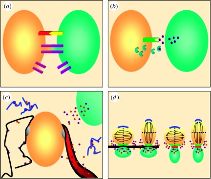
Components of the stem cell niche. (a) Cell–cell and cell–extracellular environment interactions. Membrane-associated receptors and ligands (red and yellow) mediate cell–cell contacts and cell fate decisions, including self-renewal and differentiation. In addition, gap junctions (purple), intercellular channels that allow the passage of ions and metabolites, both coordinate behaviour between coupled cells and can tether cells together. Hemi-channels allow communication between the cell and the environment. (b) Diffusible factors. Diffusible factors (purple spheres) can direct stem cells to either self-renew or generate differentiated progeny. The availability of diffusible factors that bind to receptors (green) in turn can be regulated by ligand inhibitors (blue half-moons), which can sequester these factors and prevent signalling. (c) Basal lamina and blood vessels. An ECM-rich basal lamina (black), which can be associated with blood vessels (red) or cells, has several functions in stem cell niches, including anchoring cells to the niche (grey spheres), sequestering and presenting diffusible signals (purple and brown spheres), and linking cells and the ECM. In addition, proteolytic fragments of ECM components (blue squiggles) may regulate stem cells. Endothelial cells and the vasculature are emerging as integral components of stem cell niches, where they can regulate stem cell fate decisions through either diffusible signals and/or direct cell–cell contact. (d) Oriented cell division. Both cell–cell contact (via adherens junctions, red) and tethering of cells to the basal lamina (black) can influence cell fate by orienting the plane of cell division, such that key intracellular determinants are symmetrically or asymmetrically distributed. Oriented segregation of these factors may influence cell fate. Short-range diffusible factors (purple and brown spheres) secreted by stromal cells (green) that promote self-renewal only influence immediately adjacent cells, allowing differentiating daughter cells to escape the niche.
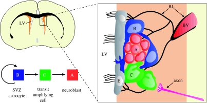
Cell types and anatomy of the adult SVZ niche. Schema of frontal section of the adult mouse brain showing the SVZ (orange) adjacent to the lateral ventricle (LV). SVZ astrocytes in this region (B, blue) are stem cells which generate migrating neuroblasts (A, red) destined for the olfactory bulb via a rapidly dividing transit-amplifying cell (C, green). Region in box is expanded at right to show the relationship of cells in this region and some elements of the SVZ niche. Multi-ciliated ependymal cells (E, grey) line the walls of the lateral ventricle. Chains of neuroblasts travel through tunnels formed by processes of SVZ astrocytes. Transit-amplifying cells are found in small clusters adjacent to the chains. Signals released from axons (pink) regulate proliferation and survival in this region. A specialized basal lamina (BL, black) extends from perivascular cells and contacts all cell types. Endothelial cells, blood vessels (BV) and the basal lamina are all likely key components of the niche.
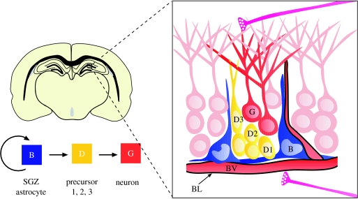
Cell types and anatomy of the adult SGZ niche. Schema of frontal section of the adult mouse brain showing the SGZ at the interface between the hilus (area below blood vessel) and the granule cell layer (light pink cells) of the dentate gyrus. SGZ astrocytes (B, blue) divide to generate intermediate precursors (type D cells; nomenclature according to Seri et al. 2004, yellow), which progressively generate more differentiated progeny (type D1→type D2→type D3), which mature into granule neurons (G, red). Neurogenesis occurs in pockets adjacent to blood vessels and although a specialized basal lamina has not yet been described in this region, the vascular basal lamina likely plays an important role in the niche. Afferent axons (pink) from the entorhinal cortex and axons from subcortical regions as well as from local inhibitory interneurons project to the SGZ.
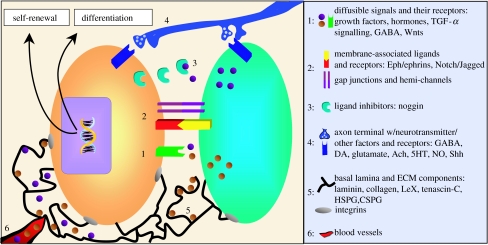
Neural stem cell regulation. Neural stem cells in the two adult neurogenic niches, the SVZ and SGZ, can be regulated by (1) diffusible factors (EGF, FGF, TGF-α, VEGF, PEDF, hormones, BMPs, ATP, Wnts and GABA) and their receptors, (2) cell–cell and cell–extracellular environment interactions via membrane-associated ligands and their receptors (Eph/ephrin and Notch/Jagged) or gap junctions and hemi-channels, (3) ligand inhibition (noggin), (4) release of neurotransmitters and other factors from axons (GABA, dopamine (DA), glutamate, acetylcholine (ACh), serotonin (5HT), NO and Shh), (5) basal lamina (sequestration and presentation of diffusible factors, such as growth factors) and ECM proteins (laminin, collagen-1, tenascin-C, LeX, heparan sulphate and chondroitin sulphate proteoglycans), and (6) endothelial cell/blood vessel-mediated cues. The cell types that are able to direct neural stem cell fate choices include cells within the niche (both support cells and stem cells and their progeny) (green), neurons (axonal projection; blue) and endothelial cells/blood vessels (red). Structural elements that likely regulate neural stem cell fate decisions include fractones (in the SVZ) and basal laminae (black lines). In addition, CSF and meningeal projections (not pictured in this figure) are likely important components of the niche. These elements may work in concert or independently to promote neural stem cell self-renewal or differentiation. The key to the right lists some of the factors known to be present in adult neurogenic niches.
References
-
- Ahn S, Joyner A.L. In vivo analysis of quiescent adult neural stem cells responding to Sonic hedgehog. Nature. 2005;437:894–897. doi:10.1038/nature03994 - DOI - PubMed
-
- Alvarez-Buylla A, Garcia-Verdugo J.M, Tramontin A.D. A unified hypothesis on the lineage of neural stem cells. Nat. Rev. Neurosci. 2001;2:287–293. doi:10.1038/35067582 - DOI - PubMed
-
- Alvarez-Buylla A, Lim D.A. For the long run: maintaining germinal niches in the adult brain. Neuron. 2004;41:683–686. doi:10.1016/S0896-6273(04)00111-4 - DOI - PubMed
-
- Alves J.A, Barone P, Engelender S, Froes M.M, Menezes J.R. Initial stages of radial glia astrocytic transformation in the early postnatal anterior subventricular zone. J. Neurobiol. 2002;52:251–265. doi:10.1002/neu.10087 - DOI - PubMed
Publication types
MeSH terms
Grants and funding
LinkOut - more resources
Full Text Sources