. 2007 Mar 30;100(6):884-93.
doi: 10.1161/01.RES.0000260802.75766.00. Epub 2007 Feb 22.

Monocyte/macrophage suppression in CD11b diphtheria toxin receptor transgenic mice differentially affects atherogenesis and established plaques

Affiliations

Monocyte/macrophage suppression in CD11b diphtheria toxin receptor transgenic mice differentially affects atherogenesis and established plaques

Victoria Stoneman et al. Circ Res. .

Abstract

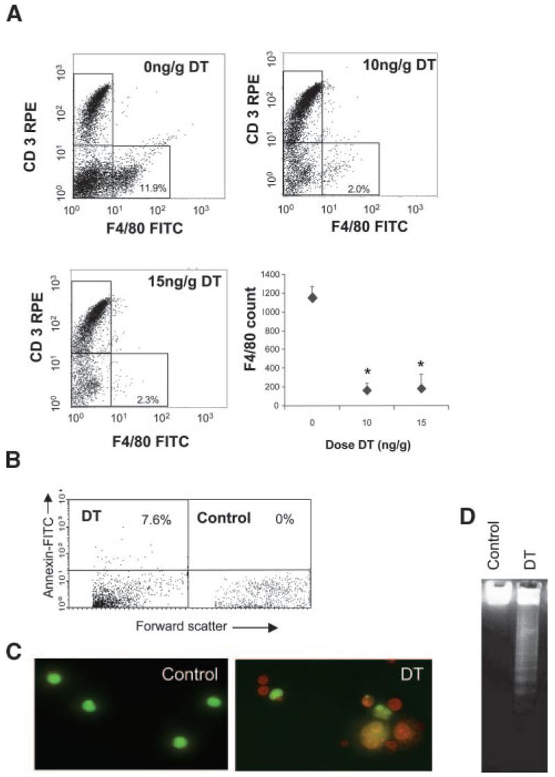
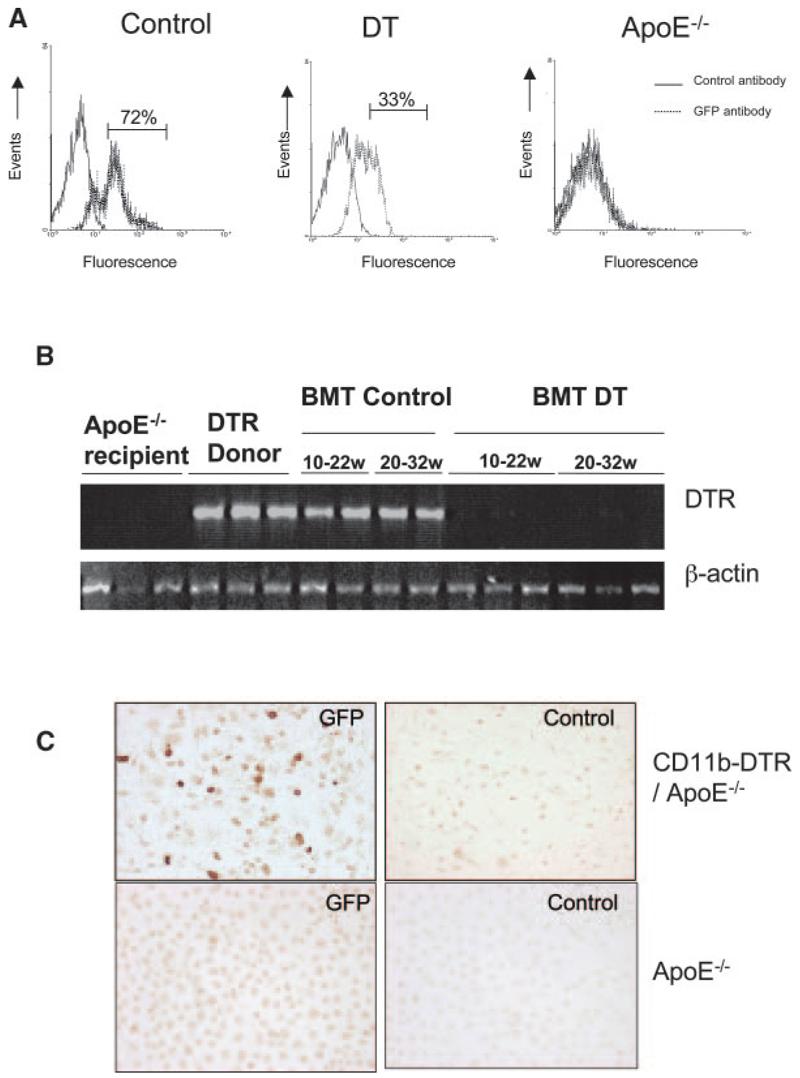
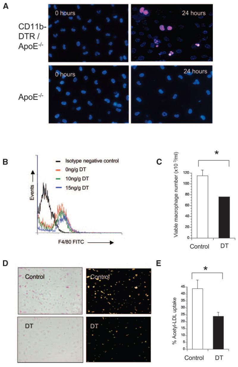
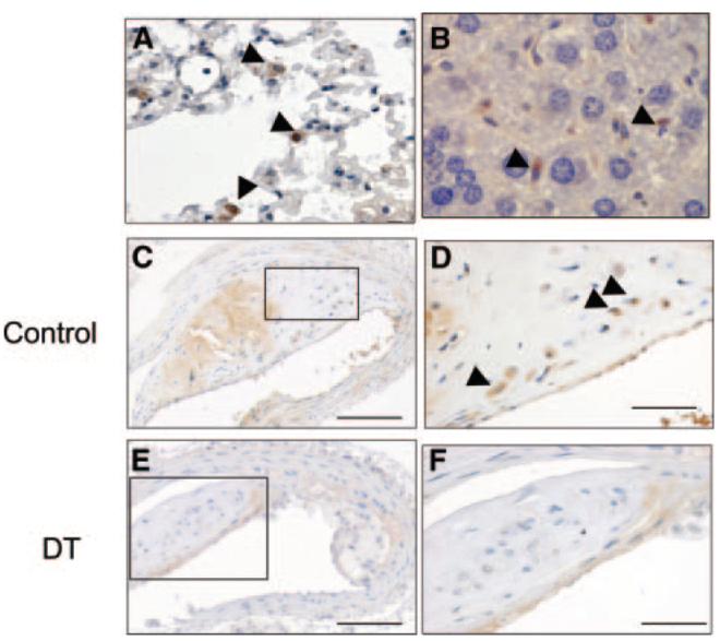
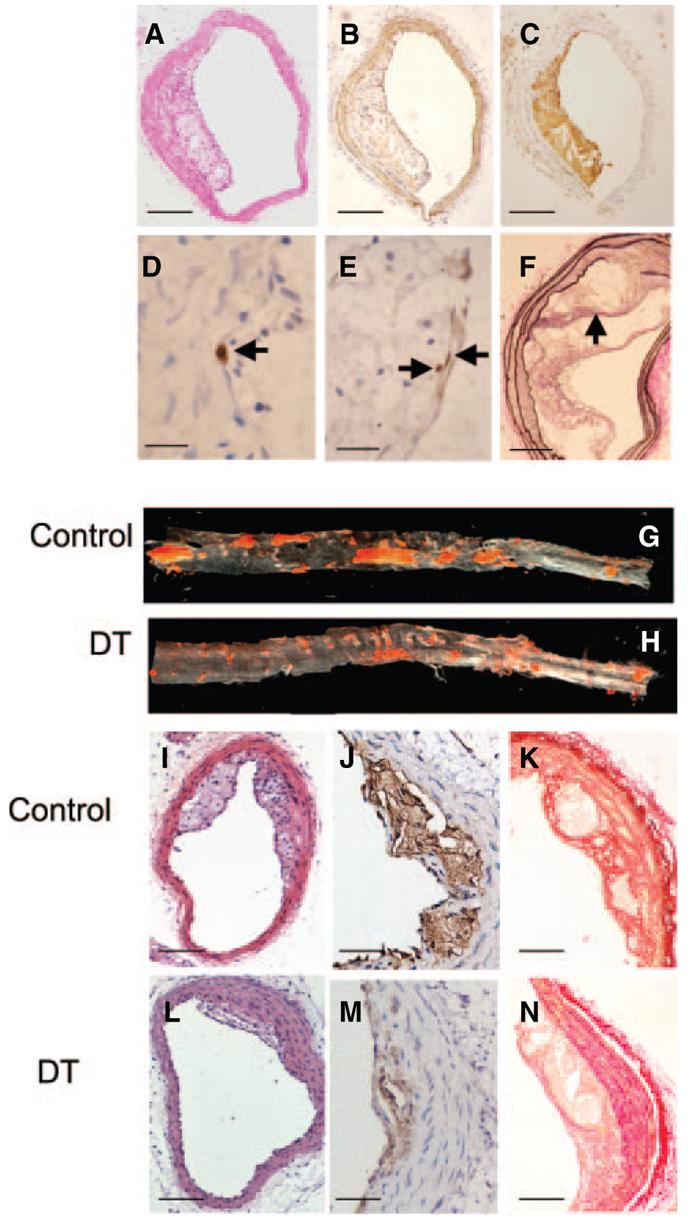
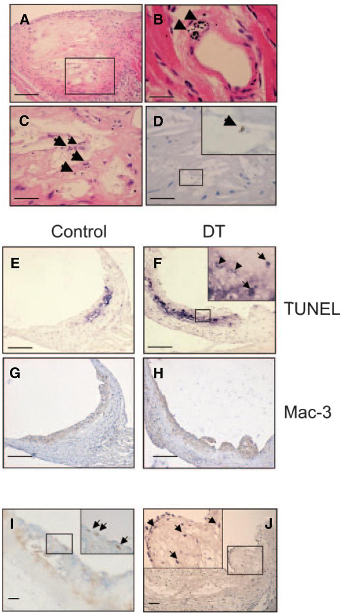
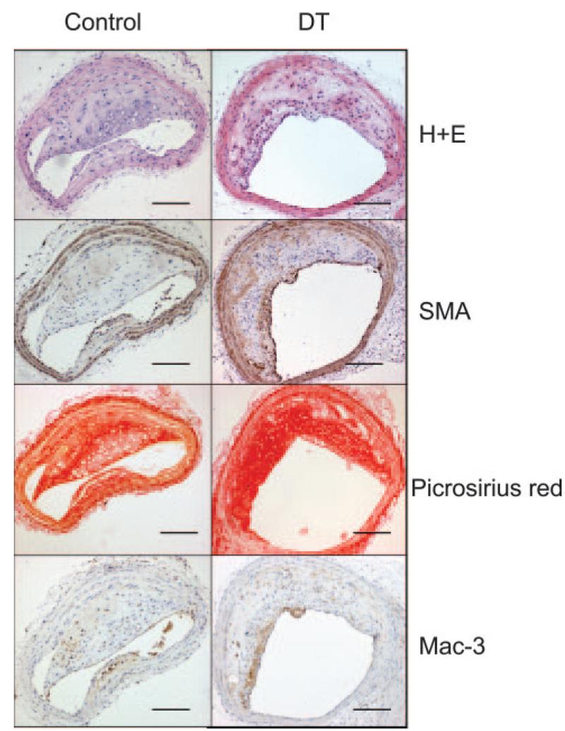
Although monocytes/macrophages are considered important in atherogenesis, their role in established plaques is unclear. For example, macrophage content is associated with plaque instability, but their loss through cell death is observed at sites of plaque rupture. To examine the role of monocytes/macrophages in atherosclerosis, we developed CD11b-diphtheria toxin (DT) receptor (DTR) transgenic mice, whereby administration of DT selectively kills monocytes/macrophages. DT treatment reduced peripheral blood monocytes and tissue macrophages and inhibited macrophage function in CD11b-DTR mice and apolipoprotein E-null (apoE(-/-)) mice transplanted with CD11b-DTR bone marrow. In atherogenesis experiments, DT markedly reduced plaque development and altered plaque composition, reducing collagen content and necrotic core formation. In mice with established plaques, acute DT treatment induced macrophage apoptosis and reduced macrophage content but did not induce plaque inflammation, thrombosis, or rupture. Furthermore, despite a 50% reduction in monocytes, chronic DT treatment of these mice did not alter plaque extent or composition, most likely because of ongoing recruitment/proliferation of monocytes with recovery of macrophage content. We conclude that monocytes/macrophages are critical to atherogenesis, but established plaques are more resistant to reductions in monocytes.


Figures

Figure 1
Figure 1
Characterization of CD11b-DTR mice. A, Representative flow-cytometric profiles from DTR-FV/B mice treated with 2 doses of 10 ng/g or 15 ng/g DT at 48-hour intervals. Blood was isolated 24 hours later, labeled with F4/80–fluorescein isothiocyanate (FITC) and CD3-R-phycoerythrin (CD3-RPE) antibodies and analyzed by flow cytometry. Absolute F4/80 counts are also shown for each dose of DT as mean±SEM. *P<0.05 (n=3) compared with controls (0 ng/g DT). B, Flow cytometry for Annexin V demonstrating increased circulating Annexin V–positive cells after DT treatment. C and D, Peritoneal macrophages isolated from these mice demonstrate apoptosis on acridine orange staining (orange cells with condensed chromatin) (C) and DNA laddering (D).
Figure 2
Figure 2
DT treatment reduces monocytes and macrophages in transplanted mice. A, ApoE−/− mice were transplanted with CD11b-DTR-EGFP/apoE−/− bone marrow at 8 weeks of age and treated with saline (control) or 10 ng/g DT 3 times per week for 5 weeks. Monocytes were separated by forward scatter/side scatter, and GFP expression was examined by flow cytometry. Nontransplanted apoE−/− mice (right) were also used as a negative control for GFP. B, PCR for DTR in blood of recipient apoE−/− mice before transplant, DTR+/+/apoE−/− donor mice and apoE−/− mice transplanted with DTR+/+/apoE−/− marrow and treated with saline control (BMT control) or DT (BMT DT) from 10 to 22 weeks or 20 to 32 weeks. C, Immunocytochemistry using anti-GFP or control antibody of peritoneal macrophages of mice transplanted with CD11b-DTR/apoE−/− marrow or control apoE−/− mice.
Figure 3
Figure 3
DT inhibits macrophage numbers and function in transplanted apoE−/− mice. A, Propidium iodide/Bisbenzamide staining of peritoneal macrophages cultured from mice transplanted with CD11b-DTR/apoE−/− marrow (top panels) compared with control apoE−/− mice (bottom panels) treated with DT for 24 hours. Typical apoptotic morphology (condensed, fragmented nuclei) is seen after DT treatment of mice transplanted with CD11b-DTR/apoE−/− marrow. B through E, ApoE−/− mice were transplanted with CD11b-DTR/apoE−/− marrow and treated with 2 doses of DT or saline control in vivo, before isolation of macrophages. F4/80+ peritoneal macrophages were quantified by flow cytometry (B). Viable peritoneal macrophages from mice receiving 10 ng/g DT or saline (control) were also quantified by acridine orange staining (C). D, Uptake of acetylated LDL determined by phase contrast (left) or fluorescence microscopy (right) in viable peritoneal macrophages from mice treated with 10 ng/g DT or saline (control). E, Percentage of viable macrophages demonstrating acetyl-LDL uptake. Data are means; error bars represent SEMs (n=3). *P<0.05.
Figure 4
Figure 4
DTR-expressing macrophages are found in multiple organs. A through D, Immunohistochemistry for GFP in lung (A), liver (B), and atherosclerotic plaque (C and D) demonstrating GFP-positive macrophages in all organs (arrowheads). C through F, ApoE−/− mice were irradiated and transplanted at 8 weeks of age with CD11b-DTR/apoE−/− marrow and 10 ng/g DT or saline control injected 3 times weekly until 22 weeks. DT reduced the percentage of GFP-positive macrophages (compare F vs control [D]). D and F, Higher-power views of areas outlined in C and E. Scale bars: 200 μm (C and E); and 50 μm (D and F).
Figure 5
Figure 5
DT treatment reduces atherosclerosis in aorta and brachiocephalic arteries. Brachiocephalic artery plaques were analyzed by hematoxylin/eosin (A) and immunohistochemistry for VSMCs (SMA) (B); macrophages (Mac3) (C); cleaved caspase 3 in cells showing a pyknotic, fragmented nucleus typical of apoptosis (arrow) (D); and Ki67 (arrows) (E). F, Van Gieson stain demonstrating buried fibrous cap (arrow). G and H, Oil red O analysis of descending thoracic and abdominal aortas from mice treated with control (saline) or 10 ng/g DT. I through N, Effects of DT on plaque composition, demonstrating reduced plaque development on hematoxylin/eosin staining (I and L) and reduced necrotic cores by Mac3 staining (J and M) after DT administration. In similar-sized lesions, DT reduced plaque collagen by Picrosirius red staining (K and N). Scale bars: 100 μm (A through C, I, and L); 50 μm (F, K, and N); 25 μm (J and M); 10 μm (D and E).
Figure 6
Figure 6
Effects of short term DT treatment on established plaques. A through D, Apoptosis in mice administered 2 doses of 10 ng/g DT or control, showing extensive apoptotic debris on hematoxylin/eosin staining of established plaque (A). B, Morphological features of apoptosis were seen in DT-treated mice, for example, in small intramyocardial artery (arrowheads). C and D, High-power views of area outlined in A showing apoptotic debris (C) and cleaved caspase 3–positive cell (D and inset). E through H, Increased TUNEL staining and reduced macrophage percentage in DT-treated vs control plaques. Inset represents high-power view of area outlined in F, showing TUNEL-positive cells (arrows) and debris (arrowheads). I, Double labeling for macrophages (blue) and Ki67 (brown) indicating superficial proliferating macrophages (arrows). J, Mac387 expression indicating presence of newly migrated macrophages (arrows). Insets represent high-power view of area outlined in (I and J). Scale bars: 200 μm (A and E through H); 50 μm (C, D, I, and J); 25 μm (B).
Figure 7
Figure 7
Effects of long-term DT treatment on established plaques. Effects of DT on composition of established plaques, demonstrating no effect on plaque development on hematoxylin/eosin (H+E) staining, VSMC content (SMA), collagen content (Picrosirius red), or macrophage content (Mac3). Scale bars=100 μm.

Comment in

References

    1. Glass CK, Witztum JL. Atherosclerosis. the road ahead. Cell. 2001;104:503–516. - PubMed
    1. Gerrity RG, Naito HK, Richardson M, Schwartz CJ. Dietary induced atherogenesis in swine. Morphology of the intima in prelesional stages. Am J Pathol. 1979;95:775–792. - PMC - PubMed
    1. Faggiotto A, Ross R, Harker L. Studies of hypercholesterolemia in the nonhuman primate. I. Changes that lead to fatty streak formation. Arteriosclerosis. 1984;4:323–340. - PubMed
    1. Charo IF, Taubman MB. Chemokines in the pathogenesis of vascular disease. Circ Res. 2004;95:858–866. - PubMed
    1. Boyle J, Bowyer D, Weissberg P, Bennett M. Human blood-derived macrophages induce apoptosis in human plaque-derived vascular smooth muscle cells by Fas ligand/Fas interactions. Art Thromb Vasc Biol. 2001;21:1402–1407. - PubMed

Publication types

MeSH terms