. 2007 Mar;153(Pt 3):814-825.
doi: 10.1099/mic.0.2006/001834-0.

The ftsA* gain-of-function allele of Escherichia coli and its effects on the stability and dynamics of the Z ring

Affiliations

The ftsA* gain-of-function allele of Escherichia coli and its effects on the stability and dynamics of the Z ring

Brett Geissler et al. Microbiology (Reading). 2007 Mar.

Abstract

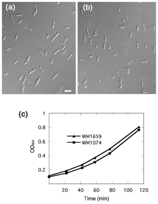
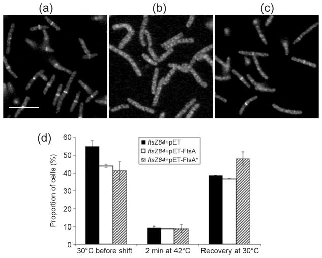
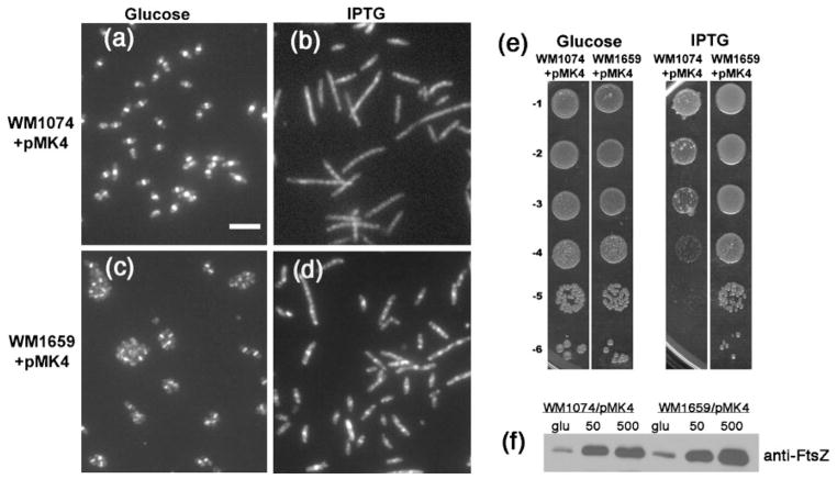
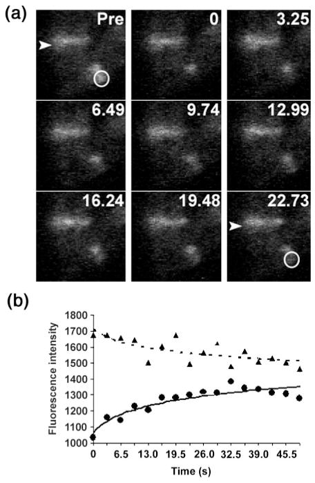
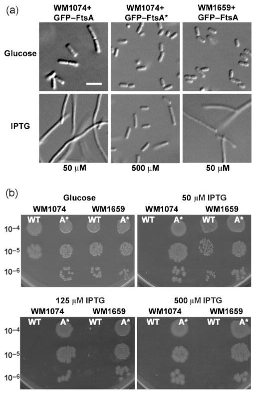
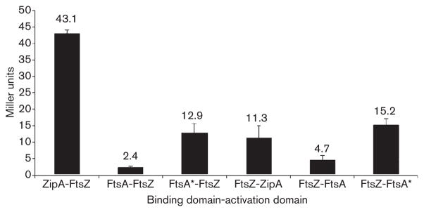
Formation of the FtsZ ring (Z ring) in Escherichia coli is the first step in the assembly of the divisome, a protein machine required for cell division. Although the biochemical functions of most divisome proteins are unknown, several, including ZipA, FtsA and FtsK, have overlapping roles in ensuring that the Z ring assembles at the cytoplasmic membrane, and that it is active. As shown previously, a single amino acid change in FtsA, R286W, also called FtsA*, bypasses the requirement for either ZipA or FtsK in cell division. In this study, the properties of FtsA* were investigated further, with the eventual goal of understanding the molecular mechanism behind the bypass. Compared to wild-type FtsA, the presence of FtsA* resulted in a modest but significant decrease in the mean length of cells in the population, accelerated the reassembly of Z rings, and suppressed the cell-division block caused by excessively high levels of FtsZ. These effects were not mediated by Z-ring remodelling, because FtsA* did not alter the kinetics of FtsZ turnover within the Z ring, as measured by fluorescence recovery after photobleaching. FtsA* was also unable to permit normal cell division at below normal levels of FtsZ, or after thermoinactivation of ftsZ84(ts). However, turnover of FtsA* in the ring was somewhat faster than that of wild-type FtsA, and overexpressed FtsA* did not inhibit cell division as efficiently as wild-type FtsA. Finally, FtsA* interacted more strongly with FtsZ compared with FtsA in a yeast two-hybrid system. These results suggest that FtsA* interacts with FtsZ in a markedly different way compared with FtsA.


Figures

Fig. 1
Fig. 1
FtsA* cells are shorter than normal. Representative fields are shown of cells of strain WM1074 (a) and its isogenic ftsA* derivative WM1659 (b), at the same magnification. Bar, 5 μm. Both strains were grown at 32 °C in LB to exponential phase, and samples were briefly spun to concentrate them five-fold prior to placing on microscope slides. Growth curves of the strains cultured at 32 °C in LB are shown in (c).
Fig. 2
Fig. 2
FtsA* promotes more rapid Z-ring formation in ftsZ84(ts) cells after thermoinactivation and recovery. (a–c) Representative fluorescence micrographs of cells with ftsZ84 +pET28a vector (WM1985) grown at 30 °C (a), shifted to 42 °C for 2 min (b), then shifted back to 30 °C for 5 min (c). Samples were taken after each shift, fixed, and processed for IFM using anti-FtsZ specific antiserum. Cultures of ftsZ84+pET-FtsA (WM1986) and ftsZ84+pET-FtsA* (WM1987) were similarly processed for IFM (data not shown). (d) Percentages of ftsZ84 cells containing pET vector (black columns), pET-FtsA (white columns) or pET-FtsA* (hatched columns) containing a clear Z ring after each step of the temperature-shift experiment. Bar, 5 μm.
Fig. 3
Fig. 3
FtsA* suppresses the toxicity caused by excess FtsZ. (a–d) Micrographs showing anti-FtsZ IFM of WM1074+pMK4 (a, b) or WM1659+pMK4 (c, d) grown at 30 °C for 4 h in LB containing either glucose (a, c) or 500 μM IPTG (b, d). Bar, 5 μm. (e) Viability plating of WM1074+pMK4 or WM1659+pMK4 on LB containing glucose (left panels) or 500 μM IPTG (right panels). (f) Immunoblot of extracts from cells described in (a–d), grown in glucose (glu), 50 μM IPTG (50) or 500 μM IPTG (500), and probed with anti-FtsZ. An equivalent amount of cells, as measured by optical density, was loaded into each lane. The FtsZ band density in the WM1074/pMK4 (500 μM IPTG) lane was low, probably because many of the cells had already been lysed by the high levels of FtsZ.
Fig. 4
Fig. 4
FRAP analysis. (a) Micrographs from one FRAP experiment with FtsZ–GFP expressed at low levels in an otherwise wild-type strain (WM2026). Three cells with Z rings are visible: two cells aligned vertically (upper left), and one horizontally (lower right) that was used for bleaching. Each panel represents a sequential time point in seconds, with the first prior to photobleaching (Pre). The arrowhead highlights one of the unbleached Z rings used as an internal control for background photobleaching during the experiment. The white circle shows the photobleached portion of another Z ring, which recovered during the time-course. (b) The fluorescence intensities (arbitrary units) for the two Z rings are plotted versus time. The FRAP data from this and all other time-courses are summarized in Table 2. ●, Bleached; ▲, no bleaching.
Fig. 5
Fig. 5
Overproduction of GFP–FtsA* is less toxic to cells than that of GFP–FtsA. (a) Micrographs of WM1074+GFP–FtsA (left column), WM1074+GFP–FtsA* (middle column) or WM1659+GFP–FtsA (right column) grown in LB containing either glucose (top row), 50 μM IPTG (bottom-left and bottom-right images) or 500 μM IPTG (bottom-middle image). Bar, 5 μm. (b) Dilution plating of WM1074 or WM1659 containing either GFP–FtsA (wild-type, WT) or GFP–FtsA* (A*) grown on glucose, or 50 μM, 125 μM or 500 μM IPTG. Serial dilutions are shown to the left of the panels.
Fig. 6
Fig. 6
Yeast two-hybrid assays of FtsZ interactions with FtsA or FtsA*. S. cerevisiae Y190 containing plasmids producing the indicated proteins fused to the GAL4 activation domain were assayed for β-galactosidase activity (Miller units) in liquid culture, as described in Methods. The values shown are the means of at least five separate assays of two different transformations, with the SEM indicated by error bars.

References

    1. Addinall SG, Bi E, Lutkenhaus J. FtsZ ring formation in fts mutants. J Bacteriol. 1996;178:3877–3884. - PMC - PubMed
    1. Anderson DE, Gueiros-Filho FJ, Erickson HP. Assembly dynamics of FtsZ rings in Bacillus subtilis and Escherichia coli and effects of FtsZ-regulating proteins. J Bacteriol. 2004;186:5775–5781. - PMC - PubMed
    1. Begg K, Nikolaichik Y, Crossland N, Donachie WD. Roles of FtsA and FtsZ in activation of division sites. J Bacteriol. 1998;180:881–884. - PMC - PubMed
    1. Bi E, Lutkenhaus J. FtsZ ring structure associated with division in E. coli. Nature. 1991;354:161–164. - PubMed
    1. Corbin BD, Geissler B, Sadasivam M, Margolin W. A Z-ring-independent interaction between a subdomain of FtsA and late septation proteins as revealed by a polar recruitment assay. J Bacteriol. 2004;186:7736–7744. - PMC - PubMed

Publication types

MeSH terms

LinkOut - more resources