. 2007 May;189(10):3868-75.
doi: 10.1128/JB.01935-06. Epub 2007 Feb 23.

Biofilms formed by nontypeable Haemophilus influenzae in vivo contain both double-stranded DNA and type IV pilin protein

Affiliations

Biofilms formed by nontypeable Haemophilus influenzae in vivo contain both double-stranded DNA and type IV pilin protein

Joseph A Jurcisek et al. J Bacteriol. 2007 May.

Abstract

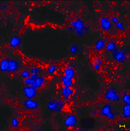
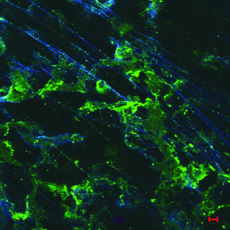
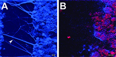
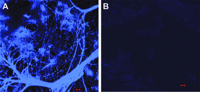
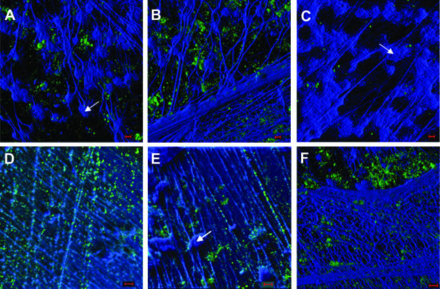
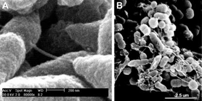
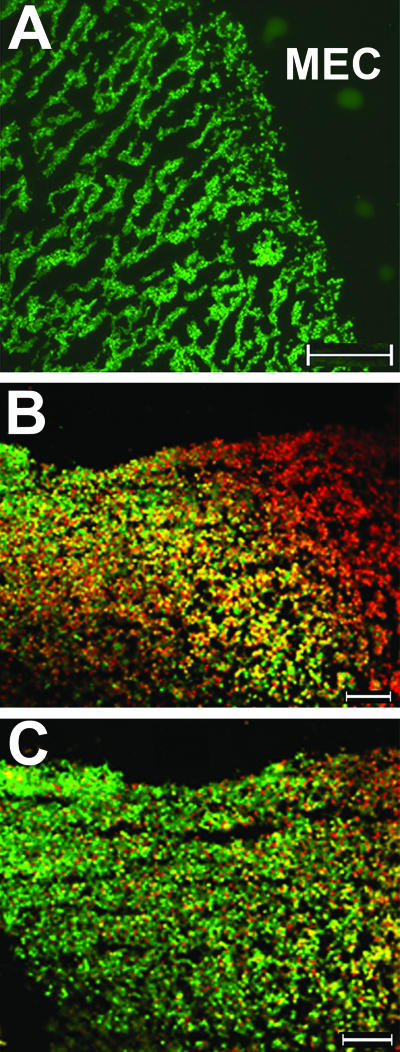
Nontypeable Haemophilus influenzae (NTHI) strains are members of the normal human nasopharyngeal flora, as well as frequent opportunistic pathogens of both the upper and lower respiratory tracts. Recently, it has been shown that NTHI can form biofilms both in vitro and in vivo. NTHI strains within in vitro-formed biofilms differentially express both epitopes of lipooligosaccharide (LOS) and the outer membrane proteins P2, P5, and P6, whereas those generated either in a 96-well plate assay in vitro or in a mammalian host have been shown to incorporate a specific glycoform of sialylated LOS within the biofilm matrix. While DNA has been identified as a key component of the biofilm matrix formed in vitro by several bacterial pathogens, here we demonstrate for the first time that in addition to sialylated LOS, the biofilm formed by NTHI in vivo contains both type IV pilin protein and a significant amount of double-stranded DNA. The DNA appeared to be arranged in a dense interlaced meshwork of fine strands as well as in individual thicker "ropes" that span water channels, suggesting that DNA could be imparting structural stability to the biofilm produced by NTHI in vivo. The presence of type IV pilin protein both appearing as small aggregates within the biofilm matrix and tracking along DNA strands supports our observations which showed that type IV pili are expressed by NTHI during experimental otitis media when these bacteria form a biofilm in the middle ear space.


Figures

FIG. 1.
FIG. 1.
The distribution of NTHI (or residual OMPs) can be seen as red fluorescence throughout a 4-day biofilm recovered from the chinchilla middle ear. OCT-embedded inferior bullae were sectioned and immunofluorescently labeled with polyclonal chinchilla anti-NTHI strain 86-028NP OMP and AlexaFluor 546-conjugated protein A (red) to detect the presence of NTHI in the biofilm. DAPI, used as a counterstain, labels the nuclei of host cells (blue fluorescence) associated with, or perhaps trafficking through, the biofilm. Bar, 5 μm.
FIG. 2.
FIG. 2.
Immunofluorescent image of an immature biofilm (4 days) labeled with rabbit anti-soluble recombinant PilA and AlexaFluor 488 goat anti-rabbit (green). Fine, widely spaced dsDNA strands are labeled with DAPI and appear blue in this image, whereas type IV pilin protein can be seen in both small aggregates and pinpoint spots that are colocalized along the length of the DNA strands. Bar, 5 μm.
FIG. 3.
FIG. 3.
Composite of high-resolution confocal images of a water channel present in a 21 day in vivo-formed biofilm. (A) Thick, rope-like strands of DAPI-labeled dsDNA can be seen as blue fluorescent filaments spanning the water channel (arrowhead). Also, dense clusters of what appear to be aggregates of bacteria are visible. (B) To confirm that these clusters were indeed NTHI, we dual labeled serial sections with both DAPI and polyclonal chinchilla anti-NTHI strain 86-028NP OMP antiserum that was detected with protein A-conjugated AlexaFluor 546, which imparts red fluorescence. One can observe clusters of NTHI (appearing red and fuchsia due to the heavy fluorescence in both the blue and red channels) populating the biofilm which borders the depicted water channel. Bars, 5 μm.
FIG. 4.
FIG. 4.
DNA present within a biofilm formed by NTHI strain 86-028NP in the chinchilla middle ear 21 days after challenge. The biofilm within the middle ear as well as the mucosal layer of the inferior bulla was embedded in OCT compound and snap frozen, and 10-μm sections were placed on slides. (A) dsDNA, labeled with DAPI, forms a complex network consisting of some thick strands, areas of aggregation, and thin web-like strands, giving a three-dimensional structure to the biofilm. (B) Serial section of the same biofilm after DNase treatment, confirming that the web-like mesh and strands seen in panel A were indeed comprised of dsDNA. Bars, 5 μm.
FIG. 5.
FIG. 5.
A biofilm produced in vitro by NTHI strain 86-028NP in a continuous-flow chamber was labeled with DAPI to detect the presence of dsDNA in the matrix when this organism was allowed to form a biofilm in the absence of eukaryotic cells. The image shows the presence of a significant amount of dsDNA in the biofilm matrix, as had been observed in those biofilms formed in vivo. These data suggest that the bacterial cells themselves are a primary source of dsDNA that is present in the NTHI-produced biofilm matrix. Bar, 1 μm.
FIG. 6.
FIG. 6.
Composite image of three-dimensional reconstructions of z-stack images from a 21-day biofilm formed in the chinchilla middle ear by NTHI strain 86-028NP and labeled for NTHI Tfp pilin protein (green fluorescence), as well as with DAPI for labeling of the dsDNA (blue fluorescence). DAPI labeling of what appear to be numerous host cell nuclei is clearly evident (A, C, and E) (arrows), as well as multiple strands forming a network of both fine, widely spaced strands (D and E) and a dense interwoven meshwork (B and F) within the biofilm. Pilin protein (or possibly NTHI that is expressing Tfp) is seen both as small aggregates (A, B and F) and tracking along fine dsDNA strand (D and E). Images are taken from different planes of sectioning within the same biofilm, where panels B and F represent the outside edge of the biofilm and panels A, C, D, and E represent more interior aspects, collectively showing the varied density but clear contribution of dsDNA to the structure of the biofilm formed by NTHI in the mammalian middle ear. Bars, 5 μm.
FIG. 7.
FIG. 7.
Composite of SEM images of biofilms formed by NTHI in in vitro cell culture systems. Structures seen extending between bacterial cells (NTHI strain 86-028NP) which formed a biofilm on NHBE cells are consistent in diameter with that reported for the type IV pilus expressed by NTHI (A). Similar structures were observed when NTHI strain 1128 was allowed to grow as a biofilm on human oropharyngeal cells (B).
FIG. 8.
FIG. 8.
Composite of fluorescent vital-stained images of OCT-embedded biofilms recovered from a chinchilla middle ear 5 or 21 days after challenge. Green fluorescence indicates the presence of viable bacteria, red fluorescence indicates dead bacteria, and dark areas (no bacterial cells) represent water channels within the biofilm. (A) Higher-magnification image of a 5-day middle ear biofilm (MEC, middle ear cavity). (B) Lower-magnification image of a day 21 biofilm which appears to contain mostly dead bacteria and some host cells. (C) Serial section of the day 21 biofilm shown in panel B after DNase treatment, demonstrating the presence of largely viable bacteria after extracellular DNA had been removed. Bars, 100 μm.

References

    1. Aas, F. E., M. Wolfgang, S. Frye, S. Dunham, C. Lovold, and M. Koomey. 2002. Competence for natural transformation in Neisseria gonorrhoeae: components of DNA binding and uptake linked to type IV pilus expression. Mol. Microbiol. 46:749-760. - PubMed
    1. Allesen-Holm, M., K. B. Barken, L. Yang, M. Klausen, J. S. Webb, S. Kjelleberg, S. Molin, M. Givskov, and T. Tolker-Nielsen. 2006. A characterization of DNA release in Pseudomonas aeruginosa cultures and biofilms. Mol. Microbiol. 59:1114-1128. - PubMed
    1. Bakaletz, L. O. 2004. Developing animal models for polymicrobial diseases. Nat. Rev. Microbiol. 2:552-568. - PMC - PubMed
    1. Bakaletz, L. O. 2002. Otitis media, p. 259-298. In K. A. Brogden and J. M. Guthmiller (ed.), Polymicrobial diseases. ASM Press, Washington, DC. - PubMed
    1. Bakaletz, L. O., B. D. Baker, J. A. Jurcisek, A. Harrison, L. A. Novotny, J. E. Bookwalter, R. Mungur, and R. S. Munson, Jr. 2005. Demonstration of type IV pilus expression and a twitching phenotype by Haemophilus influenzae. Infect. Immun. 73:1635-1643. - PMC - PubMed

Publication types

MeSH terms