. 2007 Mar;170(3):818-29.
doi: 10.2353/ajpath.2007.060547.

Macrophage depletion impairs wound healing and increases left ventricular remodeling after myocardial injury in mice

Affiliations

Macrophage depletion impairs wound healing and increases left ventricular remodeling after myocardial injury in mice

Machteld J van Amerongen et al. Am J Pathol. 2007 Mar.

Abstract

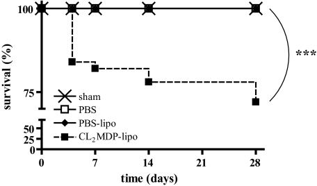
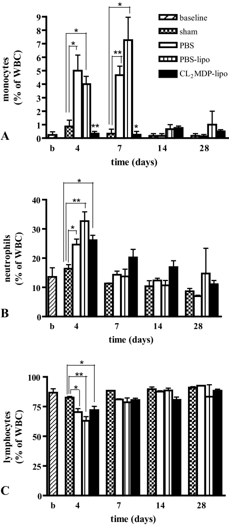
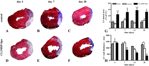
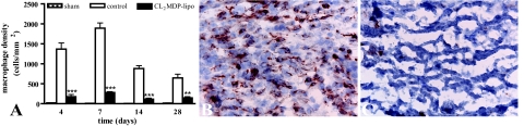
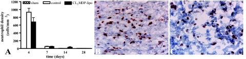
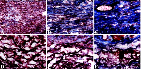
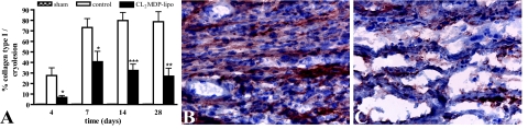
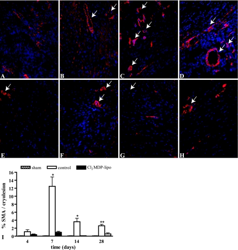
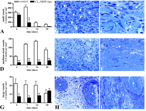
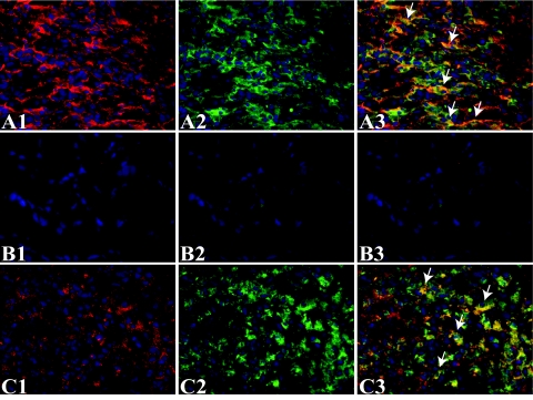
Macrophages have been suggested to be beneficial for myocardial wound healing. We investigated the role of macrophages in myocardial wound healing by inhibition of macrophage infiltration after myocardial injury. We used a murine cryoinjury model to induce left ventricular damage. Infiltrating macrophages were depleted during the 1st week after cryoinjury by serial intravenous injections of clodronate-containing liposomes. After injury, the presence of macrophages, which secreted high levels of transforming growth factor-beta and vascular endothelial growth factor-A, led to rapid removal of cell debris and replacement by granulation tissue containing inflammatory cells and blood vessels, followed by myofibroblast infiltration and collagen deposition. In macrophage-depleted hearts, nonresorbed cell debris was still observed 4 weeks after injury. Secretion of transforming growth factor-beta and vascular endothelial growth factor-A as well as neovascularization, myofibroblast infiltration, and collagen deposition decreased. Moreover, macrophage depletion resulted in a high mortality rate accompanied by increased left ventricular dilatation and wall thinning. In conclusion, infiltrating macrophage depletion markedly impairs wound healing and increases remodeling and mortality after myocardial injury, identifying the macrophage as a key player in myocardial wound healing. Based on these findings, we propose that increasing macrophage numbers early after myocardial infarction could be a clinically relevant option to promote myocardial wound healing and subsequently to reduce remodeling and heart failure.


Figures

Figure 1
Figure 1
Kaplan-Meier analysis of survival of sham-operated mice (n = 20) and of cryoinjured mice intravenously injected with PBS (n = 40), PBS liposomes (n = 12), and CL2MDP liposomes (n = 47). Significant differences are indicated as ***P < 0.001.
Figure 2
Figure 2
Differential white blood cell counts in the peripheral blood of untreated, unoperated mice (baseline), sham-operated mice and cryoinjured mice intravenously injected with PBS, PBS liposomes, and CL2MDP liposomes. A: In the Cl2MDP liposome-treated mice, monocyte levels were significantly reduced on days 4 and 7 after cryoinjury compared with PBS and PBS liposome-injected mice. Levels of neutrophils (B) and lymphocytes (C) were similar in mice intravenously injected with PBS, PBS liposomes, and CL2MDP liposomes at all time points. Significant differences are indicated as *P < 0.05, **P < 0.01. Bars represent mean ± SEM.
Figure 3
Figure 3
Left ventricular lumen area and wall thickness in the hearts of sham-operated mice, control mice, and mice intravenously injected with CL2MDP liposomes. A–F: Representative Masson’s trichrome stained cross sections of the left ventricles of control (A–C) and CL2MDP liposome-treated (D–F) mice on days 4 (A and D), 7 (B and E), and 28 (C and F) after cryoinjury. Original magnification, ×15. G: LV lumen area. H: LV wall thickness. CL2MDP liposome-mediated macrophage depletion results in a significant wall thinning and increase in LV lumen area compared with controls and sham-operated mice. Significant differences compared with controls are indicated as *P < 0.05, **P < 0.01, ***P < 0.001. Bars represent mean ± SEM.
Figure 4
Figure 4
Macrophage density in the myocardium of sham-operated mice and in the myocardial cryolesions of control mice and mice intravenously injected with CL2MDP liposomes. A: Quantitative analysis of the macrophage density in the cryolesions. Significant differences compared with controls are indicated as **P < 0.01, ***P < 0.001. Bars represent mean ± SEM. B and C: F4/80 staining in the cryolesion of a control (B) and a CL2MDP liposome-injected (C) mouse 7 days after injury. High numbers of macrophages are present in the cryolesion during the 1st week after injury, whereas they are significantly reduced after CL2MDP liposome injection. Original magnification, ×400.
Figure 5
Figure 5
Neutrophil density in the myocardium of sham-operated mice and in the myocardial cryolesions of control mice and mice intravenously injected with CL2MDP liposomes. A: Quantitative analysis of the neutrophil density in the cryolesions. Bars represent mean ± SEM. B and C: Anti-neutrophil staining in the cryolesion of a control (B) and a CL2MDP liposome-injected (C) mouse 4 days after injury. Neutrophil density is similar in both groups. Original magnification, ×400.
Figure 6
Figure 6
Cardiomyocyte clearance and collagen deposition in the myocardial cryolesions of control mice (A–C) and mice intravenously injected with CL2MDP liposomes (D–F) stained by Masson’s trichrome (blue, collagen; red, myocardial cells) on days 4 (A and D), 7 (B and E), and 28 (C and F) after cryoinjury. Four days after cryoinjury extensive inflammatory cell infiltration and a complete clearance of necrotic cardiomyocytes can be observed in the cryolesions of control mice (A). Furthermore, small collagen fibrils are present throughout the cryolesion. After day 4, the amount of collagen progressively increases (B and C), resulting in a highly collagenous scar on day 28. In contrast, on day 4, the cryolesion of macrophage-depleted mice shows only low numbers of inflammatory cells and numerous remnants of necrotic cardiomyocytes (D). These remnants can still be observed on days 14 (E) and 28 (F) after cryoinjury. Moreover, the amount of deposited collagen is low compared with controls at all time points. Magnification, ×400.
Figure 7
Figure 7
Collagen type I expression in the myocardium of sham-operated mice and in the myocardial cryolesions of control mice and mice intravenously injected with CL2MDP liposomes. A: Quantitative analysis of collagen type I deposition in the cryolesions. Significant differences compared with controls are indicated as *P < 0.05, **P < 0.01, ***P < 0.001. Bars represent mean ± SEM. B and C: Collagen type I staining in the cryolesion of a control mouse (B) and a CL2MDP liposome-injected mouse (C) 7 days after injury. Collagen type I deposition is low in liposome-mediated macrophage-depleted mice compared with controls. Original magnification, ×400.
Figure 8
Figure 8
α-SMA staining in the myocardial cryolesions. A–H: α-SMA staining (red) in the myocardium of sham-operated mice and in the cryolesion of control mice (A–D) and mice intravenously injected with CL2MDP liposomes (E–H) 4 (A and E), 7 (B and F), 14 (C and G), and 28 (D and H) days after injury. Original magnification, ×200. Spindle-shaped α-SMA-positive myofibroblasts accumulated in the border zone of the cryolesion 4 days after cryoinjury (A) and were present throughout the cryolesion on day 7 (B). On days 14 (C) and 28 (D), α-SMA staining was predominantly localized in smooth muscle cells of blood vessels. Only low numbers of myofibroblasts infiltrated into the cryolesion of macrophage-depleted mice (E–H). Furthermore, fewer α-SMA-positive vascular structures were observed in the cryolesions of macrophage-depleted mice compared with controls on days 14 and 28 (G and H). Arrows indicate examples of α-SMA-positive blood vessels, which were excluded from semiquantitative analysis. Nuclei are stained by DAPI (blue). I: Quantitative analysis of the α-SMA staining in the cryolesions. Significant differences compared with controls are indicated as *P < 0.01, **P < 0.001. Bars represent mean ± SEM.
Figure 9
Figure 9
Vascularization in myocardial cryolesions of control mice and mice intravenously injected with CL2MDP liposomes. A, D, and G: Number of small (A), medium (D), and large (G) vessels present in the cryolesion. Significant differences compared with controls are indicated as *P < 0.05, **P < 0.01. Bars represent mean ± SEM. B and C: Micrographs of small vessels (selection indicated by arrows) in the cryolesion of a toluidine blue-stained, 2-μm-thick T-7100 section of a control mouse (B) and a mouse intravenously injected with CL2MDP liposomes (C) 7 days after injury. In some small vessels, erythrocytes can be observed (light blue). The number of small vessels is lower in liposome-mediated macrophage-depleted mice compared with controls. Magnification, ×1000. E and F: Medium-sized vessels (selection indicated by arrowheads) in the cryolesion of a control mouse (E) and a mouse intravenously injected with CL2MDP liposomes (F) 14 days after injury. The number of medium-sized vessels is lower in liposome-mediated macrophage-depleted mice compared with controls. Magnification, ×400. H and I: Large vessels (asterisks) in the cryolesion of a control mouse (H) and a mouse intravenously injected with CL2MDP liposomes (I) 28 days after injury. Magnification, ×400.
Figure 10
Figure 10
Macrophage TGF-β and VEGF-A expression in myocardial cryolesions of control mice and mice intravenously injected with CL2MDP liposomes 7 days after injury. A and B: Immunofluorescent double staining using an anti-TGF-β antibody (A1 and B1, red) and an anti-F4/80 antibody for macrophages (A2 and B2, green) after PBS (A) or CL2MDP liposome (B) injection. Arrows in the merged micrographs (A3 and B3) indicate a selection of co-localization of TGF-β and macrophages. C: Immunofluorescent double staining using an anti-VEGF-A antibody (C1, red) and an anti-F4/80 antibody (C2, green) for macrophages after PBS (A) injection. Arrows in the merged micrograph (C3) indicate a selection of co-localization of VEGF-A and macrophages. A high level of TGF-β and VEGF-A is present in the cryolesion (A1 and C1). The merged micrographs show co-localization of TGF-β (A3) and VEGF-A (C3) with macrophages in the cryolesion. TGF-β (B) and VEGF-A (data not shown) expression was markedly reduced in the cryolesions of macrophage-depleted mice. Magnification, ×400.

Similar articles

Cited by

References

    1. Diez-Roux G, Lang RA. Macrophages induce apoptosis in normal cells in vivo. Development. 1997;124:3633–3638. - PubMed
    1. Leibovich SJ, Wiseman DM. Macrophages, wound repair and angiogenesis. Prog Clin Biol Res. 1988;266:131–145. - PubMed
    1. Mustoe TA, Pierce GF, Thomason A, Gramates P, Sporn MB, Deuel TF. Accelerated healing of incisional wounds in rats induced by transforming growth factor-beta. Science. 1987;237:1333–1336. - PubMed
    1. Leibovich SJ, Danon D. Promotion of wound repair in mice by application of glucan. J Reticuloendothel Soc. 1980;27:1–11. - PubMed
    1. Danon D, Kowatch MA, Roth GS. Promotion of wound repair in old mice by local injection of macrophages. Proc Natl Acad Sci USA. 1989;86:2018–2020. - PMC - PubMed

Substances