. 2007 Mar;170(3):830-42.
doi: 10.2353/ajpath.2007.060455.

Involvement of hypoxia-inducible transcription factors in polycystic kidney disease

Affiliations

Involvement of hypoxia-inducible transcription factors in polycystic kidney disease

Wanja Michael Bernhardt et al. Am J Pathol. 2007 Mar.

Abstract

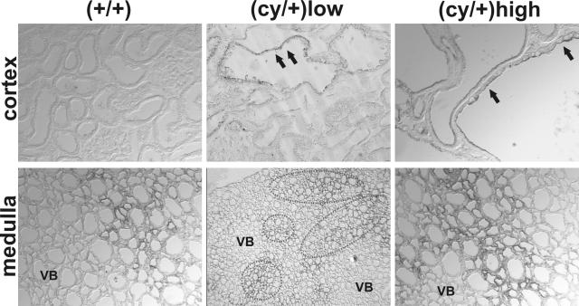
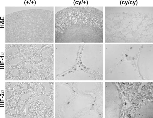
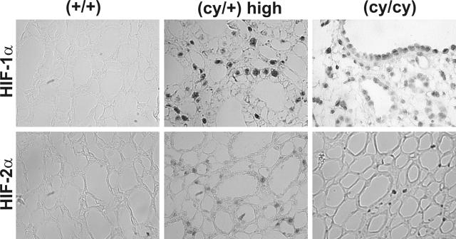
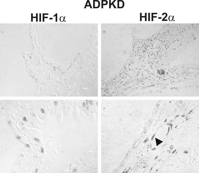
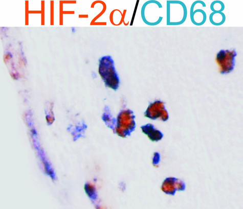
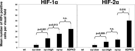
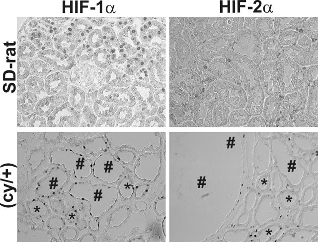
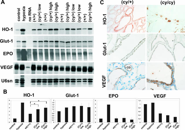
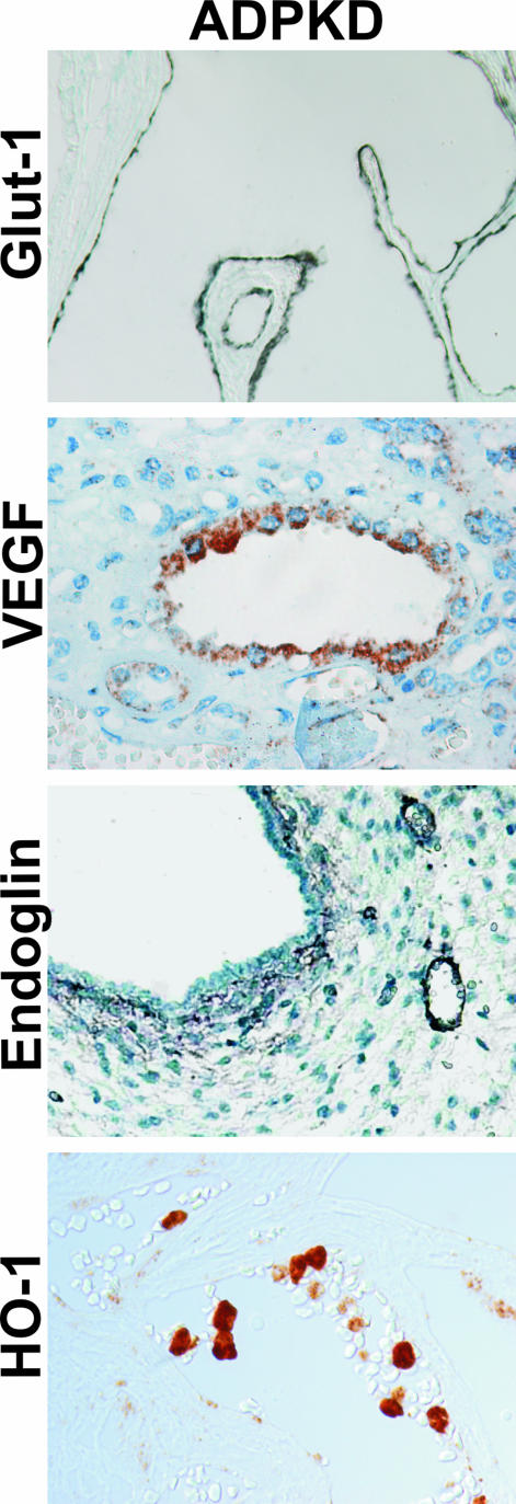
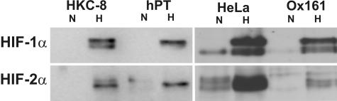
In polycystic kidney disease (PKD), erythropoietin (EPO) production and interstitial vascularization are increased compared with other kidney diseases. EPO and several angiogenic factors are controlled by hypoxia-inducible transcription factors (HIFs), which are composed of a constitutive beta-subunit and two alternative alpha-subunits (HIF-1alpha, HIF-2alpha). We hypothesized that cyst expansion may result in pericystic hypoxia and consecutive up-regulation of HIF and thus examined the expression of HIF-alpha and HIF target genes in human PKD and in a rodent PKD model. HIF-1alpha and HIF-2alpha were found to be up-regulated in cyst epithelium and cells of cyst walls, respectively. The distinct expression pattern of the HIF-alpha isoforms closely resembles the respective pattern in normal kidneys under systemic hypoxia. Pimonidazole staining, a marker for tissue hypoxia, confirmed the existence of regional hypoxia in polycystic kidneys. Immunohistochemistry for selected target genes implicated a role for HIF-1alpha in vascular endothelial growth factor and Glut-1 activation and HIF-2alpha in endoglin and EPO stimulation. Polycystin-deficient cells showed physiological, oxygen-dependent HIF-alpha modulation, excluding a direct influence of polycystin deficiency on HIF-alpha regulation. In conclusion, HIF accumulation in human and rat PKD seems to be responsible for increased EPO production and pericystic hypervascularity and may have an impact on progression of PKD.


Figures

Figure 1
Figure 1
Evidence for hypoxia in kidneys of (cy/+) rats. Pimonidazole staining (black) of kidneys of heterozygously affected Han:SPRD rats (cy/+) revealed hypoxia in cortical cyst epithelia (arrows) in animals with low-grade [(cy/+) low] as well as high-grade PKD [(cy/+) high], whereas wild-type animals (+/+) stained negative for pimonidazole in the renal cortex. In the renal medulla, the extent of hypoxia as indicated by pimonidazole staining seems to be more severe in (cy/+) animals in comparison to wt animals. Adjacent to vascular bundles pimonidazole was not detectable (VB, vascular bundles). Original magnifications: ×200 (top, middle); ×400 (top, right).
Figure 2
Figure 2
HIF-α accumulates in the renal cortex of (cy/+) and (cy/cy) rats. Top: Representative H&E-stained kidney sections of unaffected (+/+) and heterozygously (cy/+) and homozygously affected (cy/cy) Han:SPRD rats. In (+/+) animals, HIF-α was not detectable (left). Comparable focal staining of both HIF-α-isoforms occurred in (cy/+) and (cy/cy) rat-kidneys (middle and right columns). Cell populations expressing HIF-1α and HIF-2α protein were consistently different: HIF-1α was expressed in epithelial cells of the cysts, whereas HIF-2α occurred in pericystic stromal and endothelial cells. Original magnifications: ×12.5 (top); ×400 (left, middle and bottom rows); ×1000 (middle and right).
Figure 3
Figure 3
HIF-α accumulates in the renal medulla of (cy/+) and (cy/cy) rats. As in the renal cortex, HIF-α clearly accumulates in the renal papilla of (cy/+) and (cy/cy) rats with the same cellular distribution pattern of HIF-1α and -2α, whereas no HIF-α was detectable in the renal papilla of wild-type (+/+) rats. Original magnifications, ×400.
Figure 4
Figure 4
HIF-α accumulates in ADPKD. Both HIF-α isoforms are expressed in human ADPKD. The cellular distribution of HIF-1α (epithelial cells, left) and HIF-2α [pericystic stromal cells and endothelial cells (arrowhead), right] resembles the distributional pattern in PKD-rats. Original magnifications: ×100 (top); ×1000 (bottom).
Figure 5
Figure 5
HIF-2α is expressed in neutrophils in ADPKD. Double labeling was performed for HIF-2α (brown) and the leukocyte marker CD68 (blue), revealing that HIF is stabilized in human neutrophils in the interstitium of ADPKD kidneys. Original magnifications, ×630.
Figure 6
Figure 6
Scores for HIF-α in rat and human PKD. The mean number of HIF-1α- and HIF-2α-positive cells increases with increasing severity of the disease in rats. In kidneys of wt animals, HIF-α is absent. In human ADPKD kidneys, the highest numbers for HIF-α-positive cells were found, with higher scores for HIF-2α than for HIF-1α.
Figure 7
Figure 7
Increased HIF-α accumulation in phlebotomized (cy/+) rats. Cellular specificity of HIF-α is conserved under anemic conditions, but the number of cells staining positive for HIF-α increases in anemic PKD rats (bottom). In addition to the cystic epithelium (#), HIF-1α strongly stained positive in unaffected tubular epithelial cells (∗). The number of interstitial cells reacting positive for HIF-2α also increases under anemic conditions in kidneys of (cy/+) rats. Phlebotomized healthy Sprague-Dawley rats were used as controls (top). Original magnifications, ×400.
Figure 8
Figure 8
Expression of HIF target genes in (+/+), (cy/+), and (cy/cy) rats. The mRNA levels of the HIF target genes HO-1, Glut-1, EPO, and VEGF were determined by RNase protection assay (A) and quantified by densitometry (B). RNase protection assay revealed different levels of EPO mRNA in kidneys of Han:SPRD rats independent of the genetic status (A, B), whereas HO-1 mRNA levels correlated with the grade of disease (A, B), with a significant increase in mRNA in (cy/+) and (cy/cy) kidneys compared with wild-type (+/+) animals (B). Glut-1 levels were comparable with hypoxia but were not significantly different between (+/+), (cy/+), and (cy/cy) animals (A, B). Renal RNA extracts from normoxic (control) and hypoxic normal Sprague-Dawley rats served as controls. C: Glut-1 and VEGF protein were detectable in the cytoplasm of epithelial cells of the renal cysts of heterozygous (cy/+) and homozygous (cy/cy) rats. HO-1 stained positive in cyst epithelium and in (cy/cy) kidneys in addition to interstitial leukocytes. *P < 0.05.
Figure 9
Figure 9
Expression of HIF target genes in ADPKD. Glut-1 and VEGF are strongly positive in the cytoplasm of epithelial cells of the renal cysts. Endoglin, involved in neovascularization, is expressed in pericystic and interstitial vessels. HO-1 stained positive in macrophages. Original magnifications: ×400 (Glut-1, endoglin); ×630 (VEGF, HO-1).
Figure 10
Figure 10
Normal hypoxic up-regulation of HIF-α in polycystin-1-deficient cells. Immunoblots showing no differences in HIF-α regulation between HeLa cells, HKC-8 cells and primary proximal tubular cells (hPT) and polycystin-1-deficient Ox161-cells: strong increase in HIF-1α (top row) and HIF-2α (bottom row) under hypoxic conditions.

Similar articles

Cited by

References

    1. Wilson PD. Polycystic kidney disease. N Engl J Med. 2004;350:151–164. - PubMed
    1. Gabow PA. Autosomal dominant polycystic kidney disease. N Engl J Med. 1993;329:332–342. - PubMed
    1. Cowley BD., Jr Recent advances in understanding the pathogenesis of polycystic kidney disease: therapeutic implications. Drugs. 2004;64:1285–1294. - PubMed
    1. Maïz HB, Abderrahim E, Zouaghi K. Anemia and end-stage renal disease in the developing world. Artif Organs. 2002;26:760–764. - PubMed
    1. Abbott KC, Agodoa LY. Polycystic kidney disease at end-stage renal disease in the United States: patient characteristics and survival. Clin Nephrol. 2002;57:208–214. - PubMed

Publication types

Substances