. 2007 Mar;170(3):1003-17.
doi: 10.2353/ajpath.2007.060583.

Clostridium sordellii lethal toxin kills mice by inducing a major increase in lung vascular permeability

Affiliations

Clostridium sordellii lethal toxin kills mice by inducing a major increase in lung vascular permeability

Blandine Geny et al. Am J Pathol. 2007 Mar.

Abstract

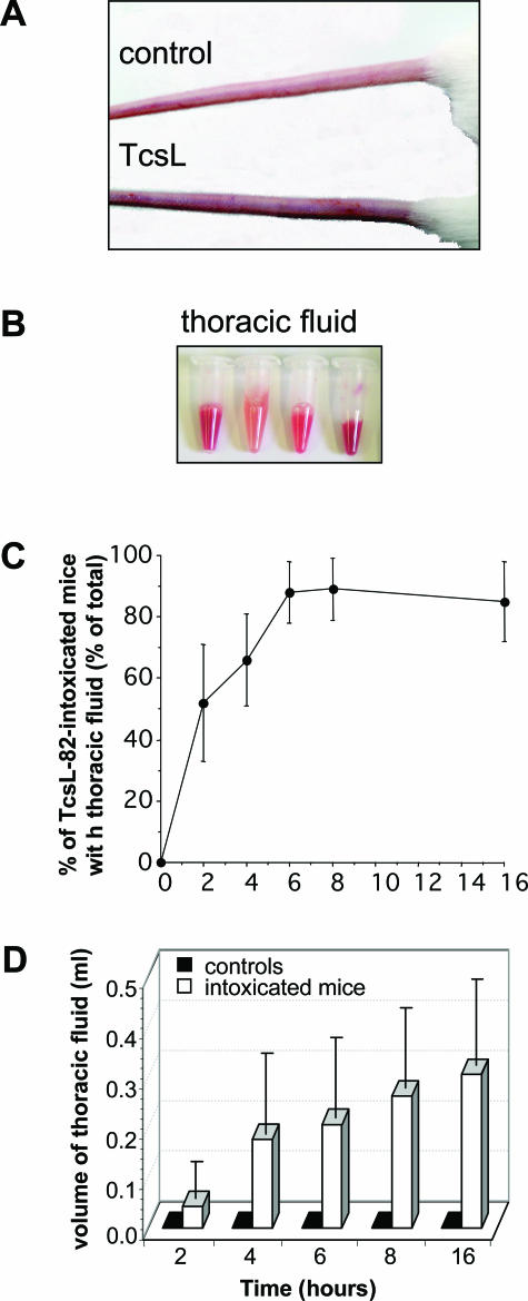
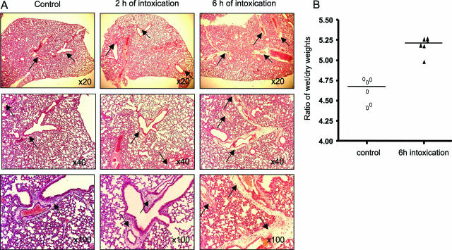
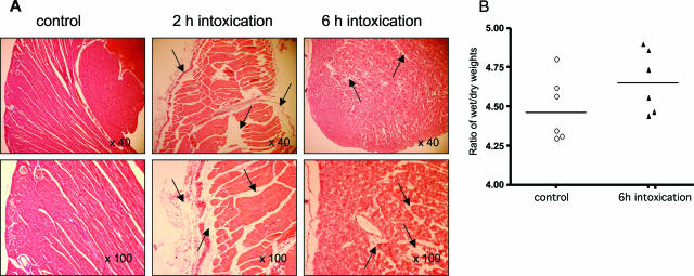
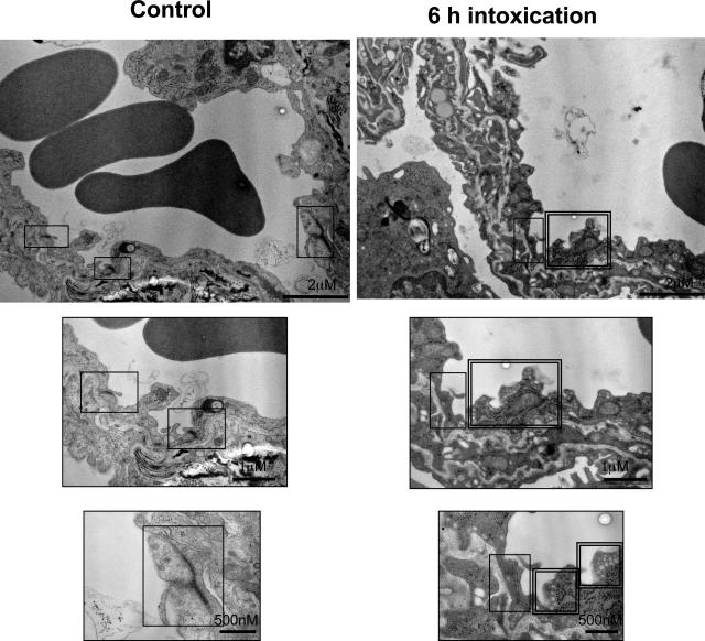
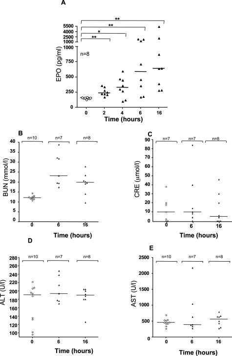
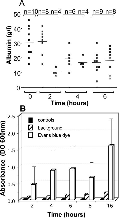
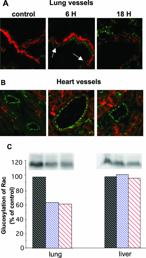
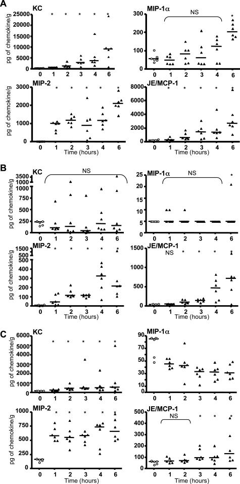
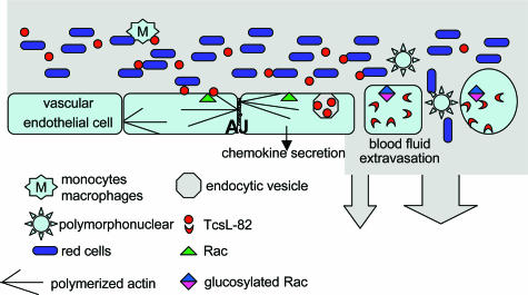
When intraperitoneally injected into Swiss mice, Clostridium sordellii lethal toxin reproduces the fatal toxic shock syndrome observed in humans and animals after natural infection. This animal model was used to study the mechanism of lethal toxin-induced death. Histopathological and biochemical analyses identified lung and heart as preferential organs targeted by lethal toxin. Massive extravasation of blood fluid in the thoracic cage, resulting from an increase in lung vascular permeability, generated profound modifications such as animal dehydration, increase in hematocrit, hypoxia, and finally, cardiorespiratory failure. Vascular permeability increase induced by lethal toxin resulted from modifications of lung endothelial cells as evidenced by electron microscopy. Immunohistochemical analysis demonstrated that VE-cadherin, a protein participating in intercellular adherens junctions, was redistributed from membrane to cytosol in lung endothelial cells. No major sign of lethal toxin-induced inflammation was observed that could participate in the toxic shock syndrome. The main effect of the lethal toxin is the glucosylation-dependent inactivation of small GTPases, in particular Rac, which is involved in actin polymerization occurring in vivo in lungs leading to E-cadherin junction destabilization. We conclude that the cells most susceptible to lethal toxin are lung vascular endothelial cells, the adherens junctions of which were altered after intoxication.


Figures

Figure 1
Figure 1
TcsL-82 induces anoxia in mice and fluid accumulation in the thoracic cavity. After intoxication for 6 hours with intraperitoneally injected TcsL-82 (15 ng/mouse), mice exhibit signs of anoxia such as darkened extremities (A) and various volumes of serohemorrhagic fluid were collected in the thoracic cage (B). In three different experiments, six to eight mice were sacrificed at various times after intraperitoneal injection of TcsL-82. For each experiment, the percentage of mice with fluid in the thoracic cage was calculated (C), and the volume of fluid present in the thoracic cage was measured (open bars) (D). Two control mice, injected with the diluent only, were euthanized at each time point. No fluid was collected from the thoracic cage of control mice (black squares). Results in C and D represent the mean ± SEM of the three different experiments.
Figure 2
Figure 2
Histopathological analysis of the lung of TcsL-82-treated Swiss mice. A: H&E staining of lungs from HBSS-injected control mice and from mice intoxicated for 2 and 6 hours with intraperitoneally injected TcsL-82 (15 ng/mouse). Lungs were examined with ×20, ×40, and ×100 lenses. Perivascular space corresponding to the area where exudate is observed after intoxication (×20 lens and higher magnifications) is very limited in control lung, increased already after 2 hours, and is massive after 6 hours of intoxication (arrows). B: Ratios of wet-to-dry weights of lung from control and intoxicated mice with TcsL-82 (15 ng/mouse, i.p.). Horizontal bars indicate the median values. The values of control versus intoxicated mice were analyzed by the Wilcoxon signed-rank test using arbitrarily the median of the control population as the hypothetical value for comparison; P = 0.0156.
Figure 3
Figure 3
Histopathological analysis of the hearts of TcsL-82-treated Swiss mice. A: H&E staining of hearts from HBSS-injected control mice as in Figure 2 and from mice intoxicated for 2 and 6 hours with intraperitoneally injected TcsL-82 (15 ng/mouse). Hearts were examined with ×40 and ×100 lenses. Exudate (arrows) is observed at the pericardium and endocardium levels and infiltrates heart muscle, which appears roughly dilacerated after 2 hours. After 6 hours of intoxication, myocardiocytes appear individualized by exudate. B: Ratios of wet-to-dry weight of lung from control and 6-hour intoxicated mice. Horizontal bars indicate the median values. The values of control versus intoxicated mice were analyzed by the Wilcoxon signed-rank test using arbitrarily the median of the control population as the hypothetical value for comparison; P = 0.047.
Figure 4
Figure 4
Transelectron microscopy analysis of lung vessels in a control mouse and a mouse intraperitoneally injected with TcsL-82 (15 ng/mouse) 6 hours before organ fixation (as in Figure 2). Top images are general views showing a highly modified aspect of the vessel after TcsL-82 intoxication. As visualized on enlarged endothelial structures (middle and bottom images), in the intoxicated mouse staining of intercellular junctions (single frame) is weaker than in control. Intracellular vesicles (double frame) are abundant in endothelial cells from intoxicated mouse. Original magnifications, ×24,000 (top).
Figure 5
Figure 5
TcsL-82-induced plasma EPO increases and does not modify renal and hepatic functions. Mice were intraperitoneally injected with 0.5 ml of HBSS containing or not TcsL-82 (15 ng/mouse). At the indicated times, eight mice were euthanized, and sera were collected. The amount of EPO present in plasma of control or TcsL-82-intoxicated mice was estimated by ELISA using a R&D kit as indicated in the Materials and Methods section. Horizontal bars indicate the mean values. The Wilcoxon signed-rank test was performed between the values of the control versus intoxicated mice at each time point using arbitrarily the median of control batch as the hypothetical value for comparison. The probability P of a significant difference in TcsL-82-intoxicated mice is indicated: *0.05 > P > 0.01, **0.01 > P > 0.001 for each time point. Renal and hepatic functions were analyzed by measuring on the one hand BUN and CRE and, on the other hand, alanine aminotransferase and aspartate aminotransferase from serum at 0, 6, and 16 hours after intraperitoneal injection of 15 ng of TcsL-82. n represents the number of sera studied for each group.
Figure 6
Figure 6
Albumin in serum and thoracic fluid and vascular permeability measurements. A: Albumin was measured in the serum (S, squares) and in the thoracic fluid (L, circles) in animals injected with TcsL-82 (15 ng/mouse) 0, 2, 4, and 6 hours before sacrifice. The number (n) of sera and thoracic fluids analyzed ranged from 6 to 10 and 4 to 8, respectively, for each time point as indicated. The mean is indicated (horizontal bars). B: Vascular permeability was estimated by measuring the amount of Evans blue present in the thoracic fluid. For that purpose, 15 to 30 minutes before mouse sacrifice, Evans blue dye was injected intravenously, and its amount present in the thoracic fluid was quantified by spectrophotometric analysis at 600 nm (open bars). The relevant background was the spectrophotometric analysis of the thoracic fluid present in mice not injected with Evans blue dye (striped bars). For each time point, two control mice were injected with the diluent only and with Evans blue 30 minutes before being euthanized. No fluid was collected from the thoracic cage of control mice (see Figure 1D), and therefore no Evans blue was measurable (black squares). The graphs are the means ± SD of three different experiments with four mice at each time point.
Figure 7
Figure 7
TcsL-82 modifies adherens junctions and actin cytoskeleton in lung vessels and glucosylates small GTPases in lung. Lung (A) and heart (B) vessel cryosections of control and TcsL-82-treated mice for 6 and 18 hours were analyzed with an anti-mouse VE-cadherin detected with a relevant secondary antibody coupled to fluorescein isothiocyanate, and with tetramethyl-rhodamine isothiocyanate phalloidin. C: Lungs and liver of mice intoxicated for 0, 6, and 16 hours with intraperitoneally injected TcsL-82 (15 ng/mouse) were homogenized and glucosylation with [14C]UDP-glucose in the presence of TcsL-82 was performed as previously reported. Autoradiograph of the gel shows the amount of in vivo glucosylated GTPases after TcsL-82 intoxication. Quantification of the radiolabeled bands was performed using Denylab 2.5.2. software (bottom; DynaLab Inc., Hong Kong). The experiments shown are representative of three different ones.
Figure 8
Figure 8
TcsL-82 increases the level of certain chemokines, primarily in lung and plasma and, to a lesser extent, in liver. Mice were injected intraperitoneally with 0.5 ml of HBSS containing or not TcsL-82 (15 ng/mouse). After the indicated times, mice were euthanized and sacrificed, blood, lung, and liver were collected. Plasma was sampled after centrifugation. After weighing, organs were homogenized and centrifuged, and the extracted supernatants were collected. KC, MIP-1α, MIP-2, and JE/MCP-1 were measured by ELISA using R&D kits in each sample from lungs (A), plasma (B), and liver (C). Chemokine measurements represent individual values of six samples for each time point. Horizontal bars represent the median value at each time point. The Wilcoxon signed-rank test was performed between the values of the control versus intoxicated mice at each time point using arbitrarily the median of the control population as the hypothetical value for comparison. The probability P of a significant difference in TcsL-82-intoxicated mice is indicated: *0.05 > P > 0.01; NS, nonsignificant.
Figure 9
Figure 9
Diagram of TcsL-82 mechanism of action on lung vascular endothelial cells. TcsL-82 after intraperitoneal injection is recognized by a still unknown receptor at the surface vascular endothelial cells. This large clostridial toxin is then endocytosed, and its enzymatic domain (N-terminal part) is translocated into the cytosol where it glucosylates Rac and Ras proteins (Ras, Rap, Ral) and depolymerizes the actin cytoskeleton, thereby modifying adherens junctions (AJ).

Similar articles

Cited by

References

    1. Cunniffe JG. Clostridium sordellii bacteraemia. J Infect. 1996;33:127–129. - PubMed
    1. Kimura AC, Higa JI, Simpson G, Vargas Y, Vugia DJ. Outbreak of necrotizing fascillitis due to Clostridium sordellii among black-tar heroin users. Clin Infect Dis. 2004;38:e87–e89. - PubMed
    1. Fischer M, Bhatnagar J, Guarner J, Reagan S, Hacker JK, Van Meter SH, Poukens V, Whiteman DB, Iton A, Cheung M, Dassey DE, Shieh WJ, Zaki SR. Fatal toxic shock syndrome associated with Clostridium sordellii after medical abortion. N Engl J Med. 2005;353:2352–2360. - PubMed
    1. Miech RP. Pathophysiology of mifepristone-induced septic shock due to Clostridium sordellii. Ann Pharmacother. 2005;39:1483–1488. - PubMed
    1. Bitti A, Mastrantonio P, Spigaglia P, Urru G, Spano AI, Moretti G, Cherchi GB. A fatal postpartum Clostridium sordellii associated toxic shock syndrome. J Clin Pathol. 1997;50:259–260. - PMC - PubMed

Publication types

MeSH terms