. 2007 Feb 15;21(4):436-49.
doi: 10.1101/gad.1509007.

A core transcriptional network for early mesoderm development in Drosophila melanogaster

Affiliations

A core transcriptional network for early mesoderm development in Drosophila melanogaster

Thomas Sandmann et al. Genes Dev. .

Abstract

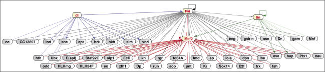
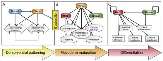
Embryogenesis is controlled by large gene-regulatory networks, which generate spatially and temporally refined patterns of gene expression. Here, we report the characteristics of the regulatory network orchestrating early mesodermal development in the fruitfly Drosophila, where the transcription factor Twist is both necessary and sufficient to drive development. Through the integration of chromatin immunoprecipitation followed by microarray analysis (ChIP-on-chip) experiments during discrete time periods with computational approaches, we identified >2000 Twist-bound cis-regulatory modules (CRMs) and almost 500 direct target genes. Unexpectedly, Twist regulates an almost complete cassette of genes required for cell proliferation in addition to genes essential for morophogenesis and cell migration. Twist targets almost 25% of all annotated Drosophila transcription factors, which may represent the entire set of regulators necessary for the early development of this system. By combining in vivo binding data from Twist, Mef2, Tinman, and Dorsal we have constructed an initial transcriptional network of early mesoderm development. The network topology reveals extensive combinatorial binding, feed-forward regulation, and complex logical outputs as prevalent features. In addition to binary activation and repression, we suggest that Twist binds to almost all mesodermal CRMs to provide the competence to integrate inputs from more specialized transcription factors.


Figures

Figure 1.
Figure 1.
Regions identified by Twist ChIP-on-chip represent functional enhancers in vivo. (A) Twist enhancers are more frequently located in intronic regions, as opposed to regions 5′ or 3′ relative to the nearest gene locus. Genes misexpressed in either twist loss-of-function or Toll10B gain-of-function mutants (B) or coexpressed with Twist (C) (information about expression patterns was compiled from the Berkeley Drosophila Genome Project in situ database, FlyBase, and published literature) are significantly enriched among genes with intronic Twist binding (right) compared with the whole Drosophila genome (left) (Fisher test, [***] p < 1e-10). (D–I) Schematic diagrams indicating the gene locus (exons in gray, introns in orange) together with Twist-enriched sequences (red bars, top: 2–4 h, bottom: 4–6 h) and the enhancer region tested (double-headed arrow). Known enhancers for T48 are indicated in green (Strutt and White 1994). (D′–I′) In situ hybridization of transcripts in wild-type embryos. H″ is reproduced from the Berkeley in situ database. (D″– I″) In situ hybridization of GFP transcripts in transgenic enhancer-GFP embryos assaying the regions indicated by double-headed arrows in D–I, respectively. All enhancers drive specific blastodermal or mesodermal expression reproducing (at least part of) the endogenous genes’ expression. All embryos are oriented with anterior facing left and the dorsal side up.
Figure 2.
Figure 2.
Twist activity is essential for the expression of many of its target genes. In situ hybridization of Twist targets in wild-type embryos (top region of panel) and twist mutant embryos (bottom). All embryos are at stage 5, with anterior to the left and dorsal to the top. The images show triple fluorescent in situ hybridization with probes against the following transcripts: twist (blue), inflated (red), and the target gene (green). As inflated is a target of Twist, lack of inflated expression serves as a functional marker for twist homozygous mutant embryos. Note, the presumptive mesoderm expression of CG12177, CG32982, CG4221, CG9005, and T48 is severely reduced or absent in twist mutant embryos. NetA expression appears largely unaffected.
Figure 3.
Figure 3.
Identification of novel enhancers for genes differentially expressed along the D–V axis. (A–G) Schematic diagrams indicating the target locus (exons in gray, introns in orange) together with Twist-enriched sequences (red bars, top: 2–4 h, bottom: 4–6 h), the location of known regulatory sequence (green bars), and the novel enhancer region tested (double-headed arrow). (A′–G′ In situ hybridization of lacZ transcripts in transgenic enhancer lines. (A′,B′) The enhancers for cactus (cact) and stumps drive expression in all or a subset of cells of the ventral mesoderm, respectively. The WntD enhancer displays repression in the central snail-expressing domain, and activation at the anterior and posterior poles (C′), while the crumbs (crb) enhancer is excluded from the entire ventral domain and is only activated in the ectoderm (D′). In addition to the recovery of known regulatory regions (E,G) novel enhancers were discovered for mir-1, vn, and sim (E′–G′). (H) Schematic overview of a transverse section through a stage 5 embryo. The nuclear Dorsal gradient (red) activates Twist expression (blue) on the ventral side of the embryo. Twist binds to CRMs associated with targets expressed in ventral (e.g., sna, stumps, cact, mir-1), lateral (m8, sim, ths, sog), and dorsal (zen, dpp) domains of the early embryo. (I) Twist regulates components of the Dorsal network; schematic model of the subcircuit leading to collaborative activation of targets in the ventral blastoderm. Twist binds to CRMs of all genes with a red border. Dashed lines indicate indirect regulation. (K) Twist also binds to CRMs regulating known components (red border) of the repressive complex associated with Dorsal.
Figure 4.
Figure 4.
Twist targets functional cassettes required for diverse developmental processes. (A) Important developmental processes are overrepresented among direct Twist target genes: Comparing the fraction of direct Twist targets (gray) annotated with specific GO-slim terms with the frequency of these terms among all annotated FlyBase genes (black) reveals significant enrichment of several GO processes (EASE resampling analysis, [***] p < 0.001, [**] p < 0.01). (B,C) Numerous FGF pathway members and important regulators of cell cycle progression are direct Twist targets: manual pathway mapping of FGF signaling (B) and cell cycle regulation in Drosophila (C). Genes with a red border were identified as direct Twist targets.
Figure 5.
Figure 5.
Twist occupies enhancers in a temporally regulated manner with Dorsal and Tinman. (A) Differentially enriched Twist-bound sequences: Roughly half of the CRMs are detected at both 2–4 h and 4–6 h (blue). About one-quarter of all detected regions are specific to either the early (green) or late (red) time periods. (B) Motifs for several characterized TFs are significantly enriched within Twist-bound CRMs: Scanning sequences bound specifically at 2–4 h (green), 4–6 h (red), or at both time periods (continuous, blue) with PWMs for known regulators in Drosophila revealed enrichment of Twist, Dorsal, Snail, and Tinman motifs within one or more of these temporal groups (Clover, [***] p < 0.001, [**] p < 0.01). (C) Dorsal co-occupies novel 2–4-h Twist-bound CRMs: ChIPs with two different α-Dorsal antisera (green, total n = 4) or preimmune serum (gray, n = 4) were performed at 2–4 h after egg laying and analyzed by quantitative real-time PCR using primers assaying predicted Dorsal sites within sequences enriched by Twist specifically at 2–4 h. Both known control sites (underlined, left) as well as all seven novel sites tested are significantly bound by Dorsal in vivo. The X-axis indicates the region tested; the Y-axis displays the level of enrichment as the ratio of enrichment using primers against the region of interest compared with primers covering a negative control region. (D) Tinman binds novel 4–6-h Twist-bound enhancers: Predicted Tinman-binding sites within continuously or late-bound sequences were assayed in α-Tinman (red) or mock (gray) ChIPs, using two independent antisera as described in C. All but one predicted site are significantly bound by Tinman in vivo. (**) p < 0.05, Wilcoxon rank test.
Figure 6.
Figure 6.
A core transcriptional network for early mesoderm development. Through the integration of ChIP-on-chip data for Twist and Mef2 with in silico predictions, ChIPs, and literature searches for Dorsal and Tinman, we identified CRMs that are cobound by at least two TFs during the same stages of development. The regulatory connections for CRMs fulfilling these criteria are shown for all genes coding for TFs. Color code: Dorsal (dark blue, known regulation; light blue, predicted interaction), Twist (gray), Mef2 (red), and Tinman (dark green, known regulation; light green, predicted interaction). The direction of regulation, if known, is indicated by pointed or bar-ended arrows. Feed-forward loops and combinatorial regulation of downstream regulators are dominant features controlling early mesoderm development.
Figure 7.
Figure 7.
Dynamic changes in the functional groups of genes targeted by Twist reflect developmental progression. (A) Before gastrulation, Twist collaborates with Dorsal to subdivide the embryo along the D–V axis and triggers target gene expression required for gastrulation. (B) Once the mesoderm has invaginated, Twist is associated with pan-mesodermal regulators (tin and Mef2) and CRMs regulating important “plug-ins” of development (Davidson 2006), providing the pluripotent cells with the competence to continue differentiation. (C) Twist expression is lost in cells activating gene batteries required for terminal differentiation (e.g., structural muscle proteins).

References

    1. Arora K., Nusslein-Volhard C., Nusslein-Volhard C. Altered mitotic domains reveal fate map changes in Drosophila embryos mutant for zygotic dorsoventral patterning genes. Development. 1992;114:1003–1024. - PubMed
    1. Barolo S., Carver L.A., Posakony J.W., Carver L.A., Posakony J.W., Posakony J.W. GFP and β-galactosidase transformation vectors for promoter/enhancer analysis in Drosophila. Biotechniques. 2000;29:726, 728, 730, 732. - PubMed
    1. Bate M., Rushton E., Currie D.A., Rushton E., Currie D.A., Currie D.A. Cells with persistent twist expression are the embryonic precursors of adult muscles in Drosophila. Development. 1991;113:79–89. - PubMed
    1. Baylies M.K., Bate M., Bate M. twist: A myogenic switch in Drosophila. Science. 1996;272:1481–1484. - PubMed
    1. Beer J., Technau G.M., Campos-Ortega J.A., Technau G.M., Campos-Ortega J.A., Campos-Ortega J.A. Lineage analysis of transplanted individual cells in embryos of Drosophila melanogaster. Roux’s Arch. Dev. Biol. 1987;196:222–230. - PubMed

Publication types

MeSH terms