. 2007 Feb 15;21(4):465-80.
doi: 10.1101/gad.406007.

Dickkopf-1 regulates gastrulation movements by coordinated modulation of Wnt/beta catenin and Wnt/PCP activities, through interaction with the Dally-like homolog Knypek

Affiliations

Dickkopf-1 regulates gastrulation movements by coordinated modulation of Wnt/beta catenin and Wnt/PCP activities, through interaction with the Dally-like homolog Knypek

Luca Caneparo et al. Genes Dev. .

Abstract

Dickkopf-1 (Dkk1) is a secreted protein that negatively modulates the Wnt/beta catenin pathway. Lack of Dkk1 function affects head formation in frog and mice, supporting the idea that Dkk1 acts as a "head inducer" during gastrulation. We show here that lack of Dkk1 function accelerates internalization and rostral progression of the mesendoderm and that gain of function slows down both internalization and convergence extension, indicating a novel role for Dkk1 in modulating these movements. The motility phenotype found in the morphants is not observed in embryos in which the Wnt/beta catenin pathway is overactivated, and that dominant-negative Wnt proteins are not able to rescue the gastrulation movement defect induced by absence of Dkk1. These data strongly suggest that Dkk1 is acting in a beta catenin independent fashion when modulating gastrulation movements. We demonstrate that the glypican 4/6 homolog Knypek (Kny) binds to Dkk1 and that they are able to functionally interact in vivo. Moreover, Dkk1 regulation of gastrulation movements is kny dependent. Kny is a component of the Wnt/planar cell polarity (PCP) pathway. We found that indeed Dkk1 is able to activate this pathway in both Xenopus and zebrafish. Furthermore, concomitant alteration of the beta catenin and PCP activities is able to mimic the morphant accelerated cell motility phenotype. Our data therefore indicate that Dkk1 regulates gastrulation movement through interaction with LRP5/6 and Kny and coordinated modulations of Wnt/beta catenin and Wnt/PCP pathways.


Figures

Figure 1.
Figure 1.
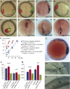
dkk1MO efficiently represses dkk1 translation, which induces severe reduction of the forebrain. (A–F) Loss of Dkk1 function represses formation of eye and telencephalon. Lateral views, anterior to the left, of heads of wild-type (A,C,E) and dkk1MO-injected (B,D,F) embryos. (G–J′) Rescue of dkk1GFP overexpression by Dkk1MO. (G–J) Lateral view of live 80% epiboly (under UV illumination, G′–J′) and prim5 (G,J) embryos after one-cell-stage injection of 50 ng/μL dkk1GFP alone (G,G′) or together with 0.4 mg/mL (H,H′), 1 mg/mL (I,I′), and 1.5 mg/mL (J,J′) Dkk1MO. With the exception of C and D, the embryos shown in this figure are alive. Bar, 100 μm.
Figure 2.
Figure 2.
Dkk1 regulates gastrulation movements. Lateral (A,B,E,F,I–Y), dorsal (C,D), and animal pole (G,H) views of control (A,C,E,G,I,K,N,Q–S), Dkk1MO-injected (B,D,F,H,J,L,O,T–V), and dkk1RNA-injected (M,P,W–Y) gastrula embryos. A–J and Q–Y show progression of the mesendoderm by markers in fixed embryos (A–J) and by observation of the GFP-expressing axial mesendoderm in live embryos (Q–Y). The black arrowhead indicates the position of the rostral-most mesendoderm. The red frame highlights the stages at which the difference in mesendoderm progression is obvious in the live Dkk1 morphants. (K–P) Lateral views of, in blue, neural plate (K–M) and epidermal (N–P) territories in wild-type (K,N), dkk1MO (L,O), and dkk1RNA (M,P) embryos. Red arrowhead shows the caudally shifted anterior edge of the neural territory.
Figure 3.
Figure 3.
More frequent mesendoderm internalization and faster rostral progression in the Dkk1 morphants and slowed-down internalization and CE defects in Dkk1 gain of function. (A–H) Dorsal (B–F), animal pole (A), and lateral (G,H) views of dkk1MO (A–C,E,F) or wild-type (D,G,H) embryos in which wild-type (red in live embryos and brown in fixed specimen) and dkk1MO (green in live embryos and dark blue in fixed specimen) cells have been transplanted inside the germ ring at onset of gastrulation. A–C show the same embryo at progressively later stages of gastrulation. Note that H shows transplanted dkk1RNA-expressing cells in dark blue. Dashed line indicates the midline. (I) Timing of internalization of 15 wild-type (blue) and dkk1MO (red) cells transplanted in the margin of early gastrula hosts. Measure of transplants in the lateral margin are represented by full dots and transplants in the axial margin (shield) are represented by empty dots. X represents time and Y represents the number of cells. (J) Measure of the AP length of the transplanted cell population at bud stage in microns (Y axis). Wild-type clones are shown in blue, dkk1MO are shown in red, and dkk1RNA are shown in green. (K) Measure of the distance of the leading transplanted cell from the embryo margin at 95% epiboly in microns (Y axis). Wild-type clones are shown in blue, dkk1MO are shown in red, and dkk1RNA are shown in green. (L) Expression of dkk1 in the marginal mesendoderm. Animal pole view of a shield stage wild-type embryo. (S) Shield (Spemann Organizer). Note the absence of transcript in the ventral margin and the graded level of expression from dorsal (S, shield) to lateral. (M,N) Cell shape and cohesion in the lateral (M) and dorsal (N) mesendoderm (arrows) as it internalizes in a live shield stage embryo. Inset in M shows a close-up of the internalizing lateral mesendoderm cell, emphasizing the mesenchymal shape of the lateral mesendoderm cells.
Figure 4.
Figure 4.
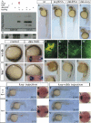
Gain of Wnt/βcatenin activity does not phenocopy the gastrulation movement defect seen in the Dkk1 morphants. (A–F) Lateral views of live prim5 (A,C,E) or dorsal views of lefty1 exression in fixed 90%–95% epiboly (B,D,F), wild-type (A,B), and mbl mutant injected with tcf3 + 3bMO (C,D), and dkk1MO (E,F) embryos. (G,H) Clone shape of transplanted wild type (G) or mbl mutant injected with tcf3 + 3bMO (H) in the host embryo at 75%–80% epiboly. (I,J) Lateral views of 75%–80% epiboly gscGFP transgenic live embryo (I) or dkk1MO coinjected with 20 pg of dominant-negative wnt8 RNA (J), showing the GFP-expressing axial mesendoderm in green under UV illumination. (K–N) Lateral views of 90% epiboly embryos showing the distribution of the wnt8-expressing cells (blue) transplanted at shield stage. The dose injected per embryo is indicated in the bottom right corner. (O) Timing of internalization of 25 wild-type (blue) and wnt8-expressing (10 pg, pink; 25 pg red; 40 pg purple) cells transplanted in the margin of early gastrula hosts. X represents time and Y represents the number of cells. (P) Measure of the distance of the leading transplanted cell from the embryo margin at 100% epiboly in microns (Y axis). Wild-type clones are shown in blue and wnt8-expressing clones are shown in pink (10 pg), red (25 pg), and purple (40 pg).
Figure 5.
Figure 5.
Knypek binds to Dkk1, is required for its propagation, and potentiates its effects. (A) Western blot of an immunoprecipitation assay. Protein extracts from cells transfected with different combinations of DNA (+) are either run untreated (last three lanes) or first immunoprecipitated with an anti-GFP antibody (first five lanes), then run on a gel. The gel is then transferred on filter and stained with an anti-Flag antibody (detecting the Flag-tagged Knypek protein) or an anti-GFP antibody (detecting the GFP [1] or dkkGFP fusion [2] proteins). (Lane 4) Red arrowshows the presence, after GFP immunoprecipitation, of Kny-Flag proteins in extracts from cells expressing both Dkk1GFP and KnyFlag. (B–E′) Lateral views, rostral to the left, of prim5 live wild-type (B), kny RNA-injected (C), dkk1RNA-injected (D), and kny + dkk1RNA-injected (E,E′) embryos. In E, the quantity of RNA injected is identical to the that used for the single injections in C and D. The phenotype shown in E and E′ are found in 31% and 64% of the injected embryos, respectively. (F–I) Animal pole views (insets) and lateral views of live or fixed (insets) wild type (F,G) and kny homozygous (H,I) injected with control (F,H) or dkk1 morpholino (G,I). Insets show expression patterns of hgg1 (rostral mesendoderm, hg) and brachyury (in the notochord, n). Arrowhead shows rostral end of the body axis. (J–M) Visualization of the dkk1GFP molecules (in green) secreted by the transplanted dkk1GFP-expressing wild-type cells (yellow) in wild-type (J,K) and kny−/− (L,M) hosts. K and M are high-magnification views of the distribution of secreted proteins away from the graft (or its absence at a distance from the clone in M). Note that the cell nuclei are visible as dark discs in unlabeled cells. The wild-type dkk1GFP-expressing cells are very intensely fluorescent when placed in the kny−/− mutant, masking the nucleus in these cells. (N–P) Lateral views, rostral to the left, of wild-type (N) or kny−/− (O,P) embryos, untreated (inset in N,O) or in which cells from a dkk1 RNA-injected donor embryo (in brown) have been transplanted in the shield at 50% epiboly (N,P). Note the shorter tail and big eye and brain in N. (P) No change in eye size is ever observed in transplanted kny−/− embryos; the brain size is generally comparable to untransplanted mutants. (Q–Z) Lateral views of live embryos (Q–V) or dorsal views of fixed gastrulae (W–Z). All embryos come from a kny+/− cross injected with either 1.8 pg of kny transcripts (Q–S,W,X) or 1.8 pg of kny and 2 pg of dkk1 transcripts (T–V,Y,Z). The number of embryos showing each illustrated phenotype is given in the bottom right corner of each picture. In W–Z, hgg1 expression shows rostral mesendoderm (hg) and brachyury, the notochord (n).
Figure 6.
Figure 6.
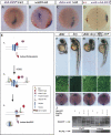
Dkk1 up-regulates the Wnt/PCP pathway. (A–C′) Lateral (A–C) and dorsal (A′–B′) views of bud stage wild type (A,A′), slb (B,B′), and slb injected with dkk1MO (C,C′) embryos, expressing hgg (in the hatching gland, hg), ntl (in the notochord, n), and dlx3b (in the neural plate border, nb). (D–G) Dkk1 blocks activin-induced animal cap elongation. Xenopus embryos were uninjected (D) or injected animally at the four- to eight-cell stage with activin (E), activin and wnt8 (F), or activin and dkk1. Animal caps were dissected from stage 9 embryos and cultured until sibling embryos had reached stage 17. (H–L) Dkk1 disrupts cell polarity. Xenopus embryos were injected at the two- to four-cell stage equatorially with mRFP mRNA, and either preprolactin (ctl) or the indicated mRNAs (top right corner). Explants of the dorsal upper blastopore lip were cut at stage 10.5, and cells were imaged by confocal microscopy. The yellow line indicates orientation of the medio-lateral axis. (L, left panel) Cell elongation was determined by the mean length-to-width ratio (LWR). (Right panel) Cell orientation was calculated as the percentage of cells with their long axis tilted >20° relative to the medio-lateral axis. (M) RT–PCR analysis of animal caps injected as in D–G for the indicated genes. (N) Dkk1 activates JNK. HA epitope-tagged JNK mRNA was injected either alone or in combination with mRNA of the indicated genes into two- to four-cell-stage embryos. HA-JNK was immunoprecipitated from extracts of stage 11 embryos and JNK phosphorylation was monitored using a phospho-specific antibody on Western blot. (ni) Noninjected control. (O–R) Dorsal views of bud stage wild-type (O,Q) and mbl (P,R) embryos uninjected (O,P) or injected with 30 pg of dkk1 RNA at the one-cell stage (Q,R), expressing hgg (in the hatching gland, hg), ntl (in the notochord, n), and dlx3b (in the neural plate border, nb).
Figure 7.
Figure 7.
Dkk1 may act as a switch between the βcatenin and PCP pathways activated by the Wnt/Fz complex. (A–C) Lateral views of 90% epiboly embryos showing the distribution of the dsh-DEP+ (A), wnt8 (B), and dsh-DEP + wnt8-expressing cells (blue) transplanted in wild-type hosts at shield stage. The dose injected per donor embryo is 5 pg for dsh-DEP+ and 12 pg for wnt8. (D) Seventy-five percent epiboly wild-type host embryo showing the rostral progression of wild-type (brown) and wnt8 + dshDEP+ (5 pg + 12 pg, blue) transplanted cells. The dashed line indicates the midline. (E) Model of the proposed mechanism by which Dkk1 may both down-regulate the Wnt/βcatenin and up-regulate the Wnt/PCP pathways. (F–I′) Live embryos injected with 8 pg of dkk1GFP (F,F′), 1.8 pg of kny-flg (G,G′), 4 pg of dkkGFP + 1.8 pg kny-flg (H,H′), and 4 pg of dkkGFP + 1.8pg of kny-flg + 1.5 pg of LRP6 (I,I′). Confocal images have been taken at 60% epiboly (F′–I′) of the GFP localization and the embryos have been left to develop until 48 hpf (F–I) to check activity of the transcripts injected. (F″–I″) Dorsal view, anterior to the top, of embryos injected with dkk1GFP (F″), kny-flg (G″), dkkGFP + kny-flg (H″), and dkkGFP + kny-flg + LRP6 (I″) at the same doses as in F–I′, showing expression of hgg (in the hatching gland, hg), ntl (in the notochord, n), and dlx3b (in the neural plate border). (J) Western blot of the immunoprecipitation assay done on extracts of 30 late-gastrula injected embryos for each condition tested. Protein extracts from the four combinations of injection (+) are either run untreated (last four lanes) or first immunoprecipitated with an anti-Flg antibody (first four lanes), then run on a gel. The gel is then transferred on filter and incubated with an anti-GFP antibody (detecting the GFP-tagged Dkk1 proteins) or an anti-Flg antibody (detecting the Flg-tagged Kny proteins. Lanes 3 and 4 show the presence, after Flg immuno-precipitation, of Dkk1-GFP proteins in extracts from embryos expressing both Dkk1GFP and KnyFlag, regardless of LRP6 overexpression. Note that a same set of injected embryos was split into one half for phenotype analysis (F–I) and the other for the coimmunoprecipitation assays (J), thereby controlling that the proteins tested for binding were active in the injected embryos.

Similar articles

Cited by

References

    1. Ahn S., Joyner A.L., Joyner A.L. Dynamic changes in the response of cells to positive hedgehog signaling during mouse limb patterning. Cell. 2004;118:505–516. - PubMed
    1. Axelrod J.D., Miller J.R., Shulman J.M., Moon R.T., Perrimon N., Miller J.R., Shulman J.M., Moon R.T., Perrimon N., Shulman J.M., Moon R.T., Perrimon N., Moon R.T., Perrimon N., Perrimon N. Differential recruitment of Dishevelled provides signaling specificity in the planar cell polarity and Wingless signaling pathways. Genes & Dev. 1998;12:2610–2622. - PMC - PubMed
    1. Belenkaya T.Y., Han C., Yan D., Opoka R.J., Khodoun M., Liu H., Lin X., Han C., Yan D., Opoka R.J., Khodoun M., Liu H., Lin X., Yan D., Opoka R.J., Khodoun M., Liu H., Lin X., Opoka R.J., Khodoun M., Liu H., Lin X., Khodoun M., Liu H., Lin X., Liu H., Lin X., Lin X. Drosophila Dpp morphogen movement is independent of dynamin-mediated endocytosis but regulated by the glypican members of heparan sulfate proteoglycans. Cell. 2004;119:231–244. - PubMed
    1. Boutros M., Paricio N., Strutt D.I., Mlodzik M., Paricio N., Strutt D.I., Mlodzik M., Strutt D.I., Mlodzik M., Mlodzik M. Dishevelled activates JNK and discriminates between JNK pathways in planar polarity and wingless signaling. Cell. 1998;94:109–118. - PubMed
    1. Brembeck F.H., Rosario M., Birchmeier W., Rosario M., Birchmeier W., Birchmeier W. Balancing cell adhesion and Wnt signaling, the key role of β-catenin. Curr. Opin. Genet. Dev. 2006;16:51–59. - PubMed

Publication types

MeSH terms

LinkOut - more resources