. 2007 Feb;19(2):549-63.
doi: 10.1105/tpc.106.049320. Epub 2007 Feb 23.

Arabidopsis irregular xylem8 and irregular xylem9: implications for the complexity of glucuronoxylan biosynthesis

Affiliations

Arabidopsis irregular xylem8 and irregular xylem9: implications for the complexity of glucuronoxylan biosynthesis

Maria J Peña et al. Plant Cell. 2007 Feb.

Abstract

Mutations of Arabidopsis thaliana IRREGULAR XYLEM8 (IRX8) and IRX9 were previously shown to cause a collapsed xylem phenotype and decreases in xylose and cellulose in cell walls. In this study, we characterized IRX8 and IRX9 and performed chemical and structural analyses of glucuronoxylan (GX) from irx8 and irx9 plants. IRX8 and IRX9 are expressed specifically in cells undergoing secondary wall thickening, and their encoded proteins are targeted to the Golgi, where GX is synthesized. 1H-NMR spectroscopy showed that the reducing end of Arabidopsis GX contains the glycosyl sequence 4-beta-D-Xylp-(1-->4)-beta-D-Xylp-(1-->3)-alpha-L-Rhap-(1-->2)-alpha-D-GalpA-(1-->4)-D-Xylp, which was previously identified in birch (Betula verrucosa) and spruce (Picea abies) GX. This indicates that the reducing end structure of GXs is evolutionarily conserved in woody and herbaceous plants. This sequence is more abundant in irx9 GX than in the wild type, whereas irx8 and fragile fiber8 (fra8) plants are nearly devoid of it. The number of GX chains increased and the GX chain length decreased in irx9 plants. Conversely, the number of GX chains decreased and the chain length heterodispersity increased in irx8 and fra8 plants. Our results suggest that IRX9 is required for normal GX elongation and indicate roles for IRX8 and FRA8 in the synthesis of the glycosyl sequence at the GX reducing end.


Figures

Figure 1.
Figure 1.
Structures of the Xylo-Oligosaccharides Generated by Endoxylanase Treatment of Arabidopsis GX. (A) 1,4-Linked β-d-xylo-oligosaccharides. (B) 1,4-Linked β-d-xylo-oligosaccharides partially substituted at O2 with glucuronic acid. (C) 1,4-Linked β-d-xylo-oligosaccharides partially substituted at O2 with 4-O-methyl glucuronic acid. (D) The glycosyl sequence (1) at the reducing end of Arabidopsis GXs. (E) The glycosyl sequence (2) of the tetraglycosyl-xylitol. The xylo-oligosaccharides are generated by treating the 1 and 4 n KOH–soluble materials with endoxylanase. The xylitol of glycosyl sequence 2 is formed from the reducing xylose of glycosyl sequence 1 when glucuronoxylans are solubilized from cell walls using alkali-containing NaBH4. NaBH4 converts the reducing xylose to xylitol. Glycosyl sequence 2 was isolated from the endoxylanase digests by reverse-phase HPLC.
Figure 2.
Figure 2.
Expression Patterns of the IRX8 and IRX9 Genes in Arabidopsis Stems and Roots. Cross sections of stems were hybridized with digoxigenin-labeled antisense RNA probes of IRX8 and IRX9. The hybridization signals were detected using alkaline phosphatase–conjugated antibodies. Transgenic plants expressing the GUS reporter gene fused with the IRX8 and IRX9 genes were examined for GUS activity. if, interfascicular fiber; sx, secondary xylem; xy, xylem. Bar in (B) = 145 μm for (B) to (L). (A) RT-PCR analysis of laser-microdissected cells from stems showing the expression of IRX8 and IRX9 together with several other secondary wall biosynthetic genes in fiber cells but not in pith cells. The expression of a ubiquitin gene was used as an internal control. (B) to (D) In situ hybridization of stem sections showing the expression of IRX8 (B) and IRX9 (C) genes in interfascicular fibers and developing xylem cells. A stem section hybridized with the IRX9 sense probe is shown as a control (D). (E) to (H) Cross sections of stems ([E] to [G]) and roots (H) of transgenic IRX8∷GUS plants showing intense GUS staining in interfascicular fibers and xylem cells undergoing secondary wall synthesis. The stem sections were from a young elongating internode (E), an internode near cessation of elongation (F), and a nonelongating internode (G). (I) to (L) Cross sections of stems ([I] to [K]) and roots (L) of transgenic IRX9∷GUS plants showing strong GUS staining in interfascicular fibers and xylem cells undergoing secondary wall synthesis. The stem sections were from a young elongating internode (I), an internode near cessation of elongation (J), and a nonelongating internode (K).
Figure 3.
Figure 3.
Subcellular Localization of Fluorescent Protein–Tagged IRX8 and IRX9 Proteins. Fluorescent protein–tagged IRX8 and IRX9 were expressed in Arabidopsis plants and carrot protoplasts, and their subcellular locations were examined with a laser confocal microscope. Bar in (A) = 11 μm for (A) to (C); bar in (D) = 21 μm for (D) to (L). (A) and (B) Confocal imaging of Arabidopsis root epidermal cells expressing GFP-tagged IRX8 (A) and GFP-tagged IRX9 (B) showing punctate fluorescence signals. (C) Confocal imaging of Arabidopsis root epidermal cells expressing GFP alone. (D) Confocal imaging of a carrot cell expressing YFP alone. (E) to (H) Differential interference contrast image (E) of a carrot cell expressing IRX8-YFP and MUR4-CFP and the corresponding IRX8-YFP signals (F), Golgi-localized MUR4-CFP signals (G), and the merged image of IRX8-YFP and MUR4-CFP signals (H). (I) to (L) Differential interference contrast image (I) of a carrot cell expressing IRX9-YFP and MUR4-CFP and the corresponding IRX9-YFP signals (J), MUR4-CFP signals (K), and the merged image of IRX9-YFP and MUR4-CFP signals (L).
Figure 4.
Figure 4.
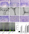
Reduced Secondary Wall Thickness in Fibers of irx8 and irx9 Plants. Roots and bottom internodes of inflorescence stems from 10-week-old plants were sectioned for examination of fibers and vessels. co, cortex; if, interfascicular fiber; ve, vessel; xf, xylary fiber; xy, xylem. Bar in (A) = 120 μm for (A) to (C); bar in (D) = 2.8 μm for (D) to (F); bar in (G) = 67 μm for (G) to (I). (A) to (C) Cross sections of interfascicular regions of stems showing thin-walled fibers in irx8 (B) and irx9 (C) compared with the wild type (A). (D) to (F) Transmission electron micrographs of walls of interfascicular fibers in the wild type (D), irx8 (E), and irx9 (F). (G) to (I) Cross sections of secondary xylem regions of roots showing thin xylary fibers and deformed vessels in irx8 (H) and irx9 (I) compared with the wild type (G). (J) Growth defects in irx8 and irx9 plants were rescued by expression of the corresponding wild-type genes. (K) Breaking force measurements showing that expression of the wild-type IRX8 and IRX9 genes restored the stem strength of irx8 and irx9, respectively, to a level comparable with that of the wild type. Each bar represents the breaking force of the inflorescence stem of an individual plant.
Figure 5.
Figure 5.
Immunolocalization of Xylan in the Stems and Roots of irx8 and irx9 Mutants. Stem and root sections from 10-week-old Arabidopsis plants were used for immunolocalization of xylan with the monoclonal antibody LM10 that was generated against plant cell wall (1,4)-β-d-xylan. Xylan signals were detected with fluorescein isothiocyanate–conjugated secondary antibodies and visualized with a laser confocal microscope, or detected with gold-conjugated secondary antibodies and visualized with a transmission electron microscope. if, interfascicular fiber; sx, secondary xylem; xy, xylem. Bar in (A) = 154 μm for (A) to (F); bar in (G) = 0.52 μm for (G) to (I). (A) to (C) Xylan immunofluorescence signals in stem sections of the wild type (A), irx8 (B), and irx9 (C). (D) to (F) Xylan immunofluorescence signals in root sections of the wild type (D), irx8 (E), and irx9 (F). (G) to (I) Immunogold labeling of xylan in fiber walls of the wild type (G), irx8 (H), and irx9 (I).
Figure 6.
Figure 6.
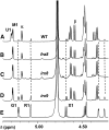
Anomeric Region of the 600-MHz 1H-NMR Spectra of the Xylo-Oligosaccharides Generated by Endoxylanase Treatment of the Material Solubilized by 1 n KOH–Containing NaBH4. (A) Wild-type xylo-oligosaccharides. (B) fra8 xylo-oligosaccharides. (C) irx8 xylo-oligosaccharides. (D) irx9 xylo-oligosaccharides. (E) Tetraglycosyl-xylitol (glycosyl sequence 2; see Figure 1) isolated from the products generated by endoxylanase treatment of irx9 GX. The anomeric region shown contains diagnostic well-resolved signals for each residue in the complex. These include the following: U1, α-d-GlcpA H1; M1, 4-O-methyl α-d-GlcpA H1; G1, α-d-GalpA H1; R1, α-l-Rhap H1; X1, 3-linked β-d-Xylp H1; G4, α-d-GalpA H4; R2, α-l-Rhap H2. α and β indicate the anomeric signals of α and β reducing xylose H1, respectively.
Figure 7.
Figure 7.
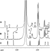
Anomeric Region of the 600-MHz 1H-NMR Spectra of Alkali-Soluble GX from the Stems of irx9 Plants. (A) 1H-NMR spectrum of GX extracted from irx9 cell walls with 0.25 n KOH in the absence of NaBH4. (B) 1H-NMR spectrum of GX extracted from irx9 cell walls with 1.0 n KOH in the presence NaBH4. (C) 1H-NMR spectrum of the purified glycosyl sequence 2 (see Figure 1). H1 resonances of reducing xylose residues are labeled with the anomeric configuration (α or β) of the residue. Changes in the positions of the resonance of specific nonreducing residues are indicated by double-headed arrows. G1, α-d-GalpA H1; R1, α-l-Rhap H1; X1, 3-linked β-d-Xylp H1; G5, α-d-GalpA H5; G4, α-d-GalpA H4; R2, α-l-Rhap H2. All of these resonances exhibit distinct anomerization effects in the spectrum of the GX solubilized without NaBH4, especially α-d-GalpA H5, which is split into two fully resolved signals labeled a and b, corresponding to the α and β forms, respectively, of the reducing xylose residue.
Figure 8.
Figure 8.
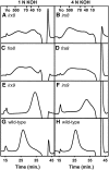
SEC Profiles Showing the Size Distribution of GX from irx8, irx9, fra8, and Wild-Type Plants. The 1 and 4 n KOH–soluble materials from wild-type and mutant plants were treated separately with endoxylanase and analyzed by SEC. Each SEC profile was then subtracted from its corresponding SEC profile of the untreated material. This resulting profile corresponds to the size distribution profile of the GX. (A) and (B) The reconstructed profiles of irx8 GX. (C) and (D) The reconstructed profiles of fra8 GX. (E) and (F) The reconstructed profiles of irx9 GX. (G) and (H) The reconstructed profiles of wild-type GX. (A), (C), (E), and (G) show data for the 1 n KOH–soluble materials. (B), (D), (F), and (H) show data for the 4 n KOH–soluble materials. The column excluded volume (Vo) and the elution positions of dextran molecular mass markers of 500, 70, 40, and 10 kD are shown.

Similar articles

Cited by

References

    1. Andersson, S.-I., Samuelson, O., Ishihara, M., and Shimizu, K. (1983). Structure of the reducing end-groups in spruce xylan. Carbohydr. Res. 111 283–288.
    1. Aspeborg, H., et al. (2005). Carbohydrate-active enzymes involved in the secondary cell wall biogenesis in hybrid aspen. Plant Physiol. 137 983–997. - PMC - PubMed
    1. Bauer, S., Vasu, P., Persson, S., Mort, A.J., and Somerville, C.R. (2006). Development and application of a suite of polysaccharide-degrading enzymes for analyzing plant cell walls. Proc. Natl. Acad. Sci. USA 103 11417–11422. - PMC - PubMed
    1. Baydoun, E.A.-H., and Brett, C.T. (1997). Distribution of xylosyltransferases and glucuronyltransferase within the Golgi apparatus in etiolated pea (Pisum sativum L) epicotyls. J. Exp. Bot. 48 1209–1214.
    1. Baydoun, E.A.-H., Waldron, K.W., and Brett, C.T. (1989). The interaction of xylosyltransferase and glucuronyltransferase involved in glucuronoxylan synthesis in pea (Pisum sativum) epicotyls. Biochem. J. 257 853–858. - PMC - PubMed

NOTE ADDED IN PROOF

    1. After acceptance of this manuscript, Persson et al. (2007) published an analysis showing altered secondary wall morphology and reduced xylan and homogalacturonan content in the cell walls of irx8 plants.
    1. Persson, S., Caffall, K.H., Freshour, G., Hilley, M.T., Bauer, S., Poindexter, P., Hahn, M.G., Mohnen, D., and Somerville, C. (2007). The Arabidopsis irregular xylem8 mutant is deficient in glucuronoxylan and homogalacturonan, which are essential for secondary cell wall integrity. Plant Cell 19 237–255. - PMC - PubMed

Publication types

MeSH terms

Associated data