

Reconstitution of bluetongue virus polymerase activity from isolated domains based on a three-dimensional structural model
- PMID: 17323325
- PMCID: PMC7161780
- DOI: 10.1002/bip.20706
Reconstitution of bluetongue virus polymerase activity from isolated domains based on a three-dimensional structural model
Abstract
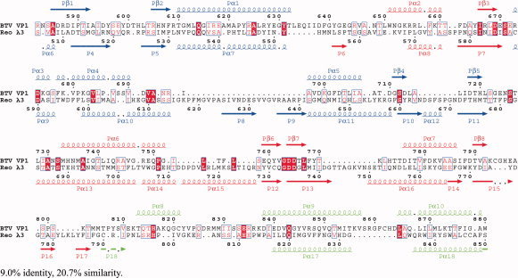
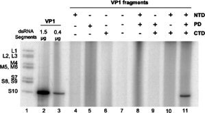
Bluetongue virus (BTV) is a double-stranded RNA virus of the Reoviridae family. The VP1 protein of BTV is the viral RNA-dependent RNA polymerase (RdRp), which is responsible for the replication of the viral genome. Currently there is no structural information available for VP1. By manual alignment of BTV, Reovirus and other viral RdRps we have generated a model for the structure of VP1, the RdRp of BTV. The structure can be divided into three domains: an N-terminal domain, a C-terminal domain, and a central polymerase domain. Mutation of the putative catalytic site in the central polymerase domain by site-directed mutagenesis abrogated in vitro replicase activity. Each of the domains was expressed individually and subsequently partially purified to obtain direct evidence for the location of polymerase activity and the nucleoside triphosphate binding site. The nucleoside triphosphate binding site was located by showing that CTP only bound to the full-length protein or to the polymerase domain and not to either of the other two domains. None of the domains had catalytic activity when tested individually or in tandem but when all three domains were mixed together the RdRp activity was reconstituted. This is the first report of the reconstitution of a functional viral RdRp in vitro from individual domains.
(c) 2007 Wiley Periodicals, Inc.
Figures
Similar articles
-
In situ structures of RNA-dependent RNA polymerase inside bluetongue virus before and after uncoating.Proc Natl Acad Sci U S A. 2019 Aug 13;116(33):16535-16540. doi: 10.1073/pnas.1905849116. Epub 2019 Jul 26. Proc Natl Acad Sci U S A. 2019. PMID: 31350350 Free PMC article.
-
Purified recombinant bluetongue virus VP1 exhibits RNA replicase activity.J Virol. 2004 Apr;78(8):3994-4002. doi: 10.1128/jvi.78.8.3994-4002.2004. J Virol. 2004. PMID: 15047815 Free PMC article.
-
Non-nucleoside Inhibitors of Zika Virus RNA-Dependent RNA Polymerase.J Virol. 2020 Oct 14;94(21):e00794-20. doi: 10.1128/JVI.00794-20. Print 2020 Oct 14. J Virol. 2020. PMID: 32796069 Free PMC article.
-
Structure-function relationships among RNA-dependent RNA polymerases.Curr Top Microbiol Immunol. 2008;320:137-56. doi: 10.1007/978-3-540-75157-1_7. Curr Top Microbiol Immunol. 2008. PMID: 18268843 Free PMC article. Review.
-
Bluetongue virus: dissection of the polymerase complex.J Gen Virol. 2008 Aug;89(Pt 8):1789-1804. doi: 10.1099/vir.0.2008/002089-0. J Gen Virol. 2008. PMID: 18632949 Free PMC article. Review.
Cited by
-
The molecular biology of Bluetongue virus replication.Virus Res. 2014 Mar;182:5-20. doi: 10.1016/j.virusres.2013.12.017. Epub 2013 Dec 25. Virus Res. 2014. PMID: 24370866 Free PMC article. Review.
-
Viral Capsid and Polymerase in Reoviridae.Subcell Biochem. 2022;99:525-552. doi: 10.1007/978-3-031-00793-4_17. Subcell Biochem. 2022. PMID: 36151388
-
A multidisciplinary approach to the identification of the protein-RNA connectome in double-stranded RNA virus capsids.Nucleic Acids Res. 2023 Jun 9;51(10):5210-5227. doi: 10.1093/nar/gkad274. Nucleic Acids Res. 2023. PMID: 37070191 Free PMC article.
-
The ins and outs of four-tunneled Reoviridae RNA-dependent RNA polymerases.Curr Opin Struct Biol. 2009 Dec;19(6):775-82. doi: 10.1016/j.sbi.2009.10.007. Epub 2009 Nov 14. Curr Opin Struct Biol. 2009. PMID: 19914820 Free PMC article. Review.
-
Bluetongue virus VP1 polymerase activity in vitro: template dependency, dinucleotide priming and cap dependency.PLoS One. 2011;6(11):e27702. doi: 10.1371/journal.pone.0027702. Epub 2011 Nov 15. PLoS One. 2011. PMID: 22110731 Free PMC article.
References
-
- Hansen, J. L. ; Long, A. M. ; Schultz, S. C. Structure 1997, 5, 1109–1122. - PubMed
-
- Ng, K. S. ; Cherney, M. M. ; Lopez Vazquez, A. ; Machin, A. ; Martin Alonso, J. M. ; Parra, F. ; James, M. N. G. J Biol Chem 2002, 277, 1381–1387. - PubMed
-
- Butcher, S. J. M. G. J. ; Makeyev, E. V. ; Bamford, D. H. ; Stuart, D. I. Nature 2001, 410, 235–240. - PubMed
-
- Ferrer‐Orta, C. ; Arias, A. ; Perez‐Luque, R. ; Escarmis, C. ; Domingo, E. ; Verdaguer, N. J Biol Chem 2004, 279, 47212–47221. - PubMed
Publication types
MeSH terms
Substances
Grants and funding
LinkOut - more resources
Full Text Sources
Research Materials
