. 2007 Jun 1;404(2):257-68.
doi: 10.1042/BJ20060960.

Insulin-induced formation of macromolecular complexes involved in activation of cyclic nucleotide phosphodiesterase 3B (PDE3B) and its interaction with PKB

Affiliations

Insulin-induced formation of macromolecular complexes involved in activation of cyclic nucleotide phosphodiesterase 3B (PDE3B) and its interaction with PKB

Faiyaz Ahmad et al. Biochem J. .

Abstract

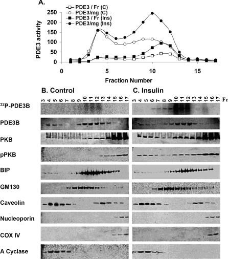
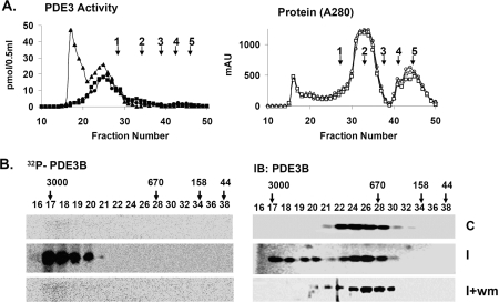
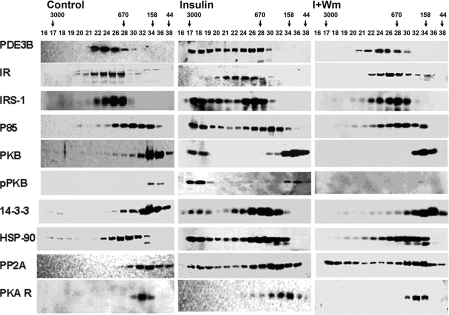
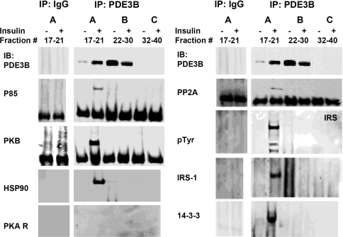
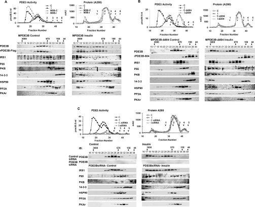
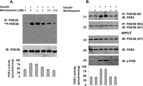
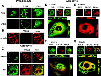
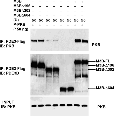
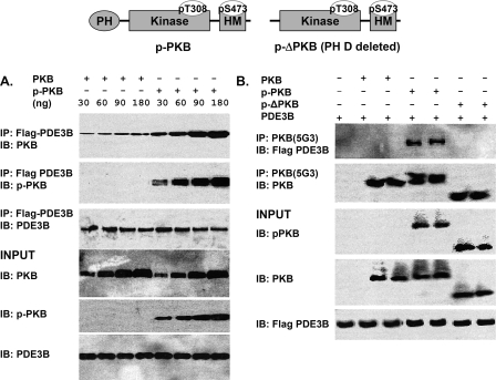
Fractionation of 3T3-L1 adipocyte membranes revealed that PDE3B (phosphodiesterase 3B) was associated with PM (plasma membrane) and ER (endoplasmic reticulum)/Golgi fractions, that insulin-induced phosphorylation/activation of PDE3B was greater in internal membranes than PM fractions, and that there was no significant translocation of PDE3B between membrane fractions. Insulin also induced formation of large macromolecular complexes, separated during gel filtration (Superose 6 columns) of solubilized membranes, which apparently contain phosphorylated/activated PDE3B and signalling molecules potentially involved in its activation by insulin, e.g. IRS-1 (insulin receptor substrate-1), IRS-2, PI3K p85 [p85-subunit of PI3K (phosphoinositide 3-kinase)], PKB (protein kinase B), HSP-90 (heat-shock protein 90) and 14-3-3. Expression of full-length recombinant FLAG-tagged murine (M) PDE3B and M3BDelta604 (MPDE3B lacking N-terminal 604 amino acids) indicated that the N-terminal region of MPDE3B was necessary for insulin-induced activation and recruitment of PDE3B. siRNA (small interfering RNA) knock-down of PDE3B indicated that PDE3B was not required for formation of insulin-induced complexes. Wortmannin inhibited insulin-induced assembly of macromolecular complexes, as well as phosphorylation/activation of PKB and PDE3B, and their co-immunoprecipitation. Another PI3K inhibitor, LY294002, and the tyrosine kinase inhibitor, Genistein, also inhibited insulin-induced activation of PDE3B and its co-immunoprecipitation with PKB. Confocal microscopy indicated co-localization of PDE3B and PKB. Recombinant MPDE3B co-immunoprecipitated, and co-eluted during Superose 12 chromatography, to a greater extent with recombinant pPKB (phosphorylated/activated PKB) than dephospho-PKB or p-DeltaPKB [pPKB lacking its PH domain (pleckstrin homology domain)]. Truncated recombinant MPDE3B proteins and pPKB did not efficiently co-immunoprecipitate, suggesting that structural determinants for their interaction reside in, or are regulated by, the N-terminal portion of MPDE3B. Recruitment of PDE3B in macromolecular complexes may be critical for regulation of specific cAMP pools and signalling pathways by insulin, e.g. lipolysis.


Figures

Figure 1
Figure 1. Fractionation of 3T3-L1 adipocyte membranes on 10–45% sucrose gradients
Adipocytes were incubated in serum-free DMEM for 16 h and then without (C) or with (Ins) 100 nM insulin (10 min, 37 °C). Some cells were incubated with [32P]Pi for 90 min before exposure to insulin. Total membranes were prepared, and 17 fractions (0.7 ml) were collected manually from continuous sucrose gradients. (A) Portions (10 μl) of fractions were assayed for PDE3 activity (expressed as pmol of cAMP hydrolysed·min−1·mg−1 or fraction−1) and protein content (results not shown). (B, C) Portions (400 μl) of fractions from adipocytes labelled with [32P]Pi incubated without (B) or with (C) insulin were analysed for [32P]PDE3B after immunoprecipitation of solubilized [32P]PDE3B with anti-PDE3B-CT antibody, separation of immunoprecipitated proteins on SDS/PAGE and phosphoimager analysis of wet gels (top row). Portions (25 μl) of fractions from adipocytes not incubated with [32P]Pi were subjected to SDS/PAGE, electrotransferred to NC membranes and immunoblotted with specific antibodies against PDE3B, PKB, pPKB, BiP (ER marker), GM130 (Golgi marker), caveolin-1, nucleoporin (nuclear marker), COX IV (mitochondria marker) and adenylate (A) cyclase (PM marker). Data shown are representative of two experiments for insulin-induced activation and [32P]PDE3B phosphorylation and Western blotting, and several experiments for activity assays.
Figure 2
Figure 2. Insulin-induced phosphorylation/activation of PDE3B: inhibition by wortmannin
Solubilized total microsomes (3 mg of protein), prepared from adipocytes incubated for 10 min without [control (C)] or with 100 nM insulin [insulin (I)], or with insulin after incubation with 100 nM wortmannin (30 min) (I+wm) were subjected to chromatography on Superose 6. Fractions were analysed as described in the Materials and methods section. (A) (Left panel) PDE3 activity [pmol of cAMP hydrolysed·min−1·(0.5 ml)−1] (●, ▲, ■) and (right panel) protein content (AU 280 nm) (○, △, □) were measured in indicated fractions from control (●, ○), insulin (▲, △) and I+wm adipocytes (■, □). Of the PDE3 activity applied, >90% was recovered in column fractions. MW standards: 1, thyroglobulin; 2, γ-globulin; 3, ovalbumin; 4, myoglobin; 5, Vitamin B12 are indicated. Au, absorption units. (B) To detect phosphorylated [32P]PDE3B, experiments like those in (A) were performed with adipocytes labelled with [32P]Pi. PDE3B was immunoprecipitated with anti-PDE3B-CT antibody (25 μl) from samples (400 μl) of fractions, separated by SDS/PAGE, and 32P-labelled PDE3B was detected in wet gels by phosphoimager (Amersham) analysis before the same gels were used for Western immunoblotting of PDE3B, using anti-PDE3B CT antibody. The results are representative of two experiments.
Figure 3
Figure 3. Insulin-induced assembly of macromolecular complex(es) containing PDE3B and signalling proteins, identified during chromatography on Superose 6
Samples (20 μl) of indicated fractions (0.5 ml) from Figure 2(A) were subjected to SDS/PAGE and immunoblotting with specific antibodies against PDE3B-CT, IR, IRS-1, PI3K p85, PKB, pPKB, 14-3-3, HSP-90, PP2A and PKARI. Results are representative of three experiments.
Figure 4
Figure 4. Insulin-induced assembly of macromolecular complex(es) containing PDE3B and signalling proteins: co-immunoprecipitation with PDE3B
Superose 6 fractions, as in Figure 3, were divided into three groups (A, B and C), and pooled for co-immunoprecipitation (group A: fractions 17–21; group B: fractions 22, 24, 26, 28 and 30; group C: fractions 32, 34, 36, 38 and 40). Pooled column fractions (150 μl from each of these fractions), were cleared, and complexes containing PDE3B were immunoprecipitated with 20 μl of anti-PDE3B CT antibodies (overnight, 4 °C). Cleared group A fractions were also immunoprecipitated with control rabbit IgG. Protein G–Sepharose-bound proteins were eluted in 100 μl of Laemmli's sample buffer. Samples (15 μl) were subjected to SDS/PAGE and immunoblotting with specific antibodies against PDE3B, PI3K p85, PKB, HSP-90 and PKARI (left panel) and PP2A, p-Tyr, IRS-1 and 14-3-3 (right panel). Some membranes used for immunoblotting lower molecular mass proteins were used also for reblotting/detecting larger proteins, including IRS-1, p-Tyr and HSP-90. Co-immunoprecipitated proteins were found largely in the higher molecular mass Superose 6 fractions (group A) after stimulation with insulin. Results were similar in three experiments. Non-specific binding of proteins was not detected in control IgG immunoprecipitates.
Figure 5
Figure 5. Analysis of insulin-induced assambly of macromolecular complex(es) after overexpression of FLAG-tagged full length PDE3B (A) and mutant truncated PDE3B-Δ604 (B), or after siRNA-mediated deplation of PDE3B (C)
(A, B) Effect of adenovirus-mediated overexpression of FLAG-tagged full-length PDE3B and mutant truncated PDE3B-Δ604 on insulin-induced assembly of macromolecular complex(es). Differentiated adipocytes were infected with Adβ-gal (control) (A, B), full-length AdMPDE3B (MPDE3B) (A) or truncated AdPDE3B-Δ604 (Δ604) (B), and incubated for 10 min without [control (C), C-MPDE3B or C-Δ604] or with 100 nM insulin [insulin (I), I-MPDE3B or I-Δ604]. (A) Total microsomes (centrifugation of 13000 g supernatant at 175000 g) from adipocytes infected with AdMPDE3B or Adβ-gal were prepared and solubilized (3 mg of proteins) as described in the Materials and methods section. (B) Because Δ604 is predominantly cytosolic, total lysates (13000 g supernatant) (3 mg of protein) prepared from adipocytes infected with AdPDE3B-604 were treated with detergent, and centrifuged. (A, B) Solubilized fractions were subjected to chromatography on Superose 6 and analysed as described in the Materials and methods section. (A, B) (Left panels) PDE3 activity [pmol of cAMP hydrolysed·min−1·(0.5 ml)−1] (●, ■, ▲, ◆) and (right panels) protein content (AU 280 nm) (○, □, △, ◇) were measured in indicated fractions from (A, B) control (●, ○), insulin (■, □) and (A) AdMPDE3B- or (B) AdPDE3B-604-infected adipocyte control (▲, △), insulin (◆, ◇); of the PDE3 activity applied, >90% was recovered in column fractions. MW standards: 1, thyroglobulin; 2, γ-globulin; 3, ovalbumin; 4, myoglobin; 5, vitamin B12. Samples (20 μl) of indicated fractions (0.5 ml) were subjected to SDS/PAGE and immunoblotting with specific antibodies against the indicated proteins. Results are representative of two experiments. (C) Effect of siRNA-mediated depletion of PDE3B on insulin-induced assembly of macromolecular complex(es). Control (untreated) adipocytes, or adipocytes transfected with non-targeting control siRNA or PDE3B siRNA, were incubated for 10 min without [control (C) or siRNA-(C)] or with 100 nM insulin [insulin (I), siRNA-(I)]. Solubilized microsomal fractions were analysed as described in the Materials and methods section and Figures 5(A) and 5(B). (Left panels) PDE3 activity [pmol of cAMP hydrolysed·min−1·(0.5 ml)−1] (●, ■, ▲, ◆) and (right panels) protein content (AU 280 nm) (○, □, △, ◇) in indicated fractions from control (●, ○), insulin (■, □), and PDE3B-siRNA transfected adipocyte control (▲, △), insulin (◆, ◇); of the PDE3 activity applied, >90% was recovered in column fractions. MW standards: 1, thyroglobulin; 2, γ-globulin; 3, ovalbumin; 4, myoglobin; 5, vitamin B12. Samples (20 μl) of indicated fractions (0.5 ml) were subjected to SDS/PAGE and immunoblotting with specific antibodies against indicated proteins from the siRNA-transfected fractions. Results are representative of two experiments.
Figure 6
Figure 6. Co-immunoprecipitation of PDE3B and PKB from solubilized membranes of insulin-treated adipocytes: inhibition by wortmannin
(A) For phosphorylation of PDE3B, adipocytes were incubated with [32P]Pi as described in the Materials and methods section, and then for 10 min without or with 100 nM insulin or with insulin after incubation (30 min) with different concentrations of wortmannin (as indicated). [32P]PDE3B was immunoprecipitated from solubilized and cleared total membrane proteins (1 mg) with 25 μl of anti-PDE3B-CT antibody. Immunoisolated [32P]PDE3B was separated by SDS/PAGE and detected by phosphoimager (Amersham) analysis of the wet gels, after which proteins were electrotransferred to NC membranes for Western immunoblotting using anti-PDE3B-CT antibody. Results shown are representative of two experiments. (B) In another experiment, adipocytes not exposed to [32P]Pi were incubated (30 min) without or with 100 nM wortmannin, and then for 10 min without or with 100 nM insulin as indicated. For co-immunoprecipitation of PDE3B and PKB, solubilized, cleared, total membrane proteins (1 mg) from adipocytes were incubated with anti-PDE3B-RD antibody (25 μl) and subjected to SDS/PAGE and Western blotting with the indicated antibodies. Input proteins: 30 μg of solubilized membrane proteins. Similar results were observed in two other experiments, using anti-PKB (monoclonal) antibodies for co-immunoprecipitation of PDE3B and PKB. (A, B) Bottom panels: effects of wortmannin on insulin-stimulated PDE3 activity (expressed as pmol·min−1·mg−1) in solubilized total membranes (10 μg) prepared from adipocytes incubated without [32P]Pi, assayed as described in the Materials and methods section; values represent means±½ range.
Figure 7
Figure 7. Subcellular localization of PDE3B, PKB and pPKB by confocal microscopy
Preadipocytes (A, B) and adipocytes (CG) were fixed, permeabilized with saponin and immunostained with indicated primary antibodies and Alexa Fluor® 488- or 594-conjugated secondary antibodies. In preadipocytes (A), PKB (cat. no. 2966, dilution 1:100) (green) was distributed throughout the cell. After incubation of cells with 100 nM insulin for 10 min (A), PKB and pPKB (cat. no. 4051, dilution 1:200) (green) were concentrated in PM areas and perinuclear structures. In preadipocytes, catenin (Ba) (cat. no. C-19220, dilution 1:200) was detected in PMs, at the periphery of the cells. No significant staining for PDE3B (Bb) was detected in preadipocytes using anti-PDE3B (peptide affinity-purified anti-PDE3B-NT antibody, dilution 1:200) (red) antibody. (C) Adipocytes stained for catenin (PM marker) or BiP (ER marker) (cat. no. G73320, dilution 1:200) in (a) and PDE3B (b) are merged in (c). Differentiated adipocytes developed invaginations (not present in preadipocytes), which stained for catenin. Unstimulated (D, E) or insulin-stimulated (F, G) adipocytes were stained for PKB or pPKB (a) and PDE3B (b) and are merged in (c). The extent of apparent co-localization of PDE3B (red) with PKB or pPKB (green) is greater in the ER region than in PM region. (Dd, Ed, Fd, Gd) Z-sections: reconstructed images from a stack of 18 sections (column IV) with 1 μm intervals showing areas of co-localization of PDE3B and PKB or pPKB in different planes. Centre XY (centre), above XZ (top) and YZ (right) planes are at indicated positions. Findings are representative of several experiments.
Figure 8
Figure 8. Interaction between truncated MPDE3B recombinants and recombinant PKB and pPKB
Recombinant FLAG-tagged WT MPDE3B (M3B), M3BΔ196, M3BΔ302 and M3BΔ604 [50 u (1 u=1 pmol of cAMP hydrolysed/min)] and recombinant pPKB (150 ng, ∼2.7 pmol, based on predicted molecular mass of ∼55000 for PKB) were incubated in buffer C containing 0.1% 2-mercaptoethanol for 30 min at 30 °C in a final volume of 200 μl. Since the different MPDE3B recombinant proteins exhibited different specific activities, all reaction mixtures were adjusted to contain similar amounts of protein by addition of protein from uninfected Sf21 cells. After dilution of reaction mixtures to 1 ml with a buffer containing 1 mg/ml BSA, samples (50 μl) were removed for analysis of Input proteins, before immunoprecipitating with 25 μl of anti-FLAG–agarose. Immunoprecipitated and input proteins were then subjected to immunoblotting with anti-PDE3B-CT and anti-PKB antibodies. Results are representative of three experiments.
Figure 9
Figure 9. Interactions between recombinant MPDE3B and recombinant PKB, pPKB and p-ΔPKB (PH domain deleted)
Top panel: linear structural model of pPKB, with PH-, kinase- and HM (hydrophobic motif)-domains and phosphorylated Thr308 and Ser473 residues noted. p-ΔPKB, in which the PH domain is removed, is also depicted. (A) Recombinant FLAG-tagged WT MPDE3B (M3B) (50 u) and recombinant PKB or pPKB at concentrations between 30 and 180 ng (∼0.54–3.2 pmol) were incubated (30 min, 30 °C) in buffer C (total volume, 200 μl). After addition of 800 μl of buffer C containing 1 mg/ml BSA, samples (40 μl) were taken for analysis of input proteins. Diluted mixtures were then cleared and incubated with immobilized anti-FLAG–agarose. Eluted proteins and input proteins were subjected to SDS/PAGE and immunoblotted with anti-PDE3B-CT, -PKB and -pPKB antibodies as indicated. Results were similar in two experiments. (B) Recombinant FLAG-tagged WT MPDE3B (M3B) (50 u) and 150 ng (∼2.7 pmol) of PKB, pPKB, or p-ΔPKB were incubated as in (A). After samples were taken for analysis of input proteins, diluted reaction mixtures were cleared, and immunoprecipitated with anti-PKB antibody. Immunoprecipitated proteins and input proteins were immunoblotted with anti-FLAG, -PKB and -pPKB antibodies as indicated. Results were similar in two experiments, and in two other experiments in which anti-FLAG–agarose was used for immunoprecipitation.
Figure 10
Figure 10. Purification of recombinant MPDE3B expressed in Sf21 cells: interactions between purified recombinant MPDE3B and recombinant PKB, pPKB and p-ΔPKB during gel filtration on Superose 12
(A) Left panel: Sf21 cell lysates infected without (lane 2) or with (lane 3) MPDE3B-baculovirus; (lane 4) FLAG-tagged MPDE3B purified from Sf21 cell lysates as described in the Materials and methods section by anti-FLAG affinity chromatography (specific activity 0.71±0.16 μmol·min−1·mg−1, n=4) or (lane 5) (from another experiment) recombinant pPKB (Upstate Biotechnology) were subjected to SDS/PAGE. Gels were stained with Simply Blue Safe stain. Right panel: lanes 6–8: proteins from gels similar to that on the left (lanes 2–4) were electrophoretically transferred to NC membranes and immunoblotted with anti-FLAG peroxidase-conjugated antibodies for immunodetection of purified MPDE3B (lanes 7 and 8). (B, C) Purified recombinant WT PDE3B (0.25 or 1 μg, ∼2 or ∼8 pmol, based on predicted molecular mass of ∼122000) and PKB, pPKB or p-ΔPKB (500 ng, ∼9 pmol, based on predicted molecular mass of ∼55000) were incubated (30 min, 30 °C) in buffer C (total volume, 200 μl) with BSA (50 μg/ml), before dilution by addition of 800 μl of buffer C with BSA (50 μg/ml). Samples (1 ml) were then applied to Superose 12 HR 10/30 equilibrated and were eluted with buffer C and BSA (50 μg/ml). Fractions (0.5 ml) were collected on Frac 950 and portions were assayed for PDE3 and PKB activities (results not shown). Protein contents were monitored at A280 by AKTA monitor UPC 900. Portions (20 μl) of indicated fractions (0.5 ml) were subjected to SDS/PAGE, and immunoblotting with antibodies specific to PKB, pPKB and PDE3B-CT (B) and p-ΔPKB and PDE3B-CT (C). The results are representative of two experiments.

References

    1. Francis S. H., Turko I. V., Corbin J. D. Cyclic nucleotide phosphodiesterases: relating structure and function. Prog. Nucleic Acid. Res. Mol. Biol. 2001;65:1–52. - PubMed
    1. Shakur Y., Holst L. S., Landstrom T. R., Movsesian M., Degerman E., Manganiello V. Regulation and function of the cyclic nucleotide phosphodiesterase (PDE3) gene family. Prog. Nucleic Acid Res. Mol. Biol. 2001;66:241–277. - PubMed
    1. Walz H. A., Harndahl L., Wierup N., Zmuda-Trzebiatowska E., Svennelid F., Manganiello V. C., Ploug T., Sundler F., Degerman E., Ahren B., et al. Early and rapid development of insulin resistance, islet dysfunction and glucose intolerance after high-fat feeding in mice overexpressing phosphodiesterase 3B. J. Endocrinol. 2006;189:629–641. - PubMed
    1. Choi Y. H., Park S., Hockman S., Zmuda-Trzebiatowska E., Svennelid F., Haluzik M., Gavrilova O., Ahmad F., Pepin L., Napolitano M., et al. Alterations in regulation of energy homeostasis in cyclic nucleotide phosphodiesterase 3B-null mice. J. Clin. Invest. 2006;116:3240–3251. - PMC - PubMed
    1. Houslay M. D., Milligan G. Tailoring cAMP-signalling responses through isoform multiplicity. Trends Biochem. Sci. 1997;22:217–224. - PubMed

Publication types