

Growth regulation of simian and human AIDS-related non-Hodgkin's lymphoma cell lines by TGF-beta1 and IL-6
- PMID: 17324269
- PMCID: PMC1810304
- DOI: 10.1186/1471-2407-7-35
Growth regulation of simian and human AIDS-related non-Hodgkin's lymphoma cell lines by TGF-beta1 and IL-6
Abstract
Background: AIDS-related non-Hodgkin's lymphoma (AIDS-NHL) is the second most frequent cancer associated with AIDS, and is a frequent cause of death in HIV-infected individuals. Experimental analysis of AIDS-NHL has been facilitated by the availability of an excellent animal model, i.e., simian Acquired Immunodeficiency Syndrome (SAIDS) in the rhesus macaque consequent to infection with simian immunodeficiency virus. A recent study of SAIDS-NHL demonstrated a lymphoma-derived cell line to be sensitive to the growth inhibitory effects of the ubiquitous cytokine, transforming growth factor-beta (TGF-beta). The authors concluded that TGF-beta acts as a negative growth regulator of the lymphoma-derived cell line and, potentially, as an inhibitory factor in the regulatory network of AIDS-related lymphomagenesis. The present study was conducted to assess whether other SAIDS-NHL and AIDS-NHL cell lines are similarly sensitive to the growth inhibitory effects of TGF-beta, and to test the hypothesis that interleukin-6 (IL-6) may represent a counteracting positive influence in their growth regulation.
Methods: Growth stimulation or inhibition in response to cytokine treatment was quantified using trypan blue exclusion or colorimetric MTT assay. Intracellular flow cytometry was used to analyze the activation of signaling pathways and to examine the expression of anti-apoptotic proteins and distinguishing hallmarks of AIDS-NHL subclass. Apoptosis was quantified by flow cytometric analysis of cell populations with sub-G1 DNA content and by measuring activated caspase-3.
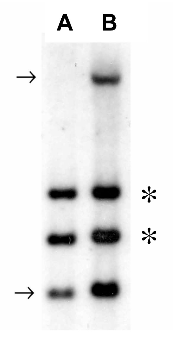
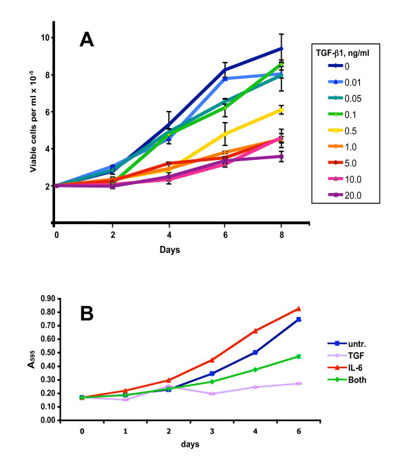
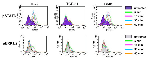
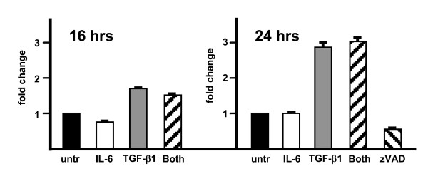
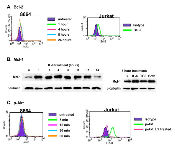
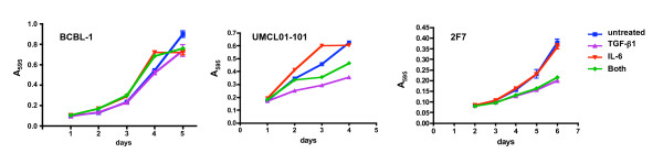
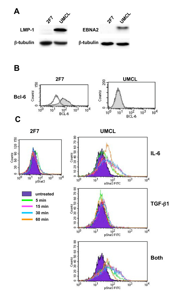
Results: Results confirmed the sensitivity of LCL8664, an immunoblastic SAIDS-NHL cell line, to TGF-beta1-mediated growth inhibition, and further demonstrated the partial rescue by simultaneous treatment with IL-6. IL-6 was shown to activate STAT3, even in the presence of TGF-beta1, and thereby to activate proliferative and anti-apoptotic pathways. By comparison, human AIDS-NHL cell lines differed in their responsiveness to TGF-beta1 and IL-6. Analysis of a recently derived AIDS-NHL cell line, UMCL01-101, indicated that it represents immunoblastic AIDS-DLCBL. Like LCL-8664, UMCL01-101 was sensitive to TGF-beta1-mediated inhibition, rescued partially by IL-6, and demonstrated rapid STAT3 activation following IL-6 treatment even in the presence of TGF-beta1.
Conclusion: These studies indicate that the sensitivity of immunoblastic AIDS- or SAIDS-DLBCL to TGF-beta1-mediated growth inhibition may be overcome through the stimulation of proliferative and anti-apoptotic signals by IL-6, particularly through the rapid activation of STAT3.
Figures
Similar articles
-
Transforming growth factor beta is a growth-inhibitory cytokine of B cell lymphoma in SIV-infected macaques.AIDS Res Hum Retroviruses. 1999 Nov 1;15(16):1477-85. doi: 10.1089/088922299309991. AIDS Res Hum Retroviruses. 1999. PMID: 10555111
-
Simian AIDS-associated lymphoma in rhesus and cynomolgus monkeys recapitulates the primary pathobiological features of AIDS-associated non-Hodgkin's lymphoma.AIDS Res Hum Retroviruses. 1999 Oct 10;15(15):1389-98. doi: 10.1089/088922299310098. AIDS Res Hum Retroviruses. 1999. PMID: 10515154
-
Rapamycin can restore the negative regulatory function of transforming growth factor beta 1 in high grade lymphomas.Cytokine. 2015 Jun;73(2):219-24. doi: 10.1016/j.cyto.2015.02.024. Epub 2015 Mar 17. Cytokine. 2015. PMID: 25794661
-
[The pathology spectrum of AIDS-related non-Hodgkin's lymphoma].Pathologica. 1998 Dec;90(6):763-70. Pathologica. 1998. PMID: 10220996 Review. Italian.
-
AIDS-related non-Hodgkin's lymphomas: from pathology and molecular pathogenesis to treatment.Hum Pathol. 2002 Apr;33(4):392-404. doi: 10.1053/hupa.2002.124723. Hum Pathol. 2002. PMID: 12055673 Review.
Cited by
-
Comparative pathobiology of macaque lymphocryptoviruses.Comp Med. 2008 Feb;58(1):57-67. Comp Med. 2008. PMID: 19793458 Free PMC article.
-
Protein kinase C-beta inhibition induces apoptosis and inhibits cell cycle progression in acquired immunodeficiency syndrome-related non-hodgkin lymphoma cells.J Investig Med. 2012 Jan;60(1):29-38. doi: 10.2310/JIM.0b013e318237eb55. J Investig Med. 2012. PMID: 21997316 Free PMC article.
-
Complete Remission of Methotrexate-Related Epstein-Barr-Virus-Associated Hodgkin-Like Lymphoma following Withdrawal of MTX Coupled with Clarithromycin Administration.Case Rep Hematol. 2012;2012:658745. doi: 10.1155/2012/658745. Epub 2012 Dec 19. Case Rep Hematol. 2012. PMID: 23316401 Free PMC article.
-
Soluble mediators of inflammation in HIV and their implications for therapeutics and vaccine development.Cytokine Growth Factor Rev. 2012 Aug-Oct;23(4-5):193-206. doi: 10.1016/j.cytogfr.2012.05.006. Epub 2012 Jun 27. Cytokine Growth Factor Rev. 2012. PMID: 22743035 Free PMC article. Review.
-
Can the vector space model be used to identify biological entity activities?BMC Genomics. 2011 Dec 22;12 Suppl 4(Suppl 4):S1. doi: 10.1186/1471-2164-12-S4-S1. Epub 2011 Dec 22. BMC Genomics. 2011. PMID: 22369514 Free PMC article.
References
-
- Goedert JJ. The epidemiology of acquired immunodeficiency syndrome malignancies. Semin Oncol. 2000;27:390–401. - PubMed
-
- Levine AM, Seneviratne L, Espina BM, Wohl AR, Tulpule A, Nathwani BN, Gill PS. Evolving characteristics of AIDS-related lymphoma. Blood. 2000;96:4084–4090. - PubMed
-
- Carbone A, Gloghini A. AIDS-related lymphomas: from pathogenesis to pathology. Br J Haematol. 2005;130:662–670. - PubMed
-
- Gaidano G, Capello D, Carbone A. The molecular basis of acquired immunodeficiency syndrome-related lymphomagenesis. Semin Oncol. 2000;27:431–441. - PubMed
-
- Diamond C, Taylor TH, Aboumrad T, Anton-Culver H. Changes in acquired immunodeficiency syndrome-related non-Hodgkin lymphoma in the era of highly active antiretroviral therapy: incidence, presentation, treatment, and survival. Cancer. 2000;106:128–135. - PubMed
Publication types
MeSH terms
Substances
Grants and funding
LinkOut - more resources
Full Text Sources
Research Materials
Miscellaneous
