. 2007 Jun;27(11):4179-97.
doi: 10.1128/MCB.01352-06. Epub 2007 Feb 26.

CSK controls retinoic acid receptor (RAR) signaling: a RAR-c-SRC signaling axis is required for neuritogenic differentiation

Affiliations

CSK controls retinoic acid receptor (RAR) signaling: a RAR-c-SRC signaling axis is required for neuritogenic differentiation

Nandini Dey et al. Mol Cell Biol. 2007 Jun.

Abstract

Herein, we report the first evidence that c-SRC is required for retinoic acid (RA) receptor (RAR) signaling, an observation that suggests a new paradigm for this family of nuclear hormone receptors. We observed that CSK negatively regulates RAR functions required for neuritogenic differentiation. CSK overexpression inhibited RA-mediated neurite outgrowth, a result which correlated with the inhibition of the SFK c-SRC. Consistent with an extranuclear effect of CSK on RAR signaling and neurite outgrowth, CSK overexpression blocked the downstream activation of RAC1. The conversion of GDP-RAC1 to GTP-RAC1 parallels the activation of c-SRC as early as 15 min following all-trans-retinoic acid treatment in LA-N-5 cells. The cytoplasmic colocalization of c-SRC and RARgamma was confirmed by immunofluorescence staining and confocal microscopy. A direct and ligand-dependent binding of RAR with SRC was observed by surface plasmon resonance, and coimmunoprecipitation studies confirmed the in vivo binding of RARgamma to c-SRC. Deletion of a proline-rich domain within RARgamma abrogated this interaction in vivo. CSK blocked the RAR-RA-dependent activation of SRC and neurite outgrowth in LA-N-5 cells. The results suggest that transcriptional signaling events mediated by RA-RAR are necessary but not sufficient to mediate complex differentiation in neuronal cells. We have elucidated a nongenomic extranuclear signal mediated by the RAR-SRC interaction that is negatively regulated by CSK and is required for RA-induced neuronal differentiation.


Figures

FIG. 1.
FIG. 1.
Effect of pan-SFK inhibitor PP1 on ATRA-induced neurite outgrowth in NB cell lines. Neurite outgrowth was semiquantified from the morphological changes in LA-N-5, LA-N-6, and SK-N-BE(2) cells treated with ATRA (10−5 M) for 7 to 9 days. PP1 (4 μM) was added 1 h prior to ATRA administration. Each bar represents the percentage of cells (out of 10 randomly chosen fields) showing neurite outgrowth at the end of the treatment. Vehicle treatment served as the control. PP1 alone did not show any neurite outgrowth. *, P < 0.001 (n = 5). Data show that PP1 blocks ATRA-induced neurite outgrowth in NB cells.
FIG. 2.
FIG. 2.
Overexpression of wild-type CSK and its effect on ATRA-induced neurite outgrowth in LA-N-5, LA-N-6, and SK-N-BE(2) cells. Overexpression (top panels) of wild-type CSK in LA-N-5, LA-N-6, and SK-N-BE(2) cells was determined by Western blot analysis. Bulk populations and individual clones of CSK-overexpressing cells were selected from parental LA-N-5 (A), LA-N-6 (B), and SK-N-BE(2) (C) cells infected with empty vectors (LXSN) and wild-type CSK. Clear lysates were resolved by 10% SDS-PAGE and probed with CSK antibody. Human β-actin was run for the loading control. Bar diagrams in the top panels show the relative densities of protein bands in arbitrary units. Data show that all the clones of LA-N-5, LA-N-6, and SK-N-BE(2) cells have significantly higher levels of wild-type CSK than their respective vectors and wild-type controls. (A) Levels of expression of CSK in LA-N-5 clones 5P, 10P, and 15P (lanes 3, 4, and 5, respectively) are compared to the endogenous levels of CSK in the empty vector (LXSN)-infected cell line (lane 2) and the wild-type (WT) cell line (lane 1). (B) Levels of expression of CSK in LA-N-6 clones 8 and 10 (lanes 3 and 4, respectively) are compared to the endogenous levels of CSK in the empty vector (LXSN)-infected cell line (lane 2) and the wild-type cell line (lane 1). (C) Levels of expression of CSK in SK-N-BE(2) clones 1 and 2 (lanes 3 and 4, respectively) are compared to the endogenous levels of CSK in the empty vector (LXSN)-infected cell line (lane 2) and the wild-type cell line (lane 1). Morphological changes were observed in LA-N-5, LA-N-6, and SK-N-BE(2) cells treated with ATRA (10−5 M) for 7 to 9 days as mentioned in Materials and Methods. The neurite outgrowth response was examined morphologically and semiquantified (bottom panels). NB cell lines were plated and grown (3 × 10⁶ cells) for 1 day, the media were removed, and differentiating media were added. The media were changed every 2 days. Differentiated cells were stained with methylene blue dye for morphological/semiquantification studies. Quantifications of neuritogenic responses to ATRA (10−5 M) for 7 to 9 days, as mentioned in Materials and Methods (bottom panels), are shown for LA-N-5 cells (parental cell line and empty vector control were compared with CSK-expressing clones 5P, 10P, and 15P) (A), LA-N-6 cells (parental cell line and empty vector control were compared with CSK-expressing clones 8 and 10) (B), and SK-N-BE(2) cells (parental cell line and empty vector control were compared with CSK-expressing clones 1 and 2) (C). Bars represent means ± standard deviations. *, P < 0.001 (n = 4). Representative photomicrographs of the neuritogenic responses to ATRA in SK-N-BE(2) cells (clone 1 is compared to vector control) are shown in C (right panel). The figure shows that the overexpression of wild-type CSK blocked ATRA-induced neurite outgrowth in LA-N-5, LA-N-6, and SK-N-BE(2) cells.
FIG. 3.
FIG. 3.
Effects of pan-SFK inhibitor PP1 or wild-type CSK overexpression on activity of the src family kinases c-SRC and FYN in LA-N-5 cells. Phosphotransferase activity towards an SRC-specific peptide was determined by IP of c-SRC, FYN, and c-YES from 100 μg of protein derived from LA-N-5 cells followed by an in vitro kinase assay as described in Materials and Methods. (A) Kinase activities of different members of SFKs in LA-N-5 cells. Bars represent changes in kinase activities from cpm values from three to four individual experiments. The changes in kinase activities were compared to the kinase activity of c-SRC (kinase activity of c-SRC represents “onefold”). *, P < 0.005. (B) Effect of PP1 treatment on c-SRC and FYN kinase activities in LA-N-5 cells. Kinase activities of c-SRC and FYN were determined from PP1 (4 μM for 1 h)-treated LA-N-5 cell lysates. Bars represent changes in kinase activities from cpm values from three to four individual experiments. **, P < 0.005; *, P < 0.05. Representative immunoblots for c-SRC and FYN are shown in the bottom panels. (C) Effect of overexpression of wild-type CSK on c-SRC and FYN kinase activities in LA-N-5 cells. Kinase activities of c-SRC and FYN were determined from the stable clone (5P) of LA-N-5 cells overexpressing wild-type CSK and compared with those of the vector control (LXSN). Bars represent changes in kinase activities from cpm values from three to four individual experiments. **, P < 0.0001; *, P < 0.005. Representative immunoblots for c-SRC and FYN are shown in the bottom panels. (D) Time course of activation of c-SRC in LA-N-5 cells following administration of ATRA. Wild-type LA-N-5 cells were treated with or without ATRA (10−5 M) under subdued-light conditions. Kinase activities of c-SRC were determined from the cell lysates following 5 min (lane 2), 15 min (lane 3), 30 min (lane 4), 60 min (lane 5), 180 min (lane 6), and 360 min (lane 7) of ATRA administration as described in Materials and Methods. Lane 1 represents the kinase activity from nontreated cells. Bars represent kinase activities from three to four individual experiments. *, P < 0.005. A representative immunoblot for c-SRC is shown in the bottom panel.
FIG. 4.
FIG. 4.
Effects of CSK overexpression on ATRA (RA)-induced activation of RAC1 in LA-N-5 cells. (A) Activation of RAC1 in ATRA-stimulated LA-N-5 cells. Wild-type LA-N-5 cells were treated with or without ATRA (10−5 M) under subdued-light conditions. The conversion of GDP-RAC1 to GTP-RAC1 was determined at 24, 48, and 72 h of ATRA administration using GST fusion proteins representing the GTP-RAC1 binding CRIB domain of the PAK-1 kinase as described in Materials and Methods. The membranes were immunoblotted for RAC1. Total RAC1 was immunoblotted for loading controls (blot in the bottom panel). Lane 5 represents the positive control. Densitometry scanning analyses (bar diagrams in the top panel) of GTP-RAC1 with or without ATRA show that ATRA administration causes the activation of RAC1 in LA-N-5 cells compared to the 48-h nontreated control. (B) Overexpression of CSK abrogates ATRA-induced RAC1 activation in LA-N-5 cells. Stable clones of LA-N-5 cells overexpressing wild-type CSK (clones 5P, 10P, and 15P) were treated with ATRA. After 48 h, lysates were evaluated by pull-down assay for the detection of activated GTP-bound RAC1 as described in Materials and Methods. The activation of RAC1 after 48 h of ATRA treatment in the empty vector control (LXSN as in lane 2) was compared with that of wild-type CSK-overexpressing clones (clones 5P, 10P, and 15P in lanes 3, 4, and 5, respectively). Total RAC1 was immunoblotted for loading controls (blot in bottom panel). Densitometry analyses (bar diagrams in top panel) of the GTP-RAC1 blot show that CSK overexpression blocked ATRA-induced activation of RAC1 in LA-N-5 cells. Both lanes 1 and 6 are positive controls. Lane 1 represents the positive control for activated RAC1 using the lysates of LA-N-5 cells (lysates treated with GTP-γS according to the manufacturer's protocol). Lane 6 represents the positive control for endogenous RAC1 protein in LA-N-5 cells (whole-cell lysates from LA-N-5 cells). For positive controls, lysates were treated with 100 μM GTP-γS at 30°C for 15 min before the addition of PAK-1 PBD glutathione agarose conjugate. (C) Time course of activation of RAC1 following ATRA administration in LA-N-5 cells. Wild-type LA-N-5 cells were treated with or without ATRA (10−5 M) under subdued-light conditions. Conversion of GDP-RAC1 to GTP-RAC1 was determined at 5 min (lane 2), 15 min (lane 3), 30 min (lane 4), 60 min (lane 5), 180 min (lane 6), and 360 min (lane 7) after ATRA administration using GST fusion proteins representing the GTP-RAC1 binding CRIB domain of PAK-1 kinase as described in Materials and Methods. Membranes were immunoblotted for RAC1. Lane 1 represents nontreated cells. Lanes 2 to 7 represent ATRA-treated cells. Lane 8 represents the positive control (as mentioned above). Total RAC1 was immunoblotted for loading controls (blot in the bottom panel). Results show that the activation of RAC1 in LA-N-5 cells occurs within 15 min of administration of ATRA.
FIG. 5.
FIG. 5.
Immunolocalization of RARγ and its colocalization with c-SRC in the cytoplasm of LA-N-5 cells. Cytoplasmic coimmunolocalization of RARγ and c-SRC in LA-N-5 cells was determined by double immunofluorescence of RARγ and c-SRC in LA-N-5 cells using mouse monoclonal antibody against c-SRC (1:50) and rabbit polyclonal antibody against RARγ (1:50). Methanol-fixed LA-N-5 cells were stained with primary antibodies specific for RARγ and c-SRC. Signals were visualized with secondary antibody conjugated to rhodamine for RARγ and fluorescein isothiocyanate for c-SRC as described in Materials and Methods. Nuclei were counterstained with DAPI. Negative controls (CON and DAPI) were prepared by incubating the cells with secondary antibody only and secondary antibody plus DAPI, respectively. Merges of RARγ with DAPI (RARγ + DAPI) and c-SRC with DAPI (c-SRC + DAPI) were obtained using the Spot Advanced program. The superimposition of the images of RARγ and DAPI and c-SRC and DAPI images (RARγ c-SRC merge) shows a cytoplasmic coimmunolocalization (arrows) of the two proteins.
FIG. 6.
FIG. 6.
Ligand-dependent binding of RARγ and c-SRC in vitro. (A) Real-time binding of RARγ and c-SRC in the presence of ATRA (RA). The interactions between RARγ and c-SRC were measured by SPR. (i) Interactions between immobilized c-SRC and individual analyte, respectively, are shown in sensorgram. A schematic representation of the sensor chip surface is represented below the sensorgram. Purified recombinant RARγ (1.85 nM) and other analytes (as shown in the figure) were injected over the sensor chip coated with c-SRC as described in Materials and Methods. ATRA (RA), 9-cis-RA, 13-cis-RA, and RARγ alone did not interact with SRC efficiently. (ii) When 20 μM of ATRA was mixed with 1.85 nM of RARγ, a significant binding was observed. (iii) No significant binding between RARγ and c-SRC was observed when ATRA was replaced by 9-cis-RA or 13-cis-RA. The specificity of the binding was detected by adding recombinant c-SRC (9.25 nM) in the analyte solution to compete for binding of RARγ. (B) Binding of RARγ and c-SRC in the presence of ATRA using Affi-Gel 15 in vitro. Affi-Gel beads coated with c-SRC (inset) were incubated in the presence of RARγ with (lanes 7, 8, and 9) and without (lanes 4, 5, and 6) ATRA in the dark at 37°C. Following the reaction, samples were immunoblotted for RARγ. Data show that the binding of RARγ and c-SRC occurs in the presence of ATRA (lanes 7, 8, and 9) compared to untreated controls (lanes 4, 5, and 6). Densitometry evaluation showed a six- to eightfold increase (*, P < 0.0005) in the binding in the presence of ATRA (lanes 7, 8, and 9) compared to that in the absence of ATRA (lanes 4, 5, and 6), as shown in the RARγ immunoblot. Uncoated Affi-Gel beads plus RARγ with (lane 2) and without (lane1) blocking and c-SRC-coated beads with blocking (lane 3) served as negative controls. Recombinant (Recomb.) RARγ was run as a positive control (lane 10). The coating of c-SRC on Affi-Gel beads was confirmed by running an immunoblot for c-SRC (inset) from the beads after coating (lane 11 of the inset) compared to the uncoated beads (lane 12 of the inset). No binding of RARγ to unconjugated Affi-Gel beads was observed in the presence of ATRA. Recombinant c-SRC (lane 13 of the inset) was used as a positive control. (C) Binding of RARγ to c-SRC in vivo. (i) Expression of FLAG-tagged RARγ and HA-tagged c-SRC in HEK293 cells. HEK293 cells were either transiently cotransfected with FLAG-tagged RARγ (0.2 μg or 0.4 μg) and HA-tagged c-SRC (0.2 μg or 0.4 μg) (lanes 2 and 3) or transfected separately with HA-tagged c-SRC (0.8 μg) (lane 4) and FLAG-tagged RARγ (0.8 μg) (lane 5). Whole-cell extracts (250 μg) obtained 24 h after transfection were resolved by 10% SDS-PAGE and immunoblotted (IB) with anti-FLAG antibody (blot in top panel) and anti-HA antibody (blot in bottom panel). Lysates from mock-transfected (pcDNA3.1 for FLAG-tagged RARγ and pCSA for HA-tagged c-SRC) HEK293 cells (lane 1) served as negative controls. Data show comparable amounts of expression of FLAG-tagged RARγ and HA-tagged c-SRC following transfection (0.8 μg) and cotransfection (0.4 μg as shown in lane 2 and 0.8 μg as shown in lane 3) of the respective DNAs. (ii) RARγ coimmunoprecipitates with c-SRC in cotransfected HEK293 cells. HEK293 cells were either transiently cotransfected with FLAG-tagged RARγ (0.4 μg) and HA-tagged c-SRC (0.4 μg) (lanes 1, 3, and 8) or transfected separately with FLAG-tagged RARγ (0.8 μg) (lanes 5, 10, and 12) and HA-tagged c-SRC (0.8 μg) (lanes 4 and 9). Whole-cell extracts (250 μg of protein) were incubated with anti-HA monoclonal antibody and then incubated with protein G-agarose beads. Preimmune control (C) for IP was performed by adding mouse IgG to cell lysates from cotransfected cells (lane 1). Immune complexes were resolved by 10% SDS-PAGE and immunoblotted with anti-FLAG antibody (blot in the top panel) and anti-HA antibody (blot in the bottom panel). Lysates from HEK293 cells (lane 6) and mock-transfected (0.4 μg of pcDNA3.1 and 0.4 μg of pCSA) HEK293 cells (lane 2) served as internal negative controls for IP. Expression levels of proteins (FLAG-tagged RARγ and HA-tagged c-SRC) in the lysates (used for IP) were tested in the same gel (lanes 7 to 11). Lanes 7 and 11 represent lysates from mock-transfected HEK 293 cells and nontransfected HEK293 cells, respectively. Lysates from cells transfected with FLAG-tagged deletion mutations (amino acids 75 to 85 [Δ75-85 RARγ]) of RARγ were included as internal controls (lane 12). (iii) c-SRC coimmunoprecipitates with RARγ in cotransfected HEK293 cells. HEK293 cells were either transiently cotransfected with FLAG-tagged RARγ (0.4 μg) and HA-tagged c-SRC (0.4 μg) (lanes 1, 3, 7, and 10) or transfected separately with FLAG-tagged RARγ (0.8 μg) (lanes 4 and 8) and HA-tagged c-SRC (0.8 μg) (lanes 5, 9, and 11). Whole-cell extracts (250 μg of protein) were incubated with anti-FLAG polyclonal antibody and then incubated with pansorbin. Preimmune control (C) for IP was performed by adding rabbit IgG to cell lysates from cotransfected cells (lane 1). Immune complexes were resolved by 10% SDS-PAGE and immunoblotted with anti-HA monoclonal antibody (top blot) and anti-FLAG monoclonal antibody (bottom blot). Lysates from mock-transfected (0.4 μg of pcDNA3.1 and 0.4 μg of pCSA) HEK293 cells (lanes 2 and 6) served as internal negative controls. Expression levels of proteins (FLAG-tagged RARγ and HA-tagged c-SRC) in the lysates (used for IP) were tested in the same gel (lanes 7 to 9). Lysates obtained from different batches of transfected cells expressing HA-tagged c-SRC and FLAG-tagged deletion mutations (amino acids 75 to 85 [Δ75-85 RARγ]) of RARγ were included as internal controls (lanes 11 and 12, respectively). (iv) The proline-rich domain-deleted mutant (Δ75-85) of RARγ does not coimmunoprecipitate with c-SRC in cotransfected HEK293 cells. HEK293 cells were either transiently cotransfected with FLAG-tagged RARγ (0.4 μg) and HA-tagged Y527-SRC (0.4 μg) (lanes 3 and 8 of the top panel); cotransfected with FLAG-tagged RARγ (0.4 μg) and HA-tagged wild-type c-SRC (0.4 μg) (lane 4 of the top panel), with the FLAG-tagged Δ75-85 mutant of RARγ (0.4 μg) and HA-tagged Y527-SRC (0.4 μg) (lane 5 of the top panel), or with the FLAG-tagged Δ75-85 mutant of RARγ (0.4 μg) and HA-tagged wild-type (WT) c-SRC (0.4 μg) (lane 6 of the top panel); or transfected separately with HA-tagged Y527-SRC (0.8 μg) (lane 9 of the top panel), with FLAG-tagged wild-type RARγ (0.8 μg) (lane 10 of the top panel), with HA-tagged wild-type c-SRC (0.8 μg) (lane 11 of the top panel), or with the FLAG-tagged Δ75-85 mutant of RARγ (0.8 μg) (lane 12 of the top panel). For IP experiments, whole-cell lysates (250 μg of protein) from the transfected cells were first incubated with anti-HA monoclonal antibody and then transfected with protein G-agarose beads as mentioned in Materials and Methods. The preimmune control (C) experiment for IP was performed by adding mouse IgG to cell lysates from cotransfected cells (lane 1). Immune complexes were resolved by 10% SDS-PAGE and immunoblotted with anti-FLAG antibody (top blot) and anti-HA antibody (middle blot). The middle panel shows the expression of HA-tagged c-SRC and HA-tagged Y527-SRC (lanes 3, 4, 5, 6, 8, 9, and 11 of the middle panel) in both immunoprecipitants and lysates. Lysates from the mock-transfected (0.4 μg of pcDNA3.1 and 0.4 μg of pCSA) HEK293 cells (lane 2) served as internal negative controls for IP. Lane 7 represents lysate from mock-transfected HEK293 cells. Lysates from cells cotransfected with FLAG-tagged RARγ and HA-tagged Y527-SRC were included as an internal control (lane 8 of the top panel). Expression of proteins (FLAG-tagged RARγ, FLAG-tagged Δ75-85 mutant of RARγ, HA-tagged c-SRC, and HA-tagged Y527-SRC) in cell lysates was tested in the same gel (lanes 9 to 12 of the top panel). The bottom blot shows the expression of FLAG-tagged RARγs (wild-type protein as shown in lanes 3 and 4 as well as mutated protein as shown in lanes 5 and 6, respectively) in cell lysates that were used for the IP studies. (v) Colocalization of exogenous EGFP-tagged c-SRC with RFP-tagged RARγ in the cytosol of HEK293 cells. HEK293 cells were transiently transfected together (photomicrographs in the bottom panel) or separately (photomicrographs in the top panel) with EGFP-tagged c-SRC and/or RFP-tagged RARγ, respectively, as described in Materials and Methods. Cotransfected cells were treated with either ATRA (10−5 M) or vehicle for ATRA (DMSO) for 1 h under subdued-light conditions. Fixed cells were processed for confocal imaging. Photomicrographs in the top panel show the subcellular distribution of c-SRC (images a and c) and RARγ (images d and f) in HEK293 cells that were transfected with EGFP-tagged c-SRC (images a, b, and c) and RFP-tagged RARγ (images d, e, and f) along with their differential interference contrast (DIC) images (images b and e) and their merged confocal images (images c and f), respectively. Scale bar, 10 μm. Photomicrographs in the bottom panel show the colocalization of c-SRC and RARγ in the cytosol of untreated (images a, b, and c) and ATRA-treated (images d, e, and f) HEK293 cells following cotransfection of EGFP-tagged c-SRC and RFP-tagged RARγ. Merged images of EGFP-tagged c-SRC and RFP-tagged RARγ from both untreated (images a and b are merged to form image c) and treated (images d and e are merged to form image f) groups show a clear change in color, indicating the colocalization of these two proteins in the cytosol of the cells. Arrowheads represent the characteristic “edge”-like c-SRC-rich adhesion structures along the plasma membrane that were observed in 100% of ATRA-treated cells. Scale bar, 10 μm. Results show (i) an exclusive cytoplasmic distribution of c-SRC, (ii) both nuclear and cytoplasmic distribution of RARγ, and (iii) cytoplasmic colocalization of c-SRC and RARγ in untreated and ATRA-treated HEK293 cells.
FIG. 6.
FIG. 6.
Ligand-dependent binding of RARγ and c-SRC in vitro. (A) Real-time binding of RARγ and c-SRC in the presence of ATRA (RA). The interactions between RARγ and c-SRC were measured by SPR. (i) Interactions between immobilized c-SRC and individual analyte, respectively, are shown in sensorgram. A schematic representation of the sensor chip surface is represented below the sensorgram. Purified recombinant RARγ (1.85 nM) and other analytes (as shown in the figure) were injected over the sensor chip coated with c-SRC as described in Materials and Methods. ATRA (RA), 9-cis-RA, 13-cis-RA, and RARγ alone did not interact with SRC efficiently. (ii) When 20 μM of ATRA was mixed with 1.85 nM of RARγ, a significant binding was observed. (iii) No significant binding between RARγ and c-SRC was observed when ATRA was replaced by 9-cis-RA or 13-cis-RA. The specificity of the binding was detected by adding recombinant c-SRC (9.25 nM) in the analyte solution to compete for binding of RARγ. (B) Binding of RARγ and c-SRC in the presence of ATRA using Affi-Gel 15 in vitro. Affi-Gel beads coated with c-SRC (inset) were incubated in the presence of RARγ with (lanes 7, 8, and 9) and without (lanes 4, 5, and 6) ATRA in the dark at 37°C. Following the reaction, samples were immunoblotted for RARγ. Data show that the binding of RARγ and c-SRC occurs in the presence of ATRA (lanes 7, 8, and 9) compared to untreated controls (lanes 4, 5, and 6). Densitometry evaluation showed a six- to eightfold increase (*, P < 0.0005) in the binding in the presence of ATRA (lanes 7, 8, and 9) compared to that in the absence of ATRA (lanes 4, 5, and 6), as shown in the RARγ immunoblot. Uncoated Affi-Gel beads plus RARγ with (lane 2) and without (lane1) blocking and c-SRC-coated beads with blocking (lane 3) served as negative controls. Recombinant (Recomb.) RARγ was run as a positive control (lane 10). The coating of c-SRC on Affi-Gel beads was confirmed by running an immunoblot for c-SRC (inset) from the beads after coating (lane 11 of the inset) compared to the uncoated beads (lane 12 of the inset). No binding of RARγ to unconjugated Affi-Gel beads was observed in the presence of ATRA. Recombinant c-SRC (lane 13 of the inset) was used as a positive control. (C) Binding of RARγ to c-SRC in vivo. (i) Expression of FLAG-tagged RARγ and HA-tagged c-SRC in HEK293 cells. HEK293 cells were either transiently cotransfected with FLAG-tagged RARγ (0.2 μg or 0.4 μg) and HA-tagged c-SRC (0.2 μg or 0.4 μg) (lanes 2 and 3) or transfected separately with HA-tagged c-SRC (0.8 μg) (lane 4) and FLAG-tagged RARγ (0.8 μg) (lane 5). Whole-cell extracts (250 μg) obtained 24 h after transfection were resolved by 10% SDS-PAGE and immunoblotted (IB) with anti-FLAG antibody (blot in top panel) and anti-HA antibody (blot in bottom panel). Lysates from mock-transfected (pcDNA3.1 for FLAG-tagged RARγ and pCSA for HA-tagged c-SRC) HEK293 cells (lane 1) served as negative controls. Data show comparable amounts of expression of FLAG-tagged RARγ and HA-tagged c-SRC following transfection (0.8 μg) and cotransfection (0.4 μg as shown in lane 2 and 0.8 μg as shown in lane 3) of the respective DNAs. (ii) RARγ coimmunoprecipitates with c-SRC in cotransfected HEK293 cells. HEK293 cells were either transiently cotransfected with FLAG-tagged RARγ (0.4 μg) and HA-tagged c-SRC (0.4 μg) (lanes 1, 3, and 8) or transfected separately with FLAG-tagged RARγ (0.8 μg) (lanes 5, 10, and 12) and HA-tagged c-SRC (0.8 μg) (lanes 4 and 9). Whole-cell extracts (250 μg of protein) were incubated with anti-HA monoclonal antibody and then incubated with protein G-agarose beads. Preimmune control (C) for IP was performed by adding mouse IgG to cell lysates from cotransfected cells (lane 1). Immune complexes were resolved by 10% SDS-PAGE and immunoblotted with anti-FLAG antibody (blot in the top panel) and anti-HA antibody (blot in the bottom panel). Lysates from HEK293 cells (lane 6) and mock-transfected (0.4 μg of pcDNA3.1 and 0.4 μg of pCSA) HEK293 cells (lane 2) served as internal negative controls for IP. Expression levels of proteins (FLAG-tagged RARγ and HA-tagged c-SRC) in the lysates (used for IP) were tested in the same gel (lanes 7 to 11). Lanes 7 and 11 represent lysates from mock-transfected HEK 293 cells and nontransfected HEK293 cells, respectively. Lysates from cells transfected with FLAG-tagged deletion mutations (amino acids 75 to 85 [Δ75-85 RARγ]) of RARγ were included as internal controls (lane 12). (iii) c-SRC coimmunoprecipitates with RARγ in cotransfected HEK293 cells. HEK293 cells were either transiently cotransfected with FLAG-tagged RARγ (0.4 μg) and HA-tagged c-SRC (0.4 μg) (lanes 1, 3, 7, and 10) or transfected separately with FLAG-tagged RARγ (0.8 μg) (lanes 4 and 8) and HA-tagged c-SRC (0.8 μg) (lanes 5, 9, and 11). Whole-cell extracts (250 μg of protein) were incubated with anti-FLAG polyclonal antibody and then incubated with pansorbin. Preimmune control (C) for IP was performed by adding rabbit IgG to cell lysates from cotransfected cells (lane 1). Immune complexes were resolved by 10% SDS-PAGE and immunoblotted with anti-HA monoclonal antibody (top blot) and anti-FLAG monoclonal antibody (bottom blot). Lysates from mock-transfected (0.4 μg of pcDNA3.1 and 0.4 μg of pCSA) HEK293 cells (lanes 2 and 6) served as internal negative controls. Expression levels of proteins (FLAG-tagged RARγ and HA-tagged c-SRC) in the lysates (used for IP) were tested in the same gel (lanes 7 to 9). Lysates obtained from different batches of transfected cells expressing HA-tagged c-SRC and FLAG-tagged deletion mutations (amino acids 75 to 85 [Δ75-85 RARγ]) of RARγ were included as internal controls (lanes 11 and 12, respectively). (iv) The proline-rich domain-deleted mutant (Δ75-85) of RARγ does not coimmunoprecipitate with c-SRC in cotransfected HEK293 cells. HEK293 cells were either transiently cotransfected with FLAG-tagged RARγ (0.4 μg) and HA-tagged Y527-SRC (0.4 μg) (lanes 3 and 8 of the top panel); cotransfected with FLAG-tagged RARγ (0.4 μg) and HA-tagged wild-type c-SRC (0.4 μg) (lane 4 of the top panel), with the FLAG-tagged Δ75-85 mutant of RARγ (0.4 μg) and HA-tagged Y527-SRC (0.4 μg) (lane 5 of the top panel), or with the FLAG-tagged Δ75-85 mutant of RARγ (0.4 μg) and HA-tagged wild-type (WT) c-SRC (0.4 μg) (lane 6 of the top panel); or transfected separately with HA-tagged Y527-SRC (0.8 μg) (lane 9 of the top panel), with FLAG-tagged wild-type RARγ (0.8 μg) (lane 10 of the top panel), with HA-tagged wild-type c-SRC (0.8 μg) (lane 11 of the top panel), or with the FLAG-tagged Δ75-85 mutant of RARγ (0.8 μg) (lane 12 of the top panel). For IP experiments, whole-cell lysates (250 μg of protein) from the transfected cells were first incubated with anti-HA monoclonal antibody and then transfected with protein G-agarose beads as mentioned in Materials and Methods. The preimmune control (C) experiment for IP was performed by adding mouse IgG to cell lysates from cotransfected cells (lane 1). Immune complexes were resolved by 10% SDS-PAGE and immunoblotted with anti-FLAG antibody (top blot) and anti-HA antibody (middle blot). The middle panel shows the expression of HA-tagged c-SRC and HA-tagged Y527-SRC (lanes 3, 4, 5, 6, 8, 9, and 11 of the middle panel) in both immunoprecipitants and lysates. Lysates from the mock-transfected (0.4 μg of pcDNA3.1 and 0.4 μg of pCSA) HEK293 cells (lane 2) served as internal negative controls for IP. Lane 7 represents lysate from mock-transfected HEK293 cells. Lysates from cells cotransfected with FLAG-tagged RARγ and HA-tagged Y527-SRC were included as an internal control (lane 8 of the top panel). Expression of proteins (FLAG-tagged RARγ, FLAG-tagged Δ75-85 mutant of RARγ, HA-tagged c-SRC, and HA-tagged Y527-SRC) in cell lysates was tested in the same gel (lanes 9 to 12 of the top panel). The bottom blot shows the expression of FLAG-tagged RARγs (wild-type protein as shown in lanes 3 and 4 as well as mutated protein as shown in lanes 5 and 6, respectively) in cell lysates that were used for the IP studies. (v) Colocalization of exogenous EGFP-tagged c-SRC with RFP-tagged RARγ in the cytosol of HEK293 cells. HEK293 cells were transiently transfected together (photomicrographs in the bottom panel) or separately (photomicrographs in the top panel) with EGFP-tagged c-SRC and/or RFP-tagged RARγ, respectively, as described in Materials and Methods. Cotransfected cells were treated with either ATRA (10−5 M) or vehicle for ATRA (DMSO) for 1 h under subdued-light conditions. Fixed cells were processed for confocal imaging. Photomicrographs in the top panel show the subcellular distribution of c-SRC (images a and c) and RARγ (images d and f) in HEK293 cells that were transfected with EGFP-tagged c-SRC (images a, b, and c) and RFP-tagged RARγ (images d, e, and f) along with their differential interference contrast (DIC) images (images b and e) and their merged confocal images (images c and f), respectively. Scale bar, 10 μm. Photomicrographs in the bottom panel show the colocalization of c-SRC and RARγ in the cytosol of untreated (images a, b, and c) and ATRA-treated (images d, e, and f) HEK293 cells following cotransfection of EGFP-tagged c-SRC and RFP-tagged RARγ. Merged images of EGFP-tagged c-SRC and RFP-tagged RARγ from both untreated (images a and b are merged to form image c) and treated (images d and e are merged to form image f) groups show a clear change in color, indicating the colocalization of these two proteins in the cytosol of the cells. Arrowheads represent the characteristic “edge”-like c-SRC-rich adhesion structures along the plasma membrane that were observed in 100% of ATRA-treated cells. Scale bar, 10 μm. Results show (i) an exclusive cytoplasmic distribution of c-SRC, (ii) both nuclear and cytoplasmic distribution of RARγ, and (iii) cytoplasmic colocalization of c-SRC and RARγ in untreated and ATRA-treated HEK293 cells.
FIG. 7.
FIG. 7.
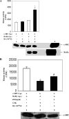
Effect of ATRA-dependent binding of RARγ to c-SRC from LA-N-5 cells on SRC kinase activity. (A) In vitro c-SRC kinase activity from LA-N-5 cells. RARγ and c-SRC were immunoprecipitated (i.p.) from LA-N-5 cell lysates using polyclonal antibodies. The immunoprecipitants were incubated with and without ATRA (10−5 M) under subdued-light conditions. Phosphotransferase activity towards an SRC-specific peptide was determined from the reaction mixture by an in vitro kinase assay. In short, endogenous c-SRC and RARγ from the normalized clear lysates of LA-N-5 cells were immunoprecipitated separately using their respective antibodies (rabbit polyclonal antibody for c-SRC and rabbit polyclonal antibody for RARγ). Individual immunoprecipitants were then used for the SRC kinase assay. The in vitro kinase assay for SRC was carried out according to the manufacturer's protocol with little modification, as described in Materials and Methods. In short, the reaction mixture representing lane 1 contained SRC kinase reaction buffer, SRC substrate peptide, immunoprecipitated c-SRC from LA-N-5 cells, and [γ-32P]ATP. The reaction mixture representing lane 2 contained SRC kinase reaction buffer, SRC substrate peptide, immunoprecipitated c-SRC from LA-N-5 cells, [γ-32P]ATP, and freshly prepared ATRA (10−5 M final concentration). The reaction mixture representing lane 3 contained SRC kinase reaction buffer, SRC substrate peptide, immunoprecipitated c-SRC from LA-N-5 cells, immunoprecipitated RARγ from LA-N-5 cells, and [γ-32P]ATP. The reaction mixture representing lane 4 contained SRC kinase reaction buffer, SRC substrate peptide, immunoprecipitated c-SRC from LA-N-5 cells, immunoprecipitated RARγ from LA-N-5 cells, [γ-32P]ATP, and freshly prepared ATRA (10−5 M final concentration). Bars represent kinase activity from three to four individual experiments. *, P < 0.05. Data show that kinase activity in the reaction mixture containing immunoprecipitated c-SRC and RARγ from LA-N-5 cells was significantly higher in the presence of ATRA (bar corresponding to lane 4) than in the nontreated control (bar corresponding to lane 3). The kinase activities from immunoprecipitated c-SRC treated with and without ATRA (bars corresponding to lanes 1 and 2, respectively) served as negative controls. Representative immunoblots for c-SRC and RARγ are shown in the bottom panels. Positive controls (recombinant proteins) for c-SRC and RARγ are shown in lane 5. (B) Effects of wild-type CSK and PP1 on kinase activity of immunoprecipitated c-SRC from LA-N-5 cells. Endogenous c-SRC and RARγ from normalized cell lysates of LA-N-5 cells were immunoprecipitated separately using their respective antibodies (rabbit polyclonal antibody for c-SRC and rabbit polyclonal antibody for RARγ). Individual immunoprecipitants were then used for the SRC kinase assay. In short, the reaction mixture representing all the lanes (lanes 1 to 3) contained SRC kinase reaction buffer, SRC substrate peptide, immunoprecipitated c-SRC from LA-N-5 cells, immunoprecipitated RARγ from LA-N-5 cells, [γ-32P]ATP, and freshly prepared ATRA (10−5 M final concentration). Immunoprecipitated c-SRC from control (LXSN) LA-N-5 cells (lane 1) and a clone (5P) of LA-N-5 cells overexpressing wild-type CSK (lane 2) were incubated in the presence of RARγ with ATRA (10−5 M) under subdued-light conditions. Immunoprecipitated c-SRC in the presence of PP1 (4 μM) was also incubated as described above (lane 3). Phosphotransferase activity towards an SRC-specific peptide was determined from the reaction mixture by an in vitro kinase assay as described in Materials and Methods. Bars represent kinase activities from three to four individual experiments. *, P < 0.005. Data show that kinase activities of immunoprecipitated c-SRC from CSK-overexpressing LA-N-5 cells and, under conditions of PP1 treatment, were significantly inhibited (bars corresponding to lanes 2 and 3, respectively) compared to the nontreated vector control (bar corresponding to lane 1).
FIG. 8.
FIG. 8.
Nuclear and extranuclear RA signaling. ATRA binds to its cognate receptor (RAR). The interaction of the ligand-bound RAR with RARE controls transcription and protein synthesis of its signature genes (classical genomic effect). The nongenomic mode of RA action involves the activation of the SRC family of nonreceptor protein tyrosine kinases, SRC, following the ligand-dependent binding of RAR to the kinase. The inset shows a schematic representation of the proline-rich motif in the functional domains of human RARγ1. A highly variable (A/B domain) amino-terminal domain, a relatively conserved DNA binding domain (DBD or C domain), a hinge region (D domain), a C-terminal ligand binding domain (LBD or E domain), and a small C-terminal domain (F domain) are schematically drawn (not to scale). The transcriptional activation regions (AF-1 and AF-2) are located in the A/B and E domains, respectively. Numbers indicate amino acid positions along the sequences in relation to the domains. Proline-rich sequences of the receptor in reference to its respective amino acid positions are indicated. The inset shows polyproline sequences in the A/B domains of RARγ1. Interestingly, RARα, RARβ, and RARγ2 have very similar proline-rich sequences in their A/B domains compared to RARγ1 (not shown in the diagram). The model proposes that as a component of its extragenomic mode of action, the RAR, upon ligand binding, undergoes a conformation change that leads to its interaction with c-SRC in the cytoplasm, leading to the activation of this kinase. The activation of SFKs activates a downstream signaling cascade, which initiates the activation of RAC1 and leads to neurite outgrowth NB cells.

Similar articles

Cited by

References

    1. Alsayed, Y., S. Uddin, N. Mahmud, F. Lekmine, D. V. Kalvakolanu, S. Minucci, G. Bokoch, and L. C. Platanias. 2001. Activation of Rac1 and the p38 mitogen-activated protein kinase pathway in response to all-trans-retinoic acid. J. Biol. Chem. 276:4012-4019. - PubMed
    1. Altucci, L., and H. Gronemeyer. 2001. The promise of retinoids to fight against cancer. Nat. Rev. Cancer 1:181-193. - PubMed
    1. Andrews, P. W. 1984. Retinoic acid induces neuronal differentiation of a cloned human embryonal carcinoma cell line in vitro. Dev. Biol. 103:285-293. - PubMed
    1. Aoki, K., T. Nakamura, K. Fujikawa, and M. Matsuda. 2005. Local phosphatidylinositol 3,4,5-trisphosphate accumulation recruits Vav2 and Vav3 to activate Rac1/Cdc42 and initiate neurite outgrowth in nerve growth factor-stimulated PC12 cells. Mol. Biol. Cell 16:2207-2217. - PMC - PubMed
    1. Aoki, K., T. Nakamura, and M. Matsuda. 2004. Spatio-temporal regulation of Rac1 and Cdc42 activity during nerve growth factor-induced neurite outgrowth in PC12 cells. J. Biol. Chem. 279:713-719. - PubMed

Publication types

MeSH terms