. 2007 May;27(9):3390-404.
doi: 10.1128/MCB.02411-06. Epub 2007 Feb 26.

Ligand binding to the androgen receptor induces conformational changes that regulate phosphatase interactions

Affiliations

Ligand binding to the androgen receptor induces conformational changes that regulate phosphatase interactions

Chun-Song Yang et al. Mol Cell Biol. 2007 May.

Abstract

We describe a mechanism for protein phosphatase 2A (PP2A) targeting to the androgen receptor (AR) and provide insight into the more general issue of kinase and phosphatase interactions with AR. Simian virus 40 (SV40) small t antigen (ST) binding to N-terminal HEAT repeats in the PP2A A subunit induces structural changes transduced to C-terminal HEAT repeats. This enables the C-terminal HEAT repeats in the PP2A A subunit, including HEAT repeat 13, to discriminate between androgen- and androgen antagonist-induced AR conformations. The PP2A-AR interaction was used to show that an AR mutant in prostate cancer cells (T877A) is activated by multiple ligands without acquiring the same conformation as that induced by androgen. The correlation between androgen binding to AR and increased phosphorylation of the activation function 1 (AF-1) region implies that changes in AR conformation or chaperone composition are causal to kinase access to phosphorylation sites. However, AF-1 phosphorylation sites are kinase accessible prior to androgen binding. This suggests that androgens can enhance the phosphorylation state of AR either by negatively regulating the ability of the ligand-binding domain to bind phosphatases or by inducing an AR conformation that is resistant to phosphatase action. SV40 ST subverts this mechanism by promoting the direct transfer of PP2A onto androgen-bound AR, resulting in multisite dephosphorylation.


Figures
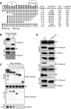
FIG. 1.
FIG. 1.
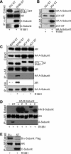
Identification of HEAT repeats in the A subunit of PP2A required for binding to AR. (A) Diagram showing the 15 HEAT repeats in the A subunit and known contact sites for ST and the C subunit. The N- and C-terminal deletions that were used to map the AR binding site are depicted. C-subunit binding to the A-subunit deletion mutant spanning HEAT repeats 6 to 15 was not determined (ND*); however, C-subunit binding to a PP2A A subunit that lacks HEAT repeats 3 to 6 was shown in a previous study (34). (B) ST binding region of the PP2A A subunit is required for PP2A A/C heterodimer binding AR. A subunits encoding HEAT repeats 3 to 15 (amino acids 84 to 589) or HEAT repeats 6 to 15 (amino acids 180 to 589) were coexpressed with AR in Cos7 cells, and the cells were incubated with 10 nM R1881 for 60 min before harvest. AR complexes were immunoprecipitated using AR441 and immunoblotted using AR21 and goat polyclonal anti-A-subunit. Deletion of HEAT repeats 3 to 5 is sufficient to disrupt ST binding to the A subunit and, therefore, ST-induced PP2A binding to AR. (C) AR binding to the PP2A A subunit is lost upon deletion of HEAT repeats 13 to 16. Flag-tagged A subunits containing the indicated HEAT repeats were coexpressed with AR as described above. IPs were carried out using AR441 or M2 anti-Flag as indicated. Antibodies used for immunoblotting were AR21, M2, and rabbit polyclonal anti-C-subunit. PP2A C-subunit binding to the A subunit is lost upon deletion of HEAT repeats 14 and 15, and AR binding to the A subunit is lost upon further deletion of HEAT repeat 13. Thus, the C-subunit and AR binding sites on the A subunit are nonidentical. (D) AR binding to the PP2A A subunit requires HEAT repeat 13. HA-tagged A subunits were coexpressed with ST in LNCaP cells, which were treated with R1881. Antibodies used were PAb430 anti-ST or 16B12 anti-HA for IP and, for immunoblotting, AR21, 16B12 anti-HA, rabbit anti-C subunit, or PAb108 anti-ST. AR binding to the PP2A A subunit is lost upon deletion of HEAT repeat 13. This deletion also results in loss of PP2A C-subunit binding to the A subunit, likely because intersubunit hydrogen binding is disrupted. Amino acids Tyr495 and Arg498 in HEAT repeat 13 form hydrogen bonds with Asn79 and Asp280 in the C subunit (5, 46). α, anti.
FIG. 2.
FIG. 2.
PP2A transfer onto AR is specific for SV40 ST. (A) The closely related ST from JCV does not mediate PP2A transfer onto AR. JCV ST and SV40 ST were expressed in LNCaP cells, which were treated with R1881. Antibodies used were AR441 for IP and, for immunoblotting, PAb108 anti-ST (for both SV40 and JCV ST), AR21, goat anti-A subunit, and rabbit anti-C subunit. (B) JCV ST interacts with the PP2A A subunit. JCV ST was expressed in LNCaP cells in the presence or absence of the HA-tagged A subunit. Cells were treated with R1881. Antibodies used were 16B12 anti-HA for IP and, for immunoblotting, 16B12 anti-HA and PAb108 anti-ST. (C) HA-tagged A subunit is transferred onto AR in response to ST and androgen. HA-tagged A subunit was coexpressed with JCV ST or SV40 ST in LNCaP cells. Cells were treated with R1881. Antibodies used were AR441 and 16B12 α-HA for IP and, for immunoblotting, PAb108 anti-ST, 16B12 anti-HA, and AR21. (D) Overexpressed PP2A B subunits do not mediate PP2A transfer onto AR. HA-tagged B-subunits (α, β, γ1, δ, and ɛ) were coexpressed with AR in 293 cells. Cells were treated with R1881. Antibodies used were 16B12 α-HA for IP and, for immunoblotting, 16B12 anti-HA, goat anti-A subunit, and AR21. These B subunits bind to the A subunit of PP2A but do not induce the conformation required for PP2A transfer onto AR. (E) PP2A B′α subunit does not mediate PP2A transfer onto AR. Flag-tagged B′α subunit was expressed in LNCaP cells. Cells were treated with R1881. Antibodies used were AR441 for IP and, for immunoblotting, M2 anti-Flag, rabbit anti-C subunit, and AR21. α, anti.
FIG. 3.
FIG. 3.
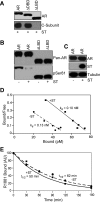
PP2A binding and dephosphorylation is dependent on the LBD of AR. (A) The LBD of AR is required for PP2A binding. Full-length AR and mutants lacking DBD (residues 559 to 616) or LBD (residue 710 to end) of AR were expressed in Cos7 cells. Cells were treated with R1881. Antibodies used were AR441 for IP and, for immunoblotting, rabbit anti-C subunit and AR21. In this reaction, ST expressed by the SV40-transformed Cos7 cells binds to endogenous A subunit and mediates transfer of the PP2A A/C heterodimer. Only the blotting for the PP2A C subunit is shown. (B) The LBD of AR is required for dephosphorylation of the AF-1 phospho-site Ser81. 293 cells were transfected with AR or the ΔLBD derivative of AR and, where indicated, ST. Cells were treated with R1881. Antibodies used were G122-434 anti-AR and AR441 (1:1) for AR IP and AR21 and rabbit anti-pSer81 for immunoblotting. ST induces PP2A binding to AR and dephosphorylation of Ser81 (lane 2) in the AF-1 region of AR (48). Upon deletion of the AR LBD, PP2A fails to bind and dephosphorylate AR (lane 4). (C) Expression levels of AR and ST used for androgen binding assays. Ad expressing AR was used to infect PC-3 cells in the presence or absence of Ad expressing SV40 ST. Antibodies used were anti-AR hinge region, PAb108 anti-ST and anti-tubulin for immunoblotting. (D) Scatchard analysis showing that PP2A binding does not affect AR affinity to androgen. PC-3 cells were infected by Ad-HisAR in the absence or presence of Ad-ST; the latter was used to promote PP2A transfer onto AR in the presence of androgen. Cells were incubated with various concentrations of [3H]R1881 (0.02 to 0.2 nM) for 60 min and then washed with PBS. Androgens were extracted and measured in a scintillation counter. Nonspecific binding was measured in the presence of a 400-fold excess of unlabeled R1881 and subtracted. Based on immunoblotting, the difference in total androgen binding likely reflects differences in AR expression levels. The results are representative of four experiments. (E) PP2A binding to AR has a small effect on androgen dissociation. PC-3 cells were infected by Ad-HisAR in the absence or presence of Ad-ST; the latter was used to promote PP2A transfer onto AR in the presence of androgen. Cells were incubated with 2 nM [3H]R1881 for 60 min, washed with PBS, and then chased with 1 μM cold R1881 for the indicated time periods. Androgens were extracted and measured in a scintillation counter. The results are representative of three experiments. AU, arbitrary units.
FIG. 4.
FIG. 4.
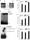
AR Complexes analyzed by EMSA and analysis of multisite phosphorylation mutants of AR. (A) AR complexes used for EMSA. PC-3 cells were infected with Ad-HisAR in the absence or presence of Ad-ST and treated with 10 nM R1881. AR complexes were isolated by IP using AR441, eluted with specific peptide in the presence of androgen, and analyzed by silver staining or immunoblotting. Antibodies used for immunoblotting were anti-AR hinge region, goat anti-A subunit, and rabbit anti-C subunit. (B) EMSA using C3(1)-ARE oligonucleotides and purified AR complexes. The arrows indicate the positions of protein-DNA complexes, which are supershifted in the presence of anti-AR antibody. When the reaction is performed using the AR-PP2A complexes, binding to the ARE DNA is reduced to 68% of that observed with AR complexes. Because a PP2A-dependent gel shift of the AR-ARE complex is not resolved under these conditions, the total mass of the PP2A heterodimer (∼100 kDa) is insufficient to give a supershift, or PP2A has dissociated from the AR-DNA complex. (C) Specificity of the antibody used for EMSA. Affinity purified anti-hinge region Ab (epitope: amino acids 656 to 669) induces supershift of a fragment encoding the AR DBD-hinge. (D to F) Transcription assay of WT and multisite mutants of AR measured in PC-3 cells using the indicated promoters. Error bars represent standard deviations. Immunoblotting was used to confirm that similar levels of WT and mutant AR proteins were expressed (not shown). 5Ala, Ser81Ala, Ser94Ala, Ser256Ala, Ser308Ala, and Ser424Ala; 6Asp, Ser81Asp, Ser94Asp, Ser256Asp, Ser308Asp, Ser424Asp, and Ser650Asp; LUC, luciferase.
FIG. 5.
FIG. 5.
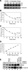
PP2A release from AR is correlated with R1881 dissociation. (A) AR-PP2A dissociation analyzed in vivo. AR-PP2A complexes were assembled in PC-3 in the presence of ST by the addition of a 2 nM concentration of radioactive [3H]R1881 (R1881 pulse). The cells were washed with PBS and then treated with 100 μM bicalutamide (Casodex chase) for the indicated time periods. AR-PP2A complexes were isolated by IP using AR441 and eluted in SDS-PAGE sample buffer, and [3H]R1881 bound to AR was measured by scintillation counting. AR and PP2A were quantified by immunoblotting using anti-AR hinge region and rabbit anti-C subunit antibodies. (B) AR-androgen dissociation rate analyzed in vivo. Androgen dissociation from AR follows first-order kinetics (R2 = 0.978). (C) Plot of the AR-PP2A dissociation data shown in panel A. The data were normalized to the amount of AR recovered in each IP and show that the loss of PP2A from AR follows first-order kinetics (R2 = 0.924). (D) Plot showing a correlation between the amount of PP2A and androgen bound to AR during the dissociation reaction (R2 = 0.986). (E) Androgen dissociation from AR (in the absence of antagonist) results in PP2A dissociation in vitro. The AR441-protein G beads containing purified AR-PP2A complexes (the initial bead fraction) were incubated with a large volume of Triton lysis buffer at room temperature for 180 min and then separated into the remaining bead fraction (bound) and the supernatant fraction (released). AR and PP2A in the initial bead fraction was compared with different gel loadings (lanes 1 to 5), the remaining bead fraction (lane 6), and the released supernatant fraction (lane 7). AU, arbitrary units.
FIG. 6.
FIG. 6.
PP2A dissociates rapidly from AR complexes assembled in the presence of ASD. (A) AR-PP2A complexes were assembled in PC-3 cells in the presence of ST by the addition of 100 nM ASD, which has a much weaker affinity (>200-fold) for AR (3). Cells were washed with PBS and then treated with 100 μM bicalutamide (Casodex chase) for indicated time periods. Antibodies used were AR441 for IP and anti-AR hinge region and rabbit anti-C subunit for immunoblotting. (B) Plot of AR-PP2A dissociation using the data shown in panel A. The data were normalized to the amount of AR recovered in each IP and show that the loss of PP2A from AR follows first-order kinetics (R2 = 0.993). AU, arbitrary units.
FIG. 7.
FIG. 7.
At least two distinct ligand-dependent conformations of AR can mediate transcription. (A) AR-dependent transcription from the PSA promoter stimulated by androgen agonists and antagonists. PC-3 cells transfected with the PSA-luciferase (PSA-Luc) reporter plasmid and either WT AR (open bars) or T877A AR (gray bars), treated with the indicated ligands, and normalized to CMV-Renilla. (B) AR-dependent transcription from the MMTV promoter stimulated by androgen agonists and antagonists. PC-3 cells transfected with the MMTV-Luc reporter plasmid and either WT AR (open bars) or T877A AR (gray bars) and assayed in the absence and presence of the indicated ligands as above. (C) PP2A binds to the DHT-bound and ASD-bound forms of WT and T877A AR. Cos7 cells were transfected with AR and treated with the indicated androgens or androgen antagonists. AR affinity purification was carried out using AR441. AR complexes were analyzed by immunoblotting using anti-AR hinge region and rabbit anti-C subunit. (D) PSA induction in LNCaP by different ligands. LNCaP was used because it expresses the T877A mutant form of AR and endogenous PSA. LNCaP cells grown in RPMI PRF medium plus 5% charcoal- and dextran-stripped fetal bovine serum were incubated with indicated ligands for 24 h. Cells were washed with PBS and extracted with SDS-PAGE sample buffer. Extracts were analyzed by immunoblotting using anti-AR hinge region, anti-tubulin, and anti-PSA antibodies.
FIG. 8.
FIG. 8.
ST mediates PP2A transfer onto AR in LNCaPs grown as xenografts in mice. (A) Characterization of LNCaP cells stably expressing ST and ST-myc. Cells were incubated with R1881 for 60 min. IP was carried out using AR441, and immunoblotting was performed with anti-AR hinge region, goat anti-A subunit, and PAb108 anti-ST. (B) Analysis of LNCaP cells stably expressing ST and ST-myc after growth as xenografts in nude mice. Tumor growth, castration, tumor homogenization, and protein extraction were carried out as described in Materials and Methods. AR complexes were purified using G122-434 anti-AR and AR441 (1:1). Antibodies used for immunoblotting were PAb108 anti-ST, anti-AR hinge region, rabbit anti-C subunit, and a panel of phospho-site-specific antibodies (48). The effect of castrate levels of androgen on ST-dependent PP2A transfer and AR dephosphorylation state was examined in mice subjected to castration (Castr) prior to tumor harvest. α, anti.
FIG. 9.
FIG. 9.
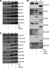
The phosphorylation state of the AF-1 is governed by conformational changes in AR that regulate phosphatase action. (A) Phosphorylation sites in AR are kinase accessible in androgen-free AR. 293 cells transfected with AR were treated with R1881 (10 nM for 1 h), OA (400 nM for 3 h), or the two combined (OA for 2 h and then OA plus R1881 for 1 h). AR complexes were isolated using G122-434 anti-AR and AR441 (1:1). Immunoblotting was performed using anti-AR hinge region and a panel of phosphoserine antibodies. Treating cells with a concentration of OA that is sufficient to inhibit PP2A and PP5 results in a high level of AF-1 phosphorylation at multiple sites. (B) OA inhibits the catalytic activity of PP2A but it does not affect PP2A binding to AR. 293 cells cotransfected with AR and ST were subjected to drug treatments and IP as described for panel A. Antibodies used for immunoblotting were anti-AR hinge region, goat anti-A subunit, rabbit anti-C subunit, and a panel of phosphoserine antibodies. The presence of OA does not affect the transfer reaction, and the concentration of OA used is capable of inhibiting PP2A in the AR complexes. (C) Deletion of LBD results in phosphorylation at several phospho-sites in the AF-1 region of AR. 293 cells were transfected with WT AR or mutant AR lacking the LBD (deletion of residues 710 to 919). Cells were treated with 10 nM R1881 for 2 h, where indicated. AR complexes were isolated using G122-434 anti-AR and AR441 (1:1). Immunoblotting was performed using anti-AR hinge region and a panel of phospho-site antibodies.
FIG. 10.
FIG. 10.
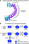
Protein conformation regulates phosphatase action on AR. (A) Model of ST-induced conformation change in the PP2A A subunit based on the structure solved by Barford and colleagues (Protein Data Bank entry 1B3U). Superimposition of noncrystallographic related copies of the PP2A A subunit emphasizes the regions of conformational flexibility, as noted previously (17). The A chain (blue and magenta; opaque) and the B chain (pink and cyan; transparent) were aligned using PyMOL (12). While the A and B chains show a high degree of alignment in HEAT repeats 4 to 12, the displacement of helices in HEAT repeats 1 to 3 and 13 to 15 in these structures suggests conformational flexibility in the N- and C-terminal regions, respectively. ST binding at the ST interaction site is proposed to induce a conformational change that is propagated N to C through the A subunit; this is manifest as an AR interaction site. In turn, AR binding is proposed to induce a conformational change that is propagated C to N that promotes release of ST. The biochemical evidence for this model is that ST induces PP2A binding to AR, but ST is fully dissociated from the PP2A-AR complex (48). (B) Two models summarizing how ligand binding could regulate the phosphorylation state of AR in cells in the absence of ST. In the kinase access model, ligand-regulated phosphorylation sites are kinase inaccessible due to AR conformation or masking by chaperones. Androgen binding induces structural changes in AR that reveal the ligand-regulated phosphorylation sites. In the phosphatase access model, AR phosphorylation is regulated by the actions of both kinases and phosphatases. Under ligand-free conditions, phosphatase action predominates, resulting in a lower level of AR phosphorylation. Ligand binding reduces phosphatase action on AR and increases AR phosphorylation. Phosphatase action on AR could be reduced because the phosphatase targeting mechanism is lost upon ligand binding. For example, if phosphatase targeting depends on AR-associated Hsp90, ligand-induced Hsp90 dissociation would reduce the AR-phosphatase interaction. Alternatively, ligand binding could generate an AR conformation that is resistant to phosphatase action by limiting phospho-site accessibility. Both kinases and phosphatases have access to ligand-free AR based on the fact that OA treatment results in the accumulation of highly phosphorylated AR. Androgen-dependent phosphorylation of AR could result from contributions from both types of mechanisms.

References

    1. Andrade, M. A., and P. Bork. 1995. HEAT repeats in the Huntington's disease protein. Nat. Genet. 11:115-116. - PubMed
    1. Chang, C. Y., and D. P. McDonnell. 2002. Evaluation of ligand-dependent changes in AR structure using peptide probes. Mol. Endocrinol. 16:647-660. - PubMed
    1. Chen, F., K. Knecht, C. Leu, S. J. Rutledge, A. Scafonas, C. Gambone, R. Vogel, H. Zhang, V. Kasparcova, C. Bai, S. Harada, A. Schmidt, A. Reszka, and L. Freedman. 2004. Partial agonist/antagonist properties of androstenedione and 4-androsten-3β,17β-diol. J. Steroid Biochem. Mol. Biol. 91:247-257. - PubMed
    1. Chen, J., J. Kim, and J. T. Dalton. 2005. Discovery and therapeutic promise of selective androgen receptor modulators. Mol. Interv. 5:173-188. - PMC - PubMed
    1. Cho, U. S., and W. Xu. 2006. Crystal structure of a protein phosphatase 2A heterotrimeric holoenzyme. Nature 445:53-57. - PubMed

Publication types

MeSH terms

LinkOut - more resources