. 2007 May;27(9):3470-80.
doi: 10.1128/MCB.00659-06. Epub 2007 Feb 26.

Targeting of the ETS factor GABPalpha disrupts neuromuscular junction synaptic function

Affiliations

Targeting of the ETS factor GABPalpha disrupts neuromuscular junction synaptic function

Debra A O'Leary et al. Mol Cell Biol. 2007 May.

Abstract

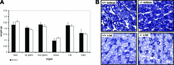
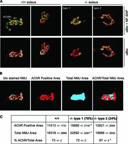
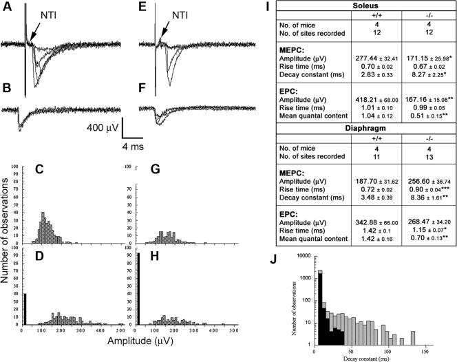
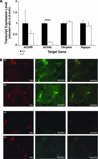
The GA-binding protein (GABP) transcription factor has been shown in vitro to regulate the expression of the neuromuscular proteins utrophin, acetylcholine esterase, and acetylcholine receptor subunits delta and epsilon through the N-box promoter motif (5'-CCGGAA-3'), but its in vivo function remains unknown. A single point mutation within the N-box of the gene encoding the acetylcholine receptor epsilon subunit has been identified in several patients suffering from postsynaptic congenital myasthenic syndrome, implicating the GA-binding protein in neuromuscular function and disease. Since conventional gene targeting results in an embryonic-lethal phenotype, we used conditional targeting to investigate the role of GABPalpha in neuromuscular junction and skeletal muscle development. The diaphragm and soleus muscles from mutant mice display alterations in morphology and distribution of acetylcholine receptor clusters at the neuromuscular junction and neurotransmission properties consistent with reduced receptor function. Furthermore, we confirmed decreased expression of the acetylcholine receptor epsilon subunit and increased expression of the gamma subunit in skeletal muscle tissues. Therefore, the GABP transcription factor aids in the structural formation and function of neuromuscular junctions by regulating the expression of postsynaptic genes.


Figures

FIG. 1.
FIG. 1.
Conditional targeting of the Gabpα locus. (A) Schematic of wild-type, targeted, floxed, and conditional-knockout Gabpα alleles. A floxed Neomycin (neo) cassette was inserted within intron 2, and a single loxP site immediately upstream of exon 2. The positions of the Southern blot probe, PCR primers and restriction sites for BamHI (B), BalI (Bl), EcoRI (E), and HindIII (H) are shown. (B) Southern blot analysis of genomic DNA from G418-resistant ES cells (clone numbers are shown), using a BamHI digest and an external intron 3 probe to detect 7-kb wild-type and 5-kb targeted fragments. (C) PCR screening of puromycin-resistant targeted ES cell clones following transient Cre transfection was performed with primers G1f and G1r, spanning Gabpα introns 1 and 2 flanking the site of insertion of the neo cassette, generating 1.8-kb wild-type (wt), 1.9-kb floxed (loxP), and 1.5-kb knockout (ko) allele products.
FIG. 2.
FIG. 2.
Specific loss of Gabpα expression in skeletal muscle. (A) PCR screening of genomic DNA extracted from organs of wild-type (+/+), heterozygous (+/−), and homozygous knockout (−/−) mice. A common 5′ primer within intron 1 (G2f) and 3′ primers within exon 2 (G2r) and intron 2 (G1r) generated products of 620 bp, 660 bp, and 740 bp, representing wild-type, floxed, and knockout alleles, respectively. The knockout is represented by an asterisk in each case where no wild-type allele was detectable. (B) Western blot analysis of Gabpα (58-kDa) and β-tubulin (50-kDa) protein levels in skeletal muscle tissues and primary skeletal muscle cell cultures from Gabpα skeletal-muscle-specific knockout mice (−/−) relative to homozygous floxed (+/+) littermates. Severely reduced Gabpα protein levels were observed in whole muscle tissue lysates of Gabpα mutant mice and complete loss of Gabpα protein expression in Gabpα mutant primary skeletal muscle cells derived from mice at postnatal days 2 and 3.
FIG. 3.
FIG. 3.
(A) Organ weights of heart and various skeletal muscle tissues (lateral gastrocnemius, medial gastrocnemius, soleus, vastus lateralis, and diaphragm) showed no statistical difference between samples from 1-month-old homozygous floxed (+/+) and Gabpα skeletal-muscle-specific knockout (−/−) mice. (B) NADH staining of transverse cryosections from 1-month-old homozygous floxed (+/+) and Gabpα skeletal-muscle-specific knockout (−/−) mice showing no significant difference in fiber size or tissue integrity between the two genotypes. Type I, IIb, and IIa fibers are shown by dark, light, and intermediate staining, respectively. The images were taken at ×200 magnification.
FIG. 4.
FIG. 4.
NMJ morphology in soleus muscles of Gabpα conditional-knockout mice. (A) Whole-mount immunostaining of single muscle fibers from 3-month-old control homozygous floxed (+/+) and homozygous Gabpα skeletal-muscle-specific knockout (−/−) mice. NMJ shape is revealed by AChR staining with Texas red-conjugated α-bungarotoxin (αBtx). Nerves and synaptic junctions were stained with a cocktail of fluorescein isothiocyanate-conjugated antibodies against neurofilament and synaptophysin (NF-SNP). The relative positions of nerves (N), synaptic junctions (SJ), and AChRs are indicated in the top left panel. NMJs of −/− mice are classified as showing less branching than those of control mice (type 1) or single ring structures (type 2). All images are at ×1,000 magnification. (B) Schematic representation of how the AChR-positive and total NMJ areas were defined, using the freehand tool in Image J image analysis software. The AChR-positive area was taken to be that area stained with α-bungarotoxin (btx) at the terminus of the synaptic nerve. The total NMJ area was taken to encompass the entire AChR-positive area, leaving no internal space between the outermost branches. (C) Quantified synaptic areas occupied by AChRs, total NMJ areas (expressed in arbitrary units ± standard errors of the mean), and percentage of the area occupied by AChRs of NMJs of 3-month-old homozygous floxed (+/+) and homozygous Gabpα skeletal-muscle-specific knockout (−/−) mice. *, P < 0.05 (Student's two-tailed t test). Types 1 and 2 are the two morphological types of NMJs observed in Gabpα mutant animals at a frequency of 76% and 24%, respectively.
FIG. 5.
FIG. 5.
Extracellular recordings from NMJs of homozygous floxed control (A to D) and Gabpα conditional knockout mice (E to H). Panels A and E show examples of evoked EPCs. Panels B and F show examples of MEPCs. In panels A, B, E, and F, five consecutive traces are superimposed. Note the invariable presence of the NTI in both control (A) and mutant (E) recordings. Frequency amplitude histograms of MEPCs (C and G) and evoked (D and H) amplitudes are shown. The histograms shown in panels C and D were constructed from a single recording site, as were the histograms shown in panels G and H. The distributions of MEPC amplitudes were similar in both control and Gabpα conditional-knockout mice. EPCs were skewed toward higher amplitudes in control mice, and the number of failures (black bars) was higher at NMJs from Gabpα mutant mice (H) compared to controls (D). (I) Comparison of biophysical properties of 3-month-old homozygous floxed Gabpα control (+/+) and Gabpα conditional-knockout (−/−) mice. *, P < 0.05; **, P < 0.01; ***, P < 0.001 (Student's two-tailed t tests). (J) Representation of MEPC decay rate constants from NMJs of Gabpα conditional-knockout (hatched bars) and homozygous floxed (black bars) mice. The histograms were constructed from a single recording site taken from either mutant or control soleus muscle. The decay rate constants for control MEPCs varied from 10 ms to 35 ms. In contrast, the decay rate for Gabpα mutant mice varied from 10 ms to 130 ms. This resulted in a significantly longer mean in decay rates at NMJs of Gabpα conditional-knockout mice than in control animals.
FIG. 6.
FIG. 6.
Expression of Gabp target genes in Gabpα conditional-knockout mice. (A) Real-time RT-PCR analysis of transcript levels of Gabp target genes, using cDNA from diaphragm muscles of four 3-month-old homozygous floxed (+/+) and homozygous Gabpα skeletal muscle-specific knockout (−/−) mice. The expression levels of each transcript are expressed as a ratio to β-actin. The ratios were adjusted so those of floxed mice were 1. The error bars represent standard errors of the mean. *****, P < 0.000005; n = 12 (two-tailed Student's t test). Levels of Rapsyn were measured as a negative control, as it is not a Gabp gene target. (B) Immunostaining of transverse cryosections from 3-month-old homozygous floxed (+/+) and Gabpα skeletal-muscle-specific knockout (−/−) mice with Texas red-conjugated α-bungarotoxin (btx) and antibodies recognizing AChRδ or AChRγ showing colocalization and increased expression of AChRγ in Gabpα mutant mice. The images were taken at ×400 magnification.

References

    1. Briguet, A., and M. A. Ruegg. 2000. The ETS transcription factor GABP is required for postsynaptic differentiation in vivo. J. Neurosci. 20:5989-5996. - PMC - PubMed
    1. Brock, J. A., and T. C. Cunnane. 1987. Relationship between the nerve action potential and transmitter release from sympathetic postganglionic nerve terminals. Nature 326:605-607. - PubMed
    1. Chan, R. Y. Y., C. Boudreau-Larivière, L. M. Angus, F. A. Mankal, and B. J. Jasmin. 1999. An intronic enhancer containing an N-box motif is required for synapse- and tissue-specific expression of the acetylcholinesterase gene in skeletal muscle fibers. Neurobiology 96:4627-4632. - PMC - PubMed
    1. Chinenov, Y., C. Coombs, and M. E. Martin. 2000. Isolation of a bi-directional promoter directing expression of the mouse GABPα and ATP synthase coupling factor 6 genes. Gene 261:311-320. - PubMed
    1. Chomczynski, P., and N. Sacchi. 1987. Single-step method of RNA isolation by acid guanidinium thiocyanate-phenol-chloroform extraction. Anal. Biochem. 162:156-159. - PubMed

Publication types

MeSH terms

Substances

LinkOut - more resources