. 2007 May;27(9):3337-52.
doi: 10.1128/MCB.01544-06. Epub 2007 Feb 26.

HOXA10 controls osteoblastogenesis by directly activating bone regulatory and phenotypic genes

Affiliations

HOXA10 controls osteoblastogenesis by directly activating bone regulatory and phenotypic genes

Mohammad Q Hassan et al. Mol Cell Biol. 2007 May.

Abstract

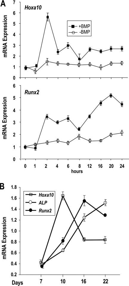
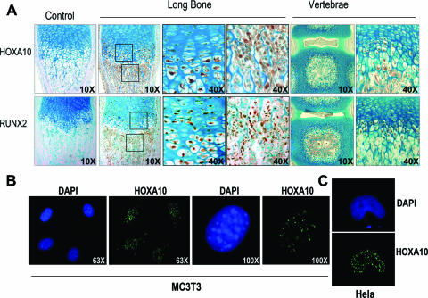
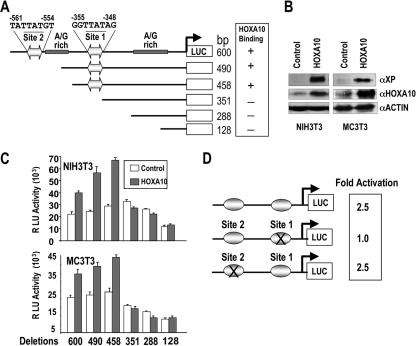
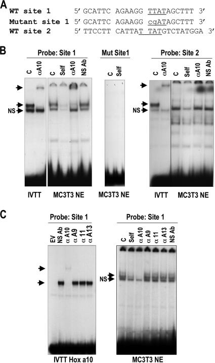
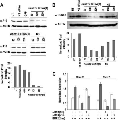
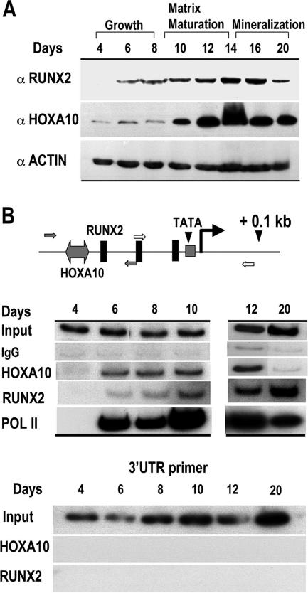
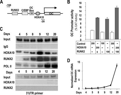
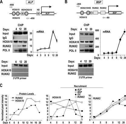
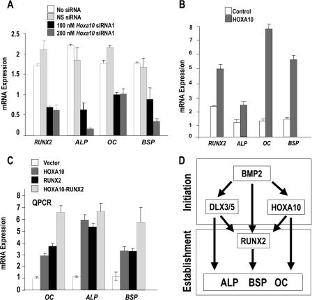
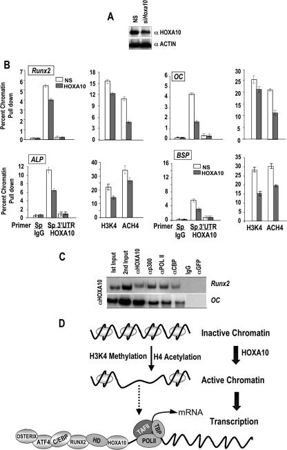
HOXA10 is necessary for embryonic patterning of skeletal elements, but its function in bone formation beyond this early developmental stage is unknown. Here we show that HOXA10 contributes to osteogenic lineage determination through activation of Runx2 and directly regulates osteoblastic phenotypic genes. In response to bone morphogenic protein BMP2, Hoxa10 is rapidly induced and functions to activate the Runx2 transcription factor essential for bone formation. A functional element with the Hox core motif was characterized for the bone-related Runx2 P1 promoter. HOXA10 also activates other osteogenic genes, including the alkaline phosphatase, osteocalcin, and bone sialoprotein genes, and temporally associates with these target gene promoters during stages of osteoblast differentiation prior to the recruitment of RUNX2. Exogenous expression and small interfering RNA knockdown studies establish that HOXA10 mediates chromatin hyperacetylation and trimethyl histone K4 (H3K4) methylation of these genes, correlating to active transcription. HOXA10 therefore contributes to early expression of osteogenic genes through chromatin remodeling. Importantly, HOXA10 can induce osteoblast genes in Runx2 null cells, providing evidence for a direct role in mediating osteoblast differentiation independent of RUNX2. We propose that HOXA10 activates RUNX2 in mesenchymal cells, contributing to the onset of osteogenesis, and that HOXA10 subsequently supports bone formation by direct regulation of osteoblast phenotypic genes.


Figures

FIG. 1.
FIG. 1.
Hoxa10 expression in relation to osteogenic differentiation. (A) Premyogenic C2C12 cells were treated with 100 ng/ml BMP2 for 24 h. Total RNA was isolated at different time points (0, 1, 2, 4, 6, 8, 12, 16, 20, and 24 h). Five micrograms of total RNA was reverse transcribed with oligo(dT) primer and amplified (RT-QPCR) by gene-specific primers. (Top) Hoxa10 expression (+BMP, −BMP) was normalized to Gapdh expression (+BMP, −BMP), and relative transcript levels were plotted; (bottom) the induction of Runx2 expression with BMP2 treatment is shown for the same time course. Error bars represent triplicate analyses of each sample from two independent experiments. (B) Temporal expression of Hoxa10 and the osteogenic markers Runx2 and ALP during MC3T3 cell growth and differentiation. cDNAs from different time points were amplified using bone-specific gene primers (Table 1). Expression values from RT-QPCR were normalized to Gapdh values. Error bars represent triplicate sample analyses from one experiment. Independent experiments exhibited similar temporal expressions (data not shown).
FIG. 2.
FIG. 2.
Coexpression of HOXA10 and RUNX2 in vivo and in vitro. (A) HOXA10 and RUNX2 are visualized in hypertrophic chondrocytes and osteoblast lineage cells in long bones and vertebrae as indicated. Immunohistochemistry was performed using anti-HOXA10 antibody and anti-RUNX2 antibody. For nonspecific controls, normal rabbit IgG was substituted for primary antibody, and blocking peptides specific for the HOXA10 and RUNX2 antibodies were used. Bone and vertebra sections are shown at ×10 and ×40 magnification as indicated for the boxed areas at the growth plate and the primary spongiosa. Trabeculae show robust expression of both proteins in surface osteoblasts. The first column shows peptide blocking controls (magnification, ×10). (B) In situ immunofluorescence examination of endogenous HOXA10 protein in MC3T3 cells to demonstrate nuclear staining with few cytoplasmic foci using the Santa Cruz antibody. (C) Nuclear localization of transfected Xpress-tagged HOXA10 is also observed for HeLa cells detected with anti-Xpress antibody (see Materials and Methods). DAPI, 4′,6′-diamidino-2-phenylindole.
FIG. 3.
FIG. 3.
HOXA10 directly regulates Runx2 promoter activity and transcription. (A) Schematic representation of the deletions of the 0.6-kb Runx2 promoter with the firefly luciferase reporter gene. The full-length 0.6-kb promoter contains two Hoxa10 putative binding sites (site 1 and site 2) having the core TTAT motifs. Deletion clones −490- and −458-Luc contain only site 1, whereas clones −351-, −288-, and −108-Luc are devoid of Hoxa10 binding sites. (B) Western blots of endogenous and exogenous HOXA10 protein levels in nonosseous (NIH 3T3) and osseous (MC3T3) cells. For detection of endogenous and exogenous HOXA10 protein, anti-HOXA10 (N20; Santa Cruz Biotech Inc.) was used. α, anti-. (C) A functional Hoxa10 resides in the proximal Runx2 promoter. Runx2 promoter deletion mutant constructs were cotransfected with 400 ng of either backbone vector (control representing basal promoter activity [open bars]) or Hoxa10 expression construct (treated [solid bars]). Runx2 promoter activities are calculated from relative luciferase values for both NIH 3T3 and MC3T3 cells. All transfections were performed in triplicate. RLU, relative light units. (D) Two nucleotide mutations (TTAT to CGAT) were constructed in the distal (site 2) and proximal (site 1) Hoxa10 binding motifs in the 0.6-kb Runx2 P1 promoter as illustrated. Hoxa10-mediated activation for WT/mutant is indicated.
FIG. 4.
FIG. 4.
Specificity of the Hoxa10 site 1 for HOXA10 interactions. (A) Oligonucleotide sequences are shown for the WT and the Hoxa10 site mutations and compared to that for the mouse consensus optimal Hoxa10 binding site (4). Underlining indicates the core binding site; lowercase indicates mutant nucleotides. (B) DNA binding activity of HOXA10 is demonstrated by EMSA. Ten femtomoles of labeled double-stranded oligonucleotides derived from the WT or mutant (Mut) probe as indicated was incubated with 2 μl of IVTT HOXA10 or 5 μg of MC3T3 nuclear extract (NE). Self-competition (Self), antibody supershift, and nonspecific antibody (NS Ab) controls are shown. The mutant double-stranded oligonucleotide for Hoxa10 does not bind nuclear proteins. The top unlabeled arrows indicate the supershifted band; the lower unlabeled arrows show the position of the HOXA10-specific band, below which is a nonspecific complex (arrows labeled NS). α, anti-; C, control. (C) IVTT of indicated HOX proteins is complexed with the Hox site 1 in Runx2 to show the specificity of the HOXA10 antibody. Nuclear extracts of MC3T3 osteoblasts were used in EMSA studies to show the specificity of the HOXA10 interaction (arrow) with the site 1 Runx2 probe. GFP, green fluorescent protein; EV, empty vector.
FIG. 5.
FIG. 5.
RNAi of Hoxa10 in MC3T3 and C2C12 cells inhibits Runx2 and osteogenic gene expression. (A) Knockdown of Hoxa10 in MC3T3-E1 cells by two sets of Hoxa10 siRNA duplexes and nonspecific controls (NS). The lower panel shows densitometric quantitation of HOXA10 knockdown when cells were isolated 72 h after siRNA treatment. (B) Preosteoblast MC3T3 cells at 30 to 50% confluence were transfected with siRNA1 specific for murine Hoxa10 at different concentrations (50, 100, and 200 nM). (Top) The Western blot shows the knockdown of RUNX2 protein upon treatment with Hoxa10-specific siRNA duplexes. UT, untransfected; NS, nonsilencing duplexes (used as controls). Densitometry shows reductions of RUNX2 protein upon knockdown of Hoxa10. (C) C2C12 cells were plated on day 0 at 30 to 40% confluence and treated with Hoxa10 siRNA or nonspecific siRNA [siRNA(NS)] (100 nM) for 48 h. After the medium was changed, BMP2 (100 ng/ml) was added to induce Hoxa10 and Runx2 for 2 h. mRNA levels were assayed by RT-QPCR. White bars represent the control for the treatment as indicated. GFP, green fluorescent protein; α, anti-.
FIG. 6.
FIG. 6.
HOXA10 protein and recruitment to the Runx2 promoter in primary calvarial cells during osteoblast growth and differentiation. (A) Western blot analyses for RUNX2 and HOXA10 protein expression are shown during stages of growth and differentiation of isolated primary calvarial osteoblasts as indicated (antibody information is provided in Materials and Methods). Protein profiles of actin demonstrate equivalent amounts of total protein loaded in the gel. (B) ChIP studies of the Runx2 proximal promoter locus during growth and differentiation. The top panel illustrates the positions of regulatory elements and primers used to amplify the Runx2 promoter-specific DNA fragments. Open arrow, RNA Pol II; solid arrow, HOXA10 and RUNX2 occupancy. For the middle panel, ChIP analysis was performed on the indicated days with antibodies for HOXA10, RUNX2, or RNA Pol II (see Materials and Methods). One percent of the soluble chromatin fraction was taken as the input fraction. IgG is shown as an antibody control. The lower panel shows the control for ChIP primer specificity.
FIG. 7.
FIG. 7.
HOXA10 is recruited to the OC promoter to directly regulate gene expression. (A) Activation of OC gene transcription by HOXA10. Schematic representation of rat proximal OC promoter segment illustrating the location of the only Hoxa10 consensus motif in the −208-bp 5′ OC fragment. Arrows are the forward (−198) and reverse (−28) OC primers used in ChIP analysis. (B) The mouse preosteoblast cell line MC3T3-E1 was cotransfected with either empty vector (control) or Hoxa10 and Runx2 expression plasmids as indicated and with the proximal OC promoter (−208 OC-CAT) by use of FuGENE6 (Roche Molecular Biologicals). Control cells contained the indicated amount of backbone plasmid (pGL3). Cells were harvested 24 h posttransfection for CAT assay. Promoter activity was calculated from values equivalent to those for six samples and expressed as percent CAT activity. (C) In vivo occupancy of HOXA10 on the OC gene. Cross-linked chromatin samples from primary rat calvarium-derived osteoblast cultures at the indicated stages of differentiation (days 4, 5, 8, 9, 12, and 20) were used for immunoprecipitation reactions with 2 μg of HOXA10, RUNX2, Pol II, and nonspecific antibody (IgG). The pull-down DNA fragments were purified and assayed by PCR. Input represents 1% of each chromatin fraction used for immunoprecipitation. The ChIP data presented are representative of multiple experiments in which all the time point data were derived from the same osteoblast preparation. The lower panel shows the control for ChIP primer specificity. (D) Expression of OC mRNA (QPCR) during differentiation of primary calvarial osteoblasts at the indicated days. C/EBP, CCAAT/enhancer-binding proteins.
FIG. 8.
FIG. 8.
Hoxa10 regulatory elements are present in the ALP (A) and BSP (B) genes. ChIP assay was performed with primary rat calvarial osteoblasts by use of the indicated antibodies at the proliferation stage (day 4) and the differentiation stages (days 12 and 20, early and mature osteoblasts in mineralized matrix). HD, homeodomain. (Top) Illustration of Hoxa10 sites and locations of primers (arrows) used to detect the association of HOXA10 with the ALP and BSP promoters; (bottom) ChIP quantitated by radioactivity with control primers shown below (left) and expression of ALP and BSP genes by RT-QPCR (right). (C) Summary profiles of cell protein levels and recruitment of HOXA10 and RUNX2 to four bone promoters. The data represent densitometric values obtained from Western blot and ChIP experiments normalized to actin and IgG values, respectively.
FIG. 9.
FIG. 9.
HOXA10 regulation of osteoblast phenotypic genes dependent on and independent of RUNX2. (A) Transcript levels of bone phenotypic markers are decreased by Hoxa10 siRNA treatment at the indicated doses. RT-QPCR mRNA expression profiles in Hoxa10 siRNA-treated (100 and 200 nM) MC3T3 cells. The relative mRNA expression profiles of the endogenous marker genes were normalized to Gapdh expression profiles. Error bars are means ± standard deviations from triplicate samples. (B) Forced expression of Hoxa10 (200 ng) for 24 h induces expression of bone phenotypic genes (RT-QPCR analyses of six samples). (C) A Runx2/ TERT-immortalized stable cell line was transfected with 400 ng cytomegalovirus-driven Hoxa10, 200 ng Runx2, or both expression plasmids for 24 to 36 h (six samples). Cell layers were harvested for total cellular RNA and analyzed for expression of indicated genes by RT-QPCR. (D) Schematic illustration of the BMP2-induced HOXA10, homeodomain proteins DLX3/DLX5, and RUNX2 gene expression representing the initiation phase of osteogenesis. Together these genes establish the osteoblast phenotype by direct and Runx2-dependent activities on target genes.
FIG. 10.
FIG. 10.
siRNA knockdown of Hoxa10 decreases histone acetylation and H3K4 methylation of osteogenic genes. (A) MC3T3 cells were treated with Hoxa10 siRNA and nonspecific control (NS) for 72 h. The knockdown effect is shown by Western blotting. (B) DNA samples from ChIP with HOXA10, nonspecific IgG, methylated histone K4 (H3K4), and acetylated histone H4 (ACH4) were amplified by gene-specific (Sp) and 3′ control UTR primers (Table 1) for the indicated gene promoters. The left panel for each gene shows the effect of Hoxa10 siRNA on HOXA10 recruitment in ChIP assay of the indicated promoter. The right panel shows the status of H3K4 methylation or H4 acetylation of the chromatin modification by Hoxa10-specific knockdown. (C) ChIP-reChIP assays were performed to identify the association of coregulatory factors p300 and CBP with HOXA10 on the Runx2 and OC promoters. A HOXA10 ChIP was performed with HOXA10 antibody, and the resulting chromatin immunoprecipitate (second input) was subjected to reChIP with the indicated antibodies (α) (secondary pull-down). Normal IgG and green fluorescent protein (GFP) antibodies provided controls. The DNA fragments for Runx2 and OC were amplified as described in Materials and Methods. (D) Schematic illustration to show how HOXA10 belongs to an epigenetic coregulatory complex for remodeling chromatin to induce transcription of osteogenic genes. We propose that HOXA10 may be among the earliest factors recruited to bone promoters. HOXA10 recruitment is followed by the coordinated occupancy of other bone-related transcription factors for maximal expression of individual genes during stages of differentiation. HD, homeodomain.

References

    1. Bae, J.-S., S. Gutierrez, R. Narla, J. Pratap, R. Devados, J. L. Stein, J. B. Lian, G. S. Stein, and A. Javed. 2007. Reconstitution of Runx2/Cbfa1 null cells identifies Runx2 functional domains required for osteoblast differentiation and responsiveness to osteogenic regulators BMP2, TGFα and 1,25(OH)2D3. J. Cell. Biochem. 100:434-449. - PubMed
    1. Bae, N. S., M. J. Swanson, A. Vassilev, and B. H. Howard. 2004. Human histone deacetylase SIRT2 interacts with the homeobox transcription factor HOXA10. J. Biochem. (Tokyo) 135:695-700. - PubMed
    1. Balint, E., D. Lapointe, H. Drissi, C. van der Meijden, D. W. Young, A. J. van Wijnen, J. L. Stein, G. S. Stein, and J. B. Lian. 2003. Phenotype discovery by gene expression profiling: mapping of biological processes linked to BMP-2-mediated osteoblast differentiation. J. Cell. Biochem. 89:401-426. - PubMed
    1. Benson, G. V., T. H. Nguyen, and R. L. Maas. 1995. The expression pattern of the murine Hoxa-10 gene and the sequence recognition of its homeodomain reveal specific properties of Abdominal B-like genes. Mol. Cell. Biol. 15:1591-1601. - PMC - PubMed
    1. Bondos, S. 2006. Variations on a theme: Hox and Wnt combinatorial regulation during animal development. Sci. STKE 2006:e38. - PubMed

Publication types

MeSH terms

Substances

LinkOut - more resources