. 2007 May;75(5):2291-6.
doi: 10.1128/IAI.01328-06. Epub 2007 Feb 26.

Th1 and Th2 cells help CD8 T-cell responses

Affiliations

Th1 and Th2 cells help CD8 T-cell responses

Melinda J Ekkens et al. Infect Immun. 2007 May.

Abstract

Help from CD4 T cells is often important for the establishment of primary and memory CD8 T-cell responses. However, it has yet to be determined whether T helper polarization affects the delivery of help and/or whether responding CD8 T cells helped by Th1 or Th2 cells express distinct effector properties. To address these issues, we compared CD8 T-cell responses in the context of Th1 or Th2 help by injecting dendritic cells copulsed with the major histocompatibility complex class I-restricted OVA peptide plus, respectively, bacterial or helminth antigens. We found that Th2 cells, like Th1 cells, can help primary and long-lived memory CD8 T-cell responses. Experiments in interleukin-12 (IL-12)-/- and IL-4-/- mice, in which polarized Th1 or Th2 responses, respectively, fail to develop, indicate that the underlying basis of CD4 help is independent of attributes acquired as a response to polarization.


Figures

FIG. 1.
FIG. 1.
CD4 help is required for the development of IFNγ-producing CD8 T cells during immunization with peptide-pulsed DCs. DCs pulsed with P. acnes or SEA in combination with OVApep were injected into naive WT mice. One week after immunization, splenocytes were harvested, and IFN-γ production was measured by ICS and ELISA after ex vivo incubation with OVApep. (A and C) Splenocytes from mice primed with OVApep+P. acnes- or OVApep+SEA-pulsed DCs were restimulated for 5 h in medium alone (−) or with OVApep (+) and then stained for intracellular IFN-γ. The FACS plots are from a representative mouse, and the numbers in the quadrants are the percentages of CD8 T cells producing IFN-γ. The values in panel A are from mice treated with a control rat IgG; the values in panel C are from mice depleted of CD4 cells by injection with GK1.5. (B) Splenocytes from untreated mice primed with DCs alone (control) or with P. acnes+OVApep- or SEA+OVApep-pulsed DCs were restimulated for 5 h with OVApep. CD8 cells were stained for intracellular IFN-γ by ICS (upper panel), or splenocytes were stimulated for 72 h with OVApep, after which the culture supernatants were assayed for IFN-γ by ELISA (lower panel). The data represent mean values ± the standard error of the mean from seven separate experiments. (D) IFN-γ levels as measured by ELISA from groups of three individual DC-immunized (shown on the x axis) control (normal rat IgG treated) or CD4 T-cell depleted mice and restimulated with medium (hatched bars), OVApep (black bars), P. acnes (gray bars), or SEA (empty bars). Little or no detectable IFN-γ was made in medium- or SEA-restimulated cultures. Values are means ± the standard deviation of three analyses. These experiments were repeated twice with similar results.
FIG. 2.
FIG. 2.
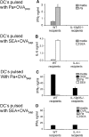
Effect of Th1 and/or Th2 polarization on CD4 help for primary CD8 T-cell responses. Naive WT, IL-12p35-deficient, or IL-4-deficient mice were injected with Ag-pulsed DCs. One week after injection, splenocytes were harvested from individual mice and cultured in medium alone, Pa, SEA, or OVApep. Th1 responses are shown in panel A, and Th2 responses are shown in panel B. Panels C and D include IFN-γ production by CD8 T cells in response to restimulation with OVA peptide. Panels C and D utilize different scales compared to panel A, since the amounts of IFN-γ produced by CD8 T cells are so much greater than those produced by Th1 cells (A). Cytokine levels in 72 h culture supernatants were measured by ELISA. Two experiments were performed with similar results.
FIG. 3.
FIG. 3.
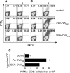
Th1 and Th2 cells provide help for memory CD8 T-cell development. Naive mice were primed with Ag-pulsed DCs. Sixty days later, mice were challenged with rLmOVA or injected with PBS. Six days after rLmOVA challenge, splenocytes were harvested and restimulated with OVApep for 5 h, and the IFN-γ and TNF-α levels were measured by ICS. Representative FACS plots show IFN-γ versus TNF-α (gated on live CD8 T cells) production by unstimulated (−) or OVApep-stimulated (+) splenocytes from unchallenged (A) and rLmOVA-challenged (B) mice. The numbers represent the percentages of CD8 T cells producing cytokines. (C) Bar graph showing mean ± the standard deviation of the numbers of splenic CD8 cells that made IFN-γ in response to in vitro stimulation with OVApep. Cells cultured in the absence of OVApep failed to make IFN-γ. These data were pooled from nine individual experiments.
FIG. 4.
FIG. 4.
Cytotoxicity of CD8 T cells helped by Th1 or Th2 cells. Naive mice were primed with Ag-pulsed DCs and >60 days later challenged with rLmOVA. At 5 days postchallenge, the mice were injected with CFSEhigh-OVApep-pulsed syngeneic target cells and with unpulsed CFSElow syngeneic control cells. One hour later individual spleens were removed, and in vivo cytotoxicity was measured directly by flow cytometry through a comparison of the ratio of CFSEhigh to CFSElow cells in individual animals. (A) Representative plots gated on CFSE-positive cells. The numbers indicate the percent lysis. (B) Mean values (± the standard error of the mean) from three mice per group for the percent lysis of target cells.

References

    1. Barber, D. L., E. J. Wherry, and R. Ahmed. 2003. Cutting edge: rapid in vivo killing by memory CD8 T cells. J. Immunol. 171:27-31. - PubMed
    1. Bennett, S. R., F. R. Carbone, F. Karamalis, R. A. Flavell, J. F. Miller, and W. R. Heath. 1998. Help for cytotoxic-T-cell responses is mediated by CD40 signalling. Nature 393:478-480. - PubMed
    1. Bevan, M. J. 2004. Helping the CD8+ T-cell response. Nat. Rev. Immunol. 4:595-602. - PubMed
    1. Buller, R. M., K. L. Holmes, A. Hugin, T. N. Frederickson, and H. C. Morse III. 1987. Induction of cytotoxic T-cell responses in vivo in the absence of CD4 helper cells. Nature 328:77-79. - PubMed
    1. Bullock, T. N., and H. Yagita. 2005. Induction of CD70 on dendritic cells through CD40 or TLR stimulation contributes to the development of CD8+ T-cell responses in the absence of CD4+ T cells. J. Immunol. 174:710-717. - PubMed

Publication types

LinkOut - more resources