. 2007 Mar 19;204(3):559-70.
doi: 10.1084/jem.20061524. Epub 2007 Feb 26.

Alpha E beta 7 integrin interaction with E-cadherin promotes antitumor CTL activity by triggering lytic granule polarization and exocytosis

Affiliations

Alpha E beta 7 integrin interaction with E-cadherin promotes antitumor CTL activity by triggering lytic granule polarization and exocytosis

Audrey Le Floc'h et al. J Exp Med. .

Abstract

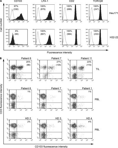
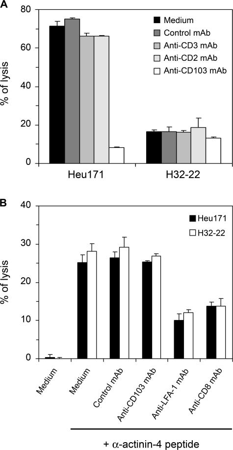
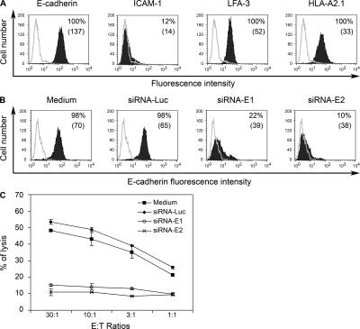
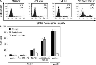
Various T cell adhesion molecules and their cognate receptors on target cells promote T cell receptor (TCR)-mediated cell killing. In this report, we demonstrate that the interaction of epithelial cell marker E-cadherin with integrin alpha(E)(CD103)beta(7), often expressed by tumor-infiltrating lymphocytes (TILs), plays a major role in effective tumor cell lysis. Indeed, we found that although tumor-specific CD103(+) TIL-derived cytotoxic T lymphocyte (CTL) clones are able to kill E-cadherin(+)/intercellular adhesion molecule 1(-) autologous tumor cells, CD103(-) peripheral blood lymphocyte (PBL)-derived counterparts are inefficient. This cell killing is abrogated after treatment of the TIL clones with a blocking anti-CD103 monoclonal antibody or after targeting E-cadherin in the tumor using ribonucleic acid interference. Confocal microscopy analysis also demonstrated that alpha(E)beta(7) is recruited at the immunological synapse and that its interaction with E-cadherin is required for cytolytic granule polarization and subsequent exocytosis. Moreover, we report that the CD103(-) profile, frequently observed in PBL-derived CTL clones and associated with poor cytotoxicity against the cognate tumor, is up-regulated upon TCR engagement and transforming growth factor beta1 treatment, resulting in strong potentiation of antitumor lytic function. Thus, CD8(+)/CD103(+) tumor-reactive T lymphocytes infiltrating epithelial tumors most likely play a major role in antitumor cytotoxic response through alpha(E)beta(7)-E-cadherin interactions.


Figures

Figure 1.
Figure 1.
Surface expression of αEβ7 integrin on tumor-infiltrating and peripheral blood T cells. (A) Expression of αEβ7, LFA-1, CD2, and TCR-α/β on Heu171 and H32-22 T cell clones. Immunofluorescence analysis was performed using anti-CD103, anti-CD11a, anti-CD2, and anti-TCRVβ8 (filled) mAbs or isotypic control (open). (B) Detection of CD103+ T cells in uncultured TILs and PBLs. Two-color flow cytometry analysis was performed using FITC-conjugated anti-CD103 and PE-conjugated anti-CD3 mAbs. Three uncultured NSCLC patient TIL and PBL samples and three healthy donor (HD) PBL samples are shown. Percentages of positive cells are indicated. Numbers in parentheses correspond to mean fluorescence intensities.
Figure 2.
Figure 2.
Role of adhesion/costimulatory molecules in T cell clone–mediated cytotoxicity. (A) Role of CD103 in Heu171 TIL clone–mediated lysis toward autologous IGR-Heu tumor cells. Cytotoxicity was determined by a conventional 4-h 51Cr-release assay at a 20:1 E/T ratio. Experiments were performed either in medium or in the presence of indicated mAb. The Heu171 and H32-22 T cell clones were preincubated for 1 h with saturating concentrations of anti-CD3, anti-CD2, anti-CD103, or a control mAb. (B) Role of LFA-1 in Heu171 and H32-22 clone–mediated lysis against peptide-pulsed autologous EBV-transformed B cell line. Cytotoxicity was determined by a conventional 4-h 51Cr-release assay at a 20:1 E/T ratio. The Heu-EBV B cell line was preincubated with mutated α-actinin-4 peptide. Experiments were performed either in medium or after preincubating effector cells with anti-CD103, anti–LFA-1, or anti-CD8 mAbs.
Figure 3.
Figure 3.
Down-regulation of E-cadherin expression on IGR-Heu results in inhibition of Heu171 cytotoxicity against its specific target. (A) Analysis of surface expression of E-cadherin (CD324), ICAM-1 (CD54), LFA-3 (CD58), and HLA-A2.1 on IGR-Heu tumor cells. Immunofluorescence analysis was performed using anti–E-cadherin, anti-CD54, anti-CD58, anti–HLA-A2.1 (filled) mAbs or an isotypic control (open). Percentages of positive cells are indicated. Numbers in parentheses correspond to mean fluorescence intensities. (B) Analysis of E-cadherin surface expression on IGR-Heu tumor cells electroporated or not with siRNA targeting E-cadherin, siRNA-E1, and siRNA-E2. Luciferase siRNA (siRNA-Luc) was used as a control. (C) Effect of E-cadherin extinction on tumor cell killing by the Heu171 TIL clone. Cytotoxicity was determined by a conventional 4-h 51Cr-release assay at the indicated E/T ratios. IGR-Heu tumor cells electroporated or not with siRNA-E1, siRNA-E2, or siRNA-Luc were used as targets.
Figure 4.
Figure 4.
Role of TGF-β1 in CD103 induction and potentiation of T cell clone–mediated lysis. (A) H32-22 T cells were cultured in medium or in the presence of TGF-β1, coated anti-CD3 mAb, or a combination of both for 96 h. CD103 expression was then assessed by immunofluorescence analysis. Percentages of positive cells are indicated. Numbers in parentheses correspond to mean fluorescence intensities. (B) Role of TGF-β1–induced expression of CD103 in potentiation of H32-22–mediated killing. Cytotoxicity was determined by a conventional 4-h 51Cr-release assay at a 20:1 E/T cell ratio. The H32-22 clone treated or not with TGF-β1 and/or coated anti-CD3 mAb and the Heu171 clone used as a control were preincubated for 1 h with saturating concentrations of anti-CD103 or a control mAb.
Figure 5.
Figure 5.
Engagement of αEβ7 in TIL–tumor cell IS is essential for granule polarization and exocytosis. (A) Recruitment of αEβ7 integrin in the IS formed between the Heu171 TIL clone and the IGR-Heu tumor cell line. Confocal microscopy analysis of CD103 localization (green fluorescence) in the contact area between the Heu171 and the IGR-Heu at the indicated time course. Nuclei were stained with TO-PRO-3 iodide (blue fluorescence). Bars, 5 μm. (B) On the left: TIL and PBL clones form stable conjugates with autologous tumor cells. Conjugates formed between the IGR-Heu and the H32-22 or Heu171 effector cells were analyzed by confocal microscopy after 15 min of co-culture. Granule polarization, as defined by the accumulation of granzyme B in the contact area between effector and tumor cells, was followed up using anti–granzyme B mAb (green fluorescence). On the right: E-cadherin siRNA does not affect conjugate formation between TIL and the cognate target but inhibits granule polarization. IGR-Heu tumor cells were pretreated with siRNA targeting E-cadherin (siRNA-E1) or luciferase (siRNA-Luc). Conjugates formed with the Heu171 TIL were then analyzed. Nuclei were stained with TO-PRO-3 iodide (blue fluorescence). Bars, 5 μm. (C) Percentages of CTLs displaying granzyme B relocalization during conjugate formation between the H32-22 and IGR-Heu or the Heu171 and IGR-Heu pretreated or not with siRNA-E1 or siRNA-Luc. Data shown represent mean ± SD of three independent experiments. Numbers of conjugates analyzed are indicated in parentheses. (D) Efficiency of conjugate formation between IGR-Heu and effector T cells was calculated by determining the E/T ratio × 100 as described in Materials and methods. Data represent the mean ± SD of quadruplicate fields including ∼140 tumor cells each. (E) CD107a induction on Heu171 cells during co-culture with the IGR-Heu pretreated or not with siRNA targeting E-cadherin or luciferase, and on H32-22 stimulated or not with a combination of TGF-β1 and coated anti-CD3 mAb, during co-culture with IGR-Heu tumor cells. Immunofluorescence analysis was performed at the indicated time course. Staining with anti–human CD8 mAb was included to identify T lymphocytes.
Figure 6.
Figure 6.
Both LFA-1 and αEβ7 integrins are engaged in TIL–tumor cell IS. (A) Confocal microscopy analysis of CD103 and LFA-1 localization (green fluorescence) in conjugates formed between the B90 TIL clone and the IGR-B2 autologous tumor cell line after 20 min of co-culture. Nuclei were stained with TO-PRO-3 iodide (blue fluorescence). Bars, 5 μm. (B) Role of LFA-1 and αEβ7 integrins in TIL-mediated lysis of the ICAM-1+/ E-cadherin+ autologous tumor. Cytotoxicity was determined by a conventional 4-h 51Cr-release assay at a 20:1 E/T ratio. The B90 TIL clone was preincubated or not for 1 h with saturating concentrations of anti–LFA-1, anti-CD103, a combination of anti–LFA-1 and anti-CD103, or a control mAb.

References

    1. Bossi, G., C. Trambas, S. Booth, R. Clark, J. Stinchcombe, and G.M. Griffiths. 2002. The secretory synapse: the secrets of a serial killer. Immunol. Rev. 189:152–160. - PubMed
    1. Monks, C.R., B.A. Freiberg, H. Kupfer, N. Sciaky, and A. Kupfer. 1998. Three-dimensional segregation of supramolecular activation clusters in T cells. Nature. 395:82–86. - PubMed
    1. Huppa, J.B., and M.M. Davis. 2003. T-cell-antigen recognition and the immunological synapse. Nat. Rev. Immunol. 3:973–983. - PubMed
    1. Stinchcombe, J.C., G. Bossi, S. Booth, and G.M. Griffiths. 2001. The immunological synapse of CTL contains a secretory domain and membrane bridges. Immunity. 15:751–761. - PubMed
    1. Kuhn, J.R., and M. Poenie. 2002. Dynamic polarization of the microtubule cytoskeleton during CTL-mediated killing. Immunity. 16:111–121. - PubMed

Publication types

MeSH terms