. 2007 Mar 19;204(3):595-603.
doi: 10.1084/jem.20061792. Epub 2007 Feb 26.

Virus-specific CD8+ T cells accumulate near sensory nerve endings in genital skin during subclinical HSV-2 reactivation

Affiliations

Virus-specific CD8+ T cells accumulate near sensory nerve endings in genital skin during subclinical HSV-2 reactivation

Jia Zhu et al. J Exp Med. .

Abstract

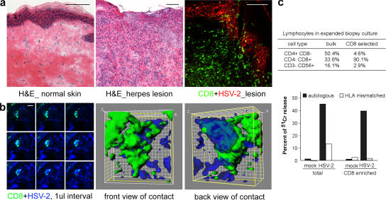
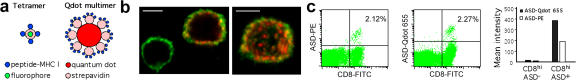
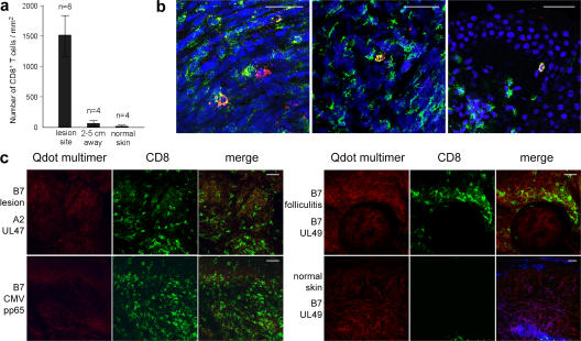
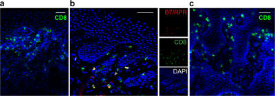
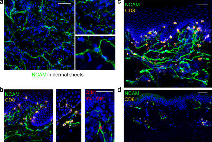
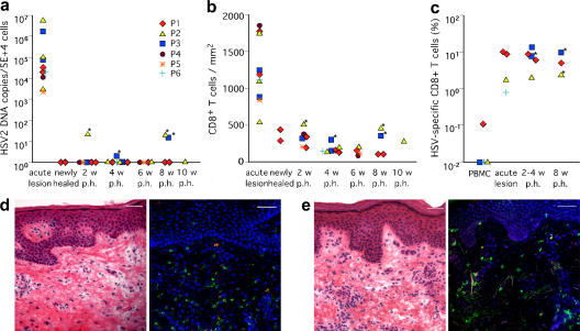
Cytotoxic CD8(+) T cells play a critical role in controlling herpes simplex virus (HSV) infection and reactivation. However, little is known about the spatiotemporal dynamics of CD8(+) T cells during HSV lesion evolution or about their involvement in immune surveillance after lesion resolution. Using quantum dot-conjugated peptide-major histocompatibility complex multimers, we investigated the in vivo localization of HSV-2-specific CD8(+) T cells in sequential biopsies of human genital skin during acute, resolving, and healed stages of HSV-2 reactivation. Our studies revealed that functionally active CD8(+) T cells selectively infiltrated to the site of viral reactivation. After lesion healing in concert with complete reepithelialization and loss of HSV DNA from skin biopsies, HSV-2-specific CD8(+) T cells persisted for more than two months at the dermal-epidermal junction, adjacent to peripheral nerve endings. In two out of the six sequentially studied individuals, HSV-2 DNA reappeared in clinically and histologically normal-appearing skin. Detection of viral DNA was accompanied by increased numbers of both HSV-specific and total CD8(+) T cells in the dermis. These findings indicate that the frequency and clinical course of HSV-2 reactivation in humans is influenced by virus-specific CD8(+) T cells that persist in peripheral mucosa and genital skin after resolution of herpes lesions.


Figures

Figure 1.
Figure 1.
Spatial and functional CD8+ CTL activity during an acute genital herpetic lesion. (a) Hematoxylin and eosin staining of normal arm (left) and lesional skin (middle). Immunofluorescence staining (right) for CD8+ cells (green) and HSV-2–infected cells (red). (b) Confocal image of CD8+ and HSV-2–infected cells in an acute lesion. Montages of a CD8+ cell (green) in proximity to HSV-2–infected cells (blue) were displayed at 1-μm intervals (left). (middle and right) The 0° (front) and 180° (back) views of a three-dimensional rendering of the interactions. (c) HSV-2–specific CD8 cytolytic activity as measured by chromium release in lymphocytes cultured from a lesion biopsy 5 d after onset, using either bulk or CD8-enriched cultured cells as effectors, and either the autologous or HLA-mismatched B lymphocyte blasts uninfected or HSV-2 infected as targets. Effector/target cell ratio = 20:1. Bars: (a) 50 μm; (b) 10 μm.
Figure 2.
Figure 2.
Qdot 655–conjugated peptide–MHC I multimer. (a) Schematic representation of the structure of Qdot 655–conjugated peptide–MHC I multimer complexes as compared with standard tetramers. (b) HSV-specific CD8+ T cells stained with an anti-CD8 antibody (green) and Qdot 655 multimer (red) for TCR recognizing the B*0702-restricted HSV-2 peptide gene UL49 (aa 49–57). A maximum projection displays surface distributions of CD8 and TCR molecules on the double-positive cell (right). Stacks of confocal images were taken with a 100× objective at 0.2-μm intervals. (c) Detection of HSV-2–specific CD8+ T cells by flow cytometry using PE-labeled tetramer and Qdot 655–conjugated multimer. Peptide-stimulated (HSV-2 gene UL46, aa 354–362) PBMC from subject P1 was incubated either with A*0101/ASD-PE (left) or with A*0101/ASD–Qdot 655 (middle). Qdot multimer A*0101/ASD–Qdot 655 shows similar specificity but increased signal intensity as compared with PE-labeled tetramer (right). Bar, 5 μm.
Figure 3.
Figure 3.
HSV-2–specific CD8+ T cell infiltration in recurrent herpes lesion. (a) Quantitation of CD8+ T cell infiltration in acute genital lesion compared with biopsies from adjacent normal genital skin or distal normal forearm skin. At least five fields were counted for each biopsy. (b) Detection of HSV-2–specific CD8+ T cells in the epidermis (left), dermis (middle), and at the dermal–epidermal junction (right) in lesional skin. Biopsies were stained with anti-CD8 antibody (green), a cocktail of Qdot multimers (red), and DAPI (blue). HSV-2–specific CD8+ T cells appear in yellow. (c) Absence of antigen-specific CD8+ T cells in herpes lesions from the same patient using HLA-mismatched (left, top) or antigen-mismatched (left, bottom) Qdot multimers. HSV-2–specific CD8+ T cells were also absent in folliculitis (right, top) or in normal skin (right, bottom), also in the same subject. Bars, 20 μm.
Figure 4.
Figure 4.
HSV-2–specific CD8+ T cells persist at the dermal–epidermal junction after the lesion heals and reepithelializes. (a) Cross-sectional view of a biopsy taken 4 wk after healing. DAPI stain (blue) illustrates normal epidermal layer. CD8+ T cells are found at the junction of the epidermis and dermis, and very few cells are located in the epidermis or in deep dermal areas. (b) Detection of HSV-2–specific CD8+ T cells (yellow) at the dermal–epidermal junction in a biopsy taken 4 wk after healing. Tissue was stained with a cocktail of B*0702 UL49/RPR and UL26/GPH multimers (red) and anti-CD8 antibody (green). HSV-2–specific CD8+ T cells appear in yellow. (c) A horizontal view demonstrates CD8+ T cells in close contact with the basal cells at the basement membrane. The tissue was obtained from a skin biopsy of the original herpetic lesion area 4 wk after its clinical resolution. A similar pattern of CD8+ T cells at the dermal–epidermal junction was seen in posthealing biopsies of all six subjects. Images represented here were from biopsies of subjects P1 (a and c) and P3 (b). Bars, 50 μm.
Figure 5.
Figure 5.
Spatial relationship between peripheral nerve endings and persisting CD8+ T cells in skin after healing. (a) Distribution of superficial nerve networks in dermal sheets. The dermal sheets were stained for NCAM (green) and counterstained with DAPI (blue). A projection of a 70-μm stack is shown from the top edge of the dermis (left). Free endings of nerve fibers (top) and terminal Schwann cells (bottom) are enlarged. (b) Close contact between CD8+ cells and cutaneous nerve endings. Biopsy tissue taken 4 wk after healing was double stained for CD8 (yellow) and NCAM (green). CD8+ cells are in close contact with the basal keratinocytes adjacent to free nerve endings at the basement membrane (left) and with terminal Schwann cells in the upper dermis (middle). A snapshot of a three-dimensional reconstruction (20-μm stack) displays spatial closeness between terminal nerve endings (green) and the cell with HSV-2 specificity (red; right). The tissue was from a biopsy taken 4 wk after healing and was stained with the A*0101/ASD and B*0702/RPR multimers and anti-NCAM antibody. (c) Similar findings are present in biopsy tissue 8 wk after healing. The snapshot shows a reconstructed three-dimensional distribution of CD8+ cells and cutaneous nerve endings. The image represents 25 μm in z. (d) Biopsy of normal skin of the upper arm was double stained for CD8 (yellow) and NCAM (green). Images represented were from biopsies of subjects P1 (b–d), P2 (b), and P6 (a). Bars, 50 μm.
Figure 6.
Figure 6.
Subclinical reactivation of HSV-2 in genital skin is associated with increased CD8+ T cell infiltration. (a–c) Quantitation of the HSV-2 genomic DNA (a), total CD8+ T cells (b), and HSV-2–specific CD8+ T cells (c) detected in skin biopsies. Each symbol represents one subject. HSV-2 DNA copies were quantitated by Taqman real-time PCR (see Materials and methods). Biopsies with reappearance of HSV-2 DNA after healing are marked with a star. (d and e) Hematoxylin and eosin and in situ staining of skin biopsies from subjects P2 (d) and P3 (e), in whom HSV-2 DNA was detected at 8 wk after healing. The epithelium is intact in both biopsies, and moderate lymphocyte infiltration is seen at the dermal–epidermal junction and in the dermis (left). In situ staining using a cocktail of A*0201 (d, right) and B*0702/RPR (e, right) Qdot multimer reveals that HSV-2–specific CD8+ T cells are among the infiltrating CD8+ cells in the dermis. Bars, 50 μm.

References

    1. Posavad, C.M., D.M. Koelle, M.F. Shaughnessy, and L. Corey. 1997. Severe genital herpes infections in HIV-infected individuals with impaired HSV-specific CD8+ cytotoxic T lymphocyte responses. Proc. Natl. Acad. Sci. USA. 94:10289–10294. - PMC - PubMed
    1. Koelle, D.M., and L. Corey. 2003. Recent progress in herpes simplex virus immunobiology and vaccine research. Clin. Microbiol. Rev. 16:96–113. - PMC - PubMed
    1. Augenbraun, M., J. Feldman, K. Chirgwin, J. Zenilman, L. Clarke, J. DeHovitz, S. Landesman, and H. Minkoff. 1995. Increased genital shedding of herpes simplex virus type 2 in HIV-seropositive women. Ann. Intern. Med. 123:845–847. - PubMed
    1. Khanna, K.M., R.H. Bonneau, P.R. Kinchington, and R.L. Hendricks. 2003. Herpes simplex virus-specific memory CD8(+) T cells are selectively activated and retained in latently infected sensory ganglia. Immunity. 18:593–603. - PMC - PubMed
    1. Liu, T., K.M. Khanna, X. Chen, D.J. Fink, and R.L. Hendricks. 2000. CD8+ T cells can block herpes simplex virus type 1 (HSV-1) reactivation from latency in sensory neurons. J. Exp. Med. 191:1459–1466. - PMC - PubMed

Publication types

MeSH terms