. 2007 Feb 26;176(5):681-94.
doi: 10.1083/jcb.200605038.

PPARgamma1 attenuates cytosol to membrane translocation of PKCalpha to desensitize monocytes/macrophages

Affiliations

PPARgamma1 attenuates cytosol to membrane translocation of PKCalpha to desensitize monocytes/macrophages

Andreas von Knethen et al. J Cell Biol. .

Abstract

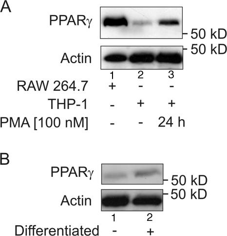
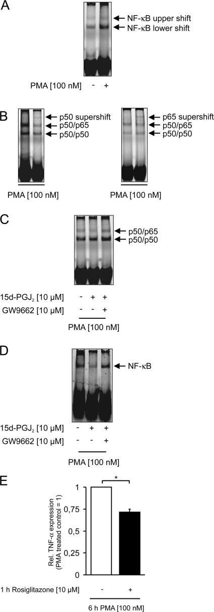
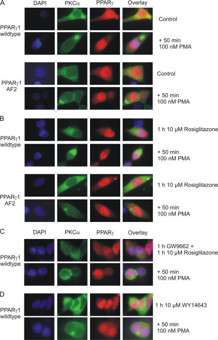
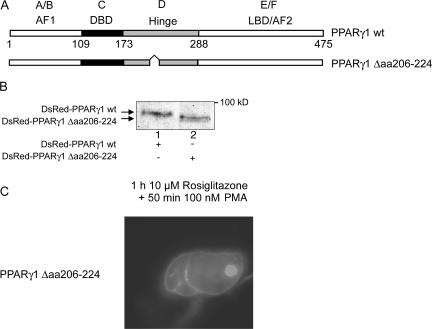
Recently, we provided evidence that PKCalpha depletion in monocytes/macrophages contributes to cellular desensitization during sepsis. We demonstrate that peroxisome proliferator-activated receptor gamma (PPARgamma) agonists dose dependently block PKCalpha depletion in response to the diacylglycerol homologue PMA in RAW 264.7 and human monocyte-derived macrophages. In these cells, we observed PPARgamma-dependent inhibition of nuclear factor-kappaB (NF-kappaB) activation and TNF-alpha expression in response to PMA. Elucidating the underlying mechanism, we found PPARgamma1 expression not only in the nucleus but also in the cytoplasm. Activation of PPARgamma1 wild type, but not an agonist-binding mutant of PPARgamma1, attenuated PMA-mediated PKCalpha cytosol to membrane translocation. Coimmunoprecipitation assays pointed to a protein-protein interaction of PKCalpha and PPARgamma1, which was further substantiated using a mammalian two-hybrid system. Applying PPARgamma1 mutation and deletion constructs, we identified the hinge helix 1 domain of PPARgamma1 that is responsible for PKCalpha binding. Therefore, we conclude that PPARgamma1-dependent inhibition of PKCalpha translocation implies a new model of macrophage desensitization.


Figures

Figure 1.
Figure 1.
PPARγ expression in monocytes/macrophages. (A) PPARγ expression was determined in lysates of RAW 264.7 macrophages and in control versus differentiated THP-1 cells. For differentiation, cells were treated for 24 h with 100 nM PMA. Western blot was performed as described in Materials and methods. (B) PPARγ expression was analyzed by Western analysis in primary human monocytes and macrophages, differentiated for 7 d with medium containing serum of AB-positive donors. Experiments were performed at least three times, and representative data are shown.
Figure 2.
Figure 2.
PPARγ agonist prestimulation inhibits PKCα depletion. RAW 264.7 macrophages (A) and PPARγ1 AF2–overexpressing RAW 264.7 cells (B) were prestimulated for 1 h with ciglitazone (1 or 10 μM), rosiglitazone (1 or 10 μM), or remained as controls, followed by the addition of 100 nM PMA for 1 h. (C) RAW 264.7 macrophages were stimulated for 1 h with 50 μg/ml CHX. Thereafter, 10 μM of rosiglitazone or ciglitazone were added for 1 h, followed by 100 nM PMA stimulation for 1 h. (D) Primary monocyte–derived macrophages were prestimulated for 1 h with 10 μM ciglitazone, 10 μM rosiglitazone, or remained as controls. Afterward, 100 nM PMA was added for 1 h. For all experiments, cells were harvested and lysed, and Western blot was performed as described in the Materials and methods. Experiments were performed at least three times, and representative data are shown.
Figure 3.
Figure 3.
PPARγ1-dependent attenuation of PKCα translocation provokes a reduction of the proinflammatory response in macrophages. (A) Activation of NF-κB in RAW 264.7 macrophages in response to PMA. RAW 264.7 macrophages were stimulated for 3 h with 100 nM PMA or left untreated as control. Afterward, cells were harvested, nuclear extracts were isolated, and NF-κB EMSA was performed as described in the Materials and methods. (B) Supershift analysis of the active NF-κB complex was described in the Materials and methods. Macrophages were stimulated with 100 nM PMA for 3 h. For supershift analysis, a p50 antibody (left, second lane) or a p65 antibody (right, second lane) was included. NF-κB activation without antibody addition (left and right, first lane) is shown. (C) 15d-PGJ2 inhibited PMA-mediated NF-κB activation. RAW 264.7 cells were pretreated with 10 μM of the endogenous PPARγ ligand 15-dPGJ2 for 1 h, followed by the addition of 100 nM PMA for 3 h (middle lane). To ensure a PPARγ-dependent effect, one sample was prestimulated before 15d-PGJ2 addition with 10 μM of the PPARγ antagonist GW9662 for 1 h (right lane). One sample remained PMA treated only as a control (left lane). Cells were harvested, nuclear protein extracts were isolated, and NF-κB EMSA was performed as described in Materials and methods. (D) PMA-mediated NF-κB activation is inhibited in human primary macrophages in response to 15d-PGJ2. Primary monocyte–derived macrophages were treated as described in C. Cells were harvested and processed, and NF-κB EMSA was performed as described in Materials and methods. (E) Inhibition of PMA-mediated PKCα activation by PPARγ reduced proinflammatory TNF-α expression. RAW 264.7 cells were treated for 1 h with 10 μM rosiglitazone or remained as controls. Afterward, cells were incubated with 100 nM PMA for 6 h, and TNF-α expression in the cell supernatant was analyzed using the CBA system. All experiments were performed at least three times. Data are the means ± the SD of the individual experiments (*, P < 0.05) or representative of three similar experiments.
Figure 4.
Figure 4.
PKCα and PPARγ localization in RAW 264.7 macrophages. To follow PKCα and PPARγ localization in RAW 264.7 macrophages, cells were seeded on slides and treated (B) for 50 min with 100 nM PMA, (C) preincubated with 10 μM rosiglitazone for 1 h followed by 100 nM PMA addition for 50 min, or (D) pretreated with 10 μM GW9662 before cells were stimulated as described in C. Afterward, cells were fixed and stained for PKCα and PPARγ as described in the Materials and methods. Cell nuclei were counterstained with DAPI. DAPI staining is shown in the first panel, PKCα staining in the second, PPARγ staining in the third, and an overlay to estimate cytosolic and nuclear region is provided in the fourth panel. All experiments were performed three times, and representative data are shown.
Figure 5.
Figure 5.
PPARγ1 inhibits PKCα translocation. HEK293 cells were cotransfected with DsRed-PPARγ1 wild type/PKCα-EGFP (A and B [top two rows] and C and D) or DsRed-PPARγ1 AF2/PKCα-EGFP (A and B, bottom two rows). To follow PKCα-EGFP translocation, 100 nM PMA was added to control cells (A, second and fourth row) or cells pretreated for 1 h with 10 μM rosiglitazone (B, second and fourth row). To verify the role of PPARγ on PKCα-EGFP translocation in DsRed-PPARγ1 wild type/PKCα-EGFP, cotransfected HEK293 cells were treated for 1 h with 10 μM of the PPARγ antagonist GW9662 before stimulation for 1 h with 10 μM rosiglitazone (C, top row) followed by 50 min of 100 nM PMA addition (C, bottom row) or preincubated for 1 h with 10 μM of the PPARα agonist WY14643 (D, top row) before activation with 100 nM PMA for 50 min (D, bottom row). Cell nuclei were counterstained with DAPI. DAPI staining is shown in the first panel, PKCα-EGFP in the second, DsRed-PPARγ in the third, and an overlay to estimate cytosolic and nuclear region is provided in the fourth panel. All experiments were performed three times, and representative data are shown.
Figure 6.
Figure 6.
PPARγ1 directly interacts with PKCα. (A) THP-1 cells were differentiated for 24 h with 50 nM PMA. To allow new synthesis of PKCα, which is depleted in response to the differentation regime, cells were further cultured for 48 h in normal medium. Afterward, cells were treated for 1 h with 10 μM rosiglitazone or remained as controls. Cells were harvested and lysed, and PKCα was immunoprecipitated as described in Materials and methods. Eluates and flowthroughs were separated by Western blotting and stained for PPARγ and PKCα as indicated. (B) COS-7 cells were transiently cotransfected with PPARγ1 wild type/PKCα-EGFP or PPARγ1 AF2/PKCα-EGFP. 24 h later, cells were treated for 1 h with 10 μM rosiglitazone or remained as controls. Cells were harvested and lysed, and PKCα-EGFP was immunoprecipitated as described in Materials and methods. Input controls, eluates, and flowthroughs were separated by Western blotting and stained for PPARγ and PKCα as indicated. All experiments were performed at least three times, and representative data are shown.
Figure 7.
Figure 7.
PPARγ directly binds to PKCα. COS-7 cells were transiently transfected with a combination of a target (PPARγ1), a bait (PKCα), and a reporter construct, as described in Materials and methods. Afterward, cells were treated with 10 μM ciglitazone, 10 μM rosiglitazone, 10 μM WY14643, or remained as controls. 6 h later, cells were harvested and lysed for a reporter analysis as described in Materials and methods. Experiments were performed at least three times in duplicate. *, P < 0.05. Data are the means ± the SD.
Figure 8.
Figure 8.
Helix 4 of the LBD/AF2 domain does not mediate PPARγ binding to PKCα. (A) Scheme of the PPARγ1 constructs. (B and C) HEK293 cells were cotransfected with DsRed-PPARγ1 wild type/PKCα-EGFP (B, first panel), DsRed-PPARγ1 L309A/PKCα-EGFP (B, second panel), DsRed-PPARγ1 N310A/PKCα-EGFP (B, third panel), DsRed-PPARγ1 G312A/PKCα- EGFP (B, fourth panel), PPARγ1 V313A/PKCα-EGFP (C, first panel), PPARγ1 L316A/PKCα-EGFP (C, second panel), PPARγ1 K317A/PKCα-EGFP (C, third panel) or DsRed-PPARγ1 Δaa309-319/PKCα-EGFP (C, fourth panel). To follow PKCα-EGFP localization, 24 h after transfection, cells were treated for 50 min with 100 nM PMA (second row), for 1 h with 10 μM rosiglitazone (third row), pretreated for 1 h with 10 μM rosiglitazone followed by the addition of 100 nM PMA for 50 min (fourth row) or remained as controls (first row). Experiments were performed three times, and representative data are shown.
Figure 9.
Figure 9.
Hinge domain mediates PPARγ1 binding to PKCα. (A) Scheme of the PPARγ1 constructs. (B) HEK293 cells were transiently transfected with one of the PPARγ1 constructs, as indicated. 24 h after transfection cells were lysed. Western blotting was performed, and blots were stained for DsRed. All experiments were performed at least three times, and representative data are shown. (C) HEK293 cells were transfected with PKCα-EGFP only (first panel) or cotransfected with DsRed-PPARγ1 wild type/PKCα-EGFP (second panel), DsRed-PPARγ1 Δ32-198/PKCα-EGFP (third panel), DsRed-PPARγ1 Δ32-250/PKCα-EGFP (fourth panel), or DsRed-PPARγ1 Δ51-406/PKCα-EGFP (fifth panel). To follow PKCα-EGFP localization, 24 h after transfection, cells were treated for 50 min with 100 nM PMA (second row), treated for 1 h with 10 μM rosiglitazone (third row), pretreated for 1 h with 10 μM rosiglitazone followed by the addition of 100 nM PMA for 50 min (fourth row), or remained controls (first row). Experiments were performed three times, and representative data are shown.
Figure 10.
Figure 10.
Hinge helix 1 mediates PPARγ1 binding to PKCα. (A) Scheme of the PPARγ1 construct. (B) HEK293 cells were transiently transfected with the DsRed-PPARγ1 wild type as control or the DsRed-PPARγ1 Δaa206-224 construct as indicated. 24 h after transfection, cells were lysed. Western blotting was performed, and blots were stained for DsRed. (C) HEK293 cells were cotransfected with DsRed-PPARγ1 Δaa206-224/PKCα-EGFP. 24 h after transfection, cells were treated for 1 h with 10 μM rosiglitazone. To follow PKCα-EGFP translocation, 100 nM PMA was added to cells, and localization of PKCα-EGFP was examined 50 min thereafter. Experiments were performed three times and representative data are shown.

Similar articles

Cited by

References

    1. Abdelrahman, M., A. Sivarajah, and C. Thiemermann. 2005. Beneficial effects of PPAR-gamma ligands in ischemia-reperfusion injury, inflammation and shock. Cardiovasc. Res. 65:772–781. - PubMed
    1. Abella, A., P. Dubus, M. Malumbres, S.G. Rane, H. Kiyokawa, A. Sicard, F. Vignon, D. Langin, M. Barbacid, and L. Fajas. 2005. Cdk4 promotes adipogenesis through PPARgamma activation. Cell Metab. 2:239–249. - PubMed
    1. Adida, A., and F. Spener. 2006. Adipocyte-type fatty acid-binding protein as inter-compartmental shuttle for peroxisome proliferator activated receptor gamma agonists in cultured cell. Biochim. Biophys. Acta. 1761:172–181. - PubMed
    1. Akaike, M., W. Che, N.L. Marmarosh, S. Ohta, M. Osawa, B. Ding, B.C. Berk, C. Yan, and J. Abe. 2004. The hinge-helix 1 region of peroxisome proliferator-activated receptor gamma1 (PPARgamma1) mediates interaction with extracellular signal-regulated kinase 5 and PPARgamma1 transcriptional activation: involvement in flow-induced PPARgamma activation in endothelial cells. Mol. Cell. Biol. 24:8691–8704. - PMC - PubMed
    1. Akiyama, T.E., C.T. Baumann, S. Sakai, G.L. Hager, and F.J. Gonzalez. 2002. Selective intranuclear redistribution of PPAR isoforms by RXR alpha. Mol. Endocrinol. 16:707–721. - PubMed

Publication types

MeSH terms