. 2007 Jan;9(1):23-35.
doi: 10.1593/neo.06658.

SPARC inhibits LPA-mediated mesothelial-ovarian cancer cell crosstalk

Affiliations

SPARC inhibits LPA-mediated mesothelial-ovarian cancer cell crosstalk

Neveen A Said et al. Neoplasia. 2007 Jan.

Abstract

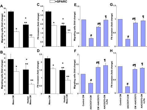
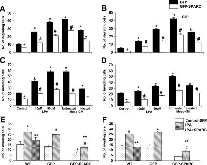
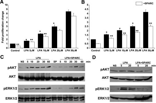
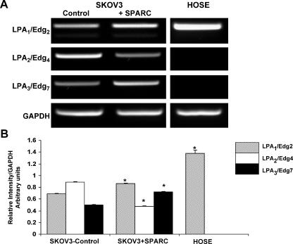
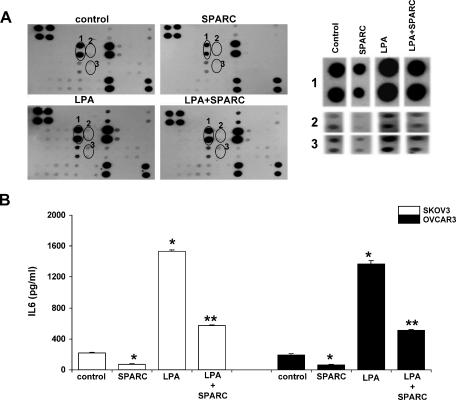
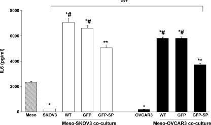
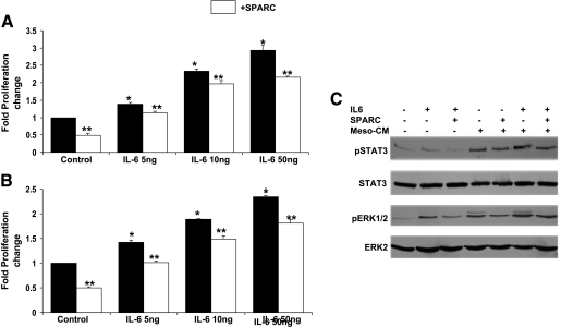
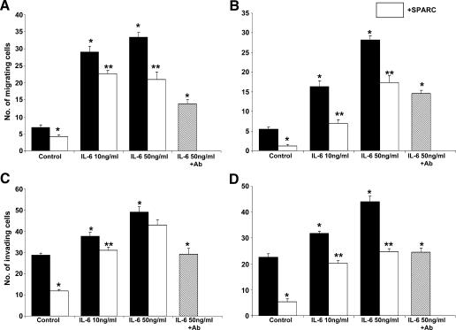
The interplay between peritoneal mesothelial cells and ovarian cancer cells is critical for the initiation and peritoneal dissemination of, and ascites formation in, ovarian cancer. The production of lysophosphatidic acid (LPA) by both peritoneal mesothelial cells and ovarian cancer cells has been shown to promote metastatic phenotype in ovarian cancer. Herein, we report that exogenous addition or ectopic overexpression of the matricellular protein SPARC (secreted protein acidic and rich in cysteine) significantly attenuated LPA-induced proliferation, chemotaxis, and invasion in both highly metastatic SKOV3 and less metastatic OVCAR3 ovarian cancer cell lines. SPARC appears to modulate these functions, at least in part, through the regulation of LPA receptor levels and the attenuation of extracellular signal-regulated kinase (ERK) 1/2 and protein kinase B/AKT signaling. Moreover, our results show that SPARC not only significantly inhibited both basal and LPA-induced interleukin (IL) 6 production in both cell lines but also attenuated IL-6-induced mitogenic, chemotactic, and proinvasive effects, in part, through significant suppression of ERK1/2 and, to a lesser extent, of signal transducers and activators of transcription 3 signaling pathways. Our results strongly suggest that SPARC exerts a dual inhibitory effect on LPA-induced mesothelial-ovarian cancer cell crosstalk through the regulation of both LPA-induced IL-6 production and function. Taken together, our findings underscore the use of SPARC as a potential therapeutic candidate in peritoneal ovarian carcinomatosis.


Figures

Figure 1
Figure 1
The conditioned medium from Meso 301 induces ovarian cancer cell motility through LPA. Subconfluent Meso 301 cells were allowed to condition in SFM for 24 hours, and the collected conditioned medium (heated or not) was used as a chemoattractant (600 µl in the bottom chambers of transwell inserts) for SKOV3 (A) and OVCAR3 (B) (1 x 10⁵ cells/100 µl of SFM-0.4% BSA in the upper chamber) in the presence or in the absence of SPARC (10 µg/ml). Results are expressed as the mean ± SEM of the fold change in cell migration induced by a complete medium in the bottom chamber as controls. FN invasion by SKOV3 (C) and OVCAR3 (D) cells was tested in response to the conditioned medium of Meso 301 (heated or not) added with the cells to the upper chamber of transwell inserts, in the presence or in the absence of SPARC (10 µg/ml). LPA production by Meso 301 was inhibited by PLA2 inhibitor (AACOCF3). The conditioned medium of Meso 301 serum-starved for 24 hours was collected in the absence (control conditioned medium) or in the presence of AACOCF3 (25 µM) added at serum starvation (AACOCF3-CM) or after the conditioned medium had been generated (conditioned medium + AACOCF3). All collected conditioned media were heat-inactivated and added to the bottom chamber of transwell inserts, and the chemotactic activity of SKOV3 (E) and OVCAR3 (F) was tested in the aforementioned conditioned medium and after replenishment with LPA (50 µM; AACOCF3-CM + LPA). Results are expressed as the mean ± SEM of the fold change of the chemotactic activity of heated conditioned medium under experimental conditions, compared to the conditioned medium (control conditioned medium) of Meso 301 heated for 24 hours. The effect of the above-mentioned conditioned medium on FN invasion by SKOV3 (G) and OVCAR3 (H) was tested when they were added with the cells to the top chambers of the inserts, whereas the bottom chambers contained a complete growth medium. Results are expressed as the mean ± SEM of the fold change of the invasive activity of SKOV3 and OVCAR3 induced by different experimental conditions, relative to that induced by the conditioned medium (control conditioned medium) of Meso 301 heated for 24 hours. Data summarized are representative of an experiment performed in triplicate and repeated thrice with similar results. *P < .05, compared to unheated Meso-CM. **P < .05, compared to heated Meso-CM. #P < .05, compared to heated Meso-CM. P < .05, compared to AACOCF3-CM.
Figure 2
Figure 2
SPARC antagonizes LPA-induced ovarian cancer cell chemotaxis and invasion. LPA (10–50 µM in SFM, added to the bottom chamber of transwell inserts) stimulated the migration of SKOV3 (A) and OVCAR3 (C) cells in a concentration-dependent manner. SPARC (10 µg/ml) added with SKOV3 and OVCAR3 cells to the top chamber of the inserts inhibited LPA-induced migration. Results are expressed as the mean ± SEM of the fold change in migrated cells under experimental conditions, compared to controls attracted by a complete growth medium placed in the bottom chamber (assigned a value of 1). LPA (10–50 µM in SFM) added to SKOV3 (B) and OVCAR3 (D) cells in the top chamber stimulated the invasion of FN-coated inserts and the migration of both cell lines toward a complete growth medium in the bottom chamber. The effect of LPA on FN invasion was significantly inhibited by SPARC. Results are expressed as the mean ± SEM of the fold change in invading cells under experimental conditions, compared to control cells exposed to 0.4% BSA in SFM (assigned a value of 1). Represented are the results of an experiment performed in triplicate and repeated thrice with similar results. *P < .05, compared to unstimulated control. **P < .05, compared to control or LPA stimulation.
Figure 3
Figure 3
Restoration of SPARC expression in ovarian cancer cells antagonizes LPA-induced and Meso-CM-induced chemotaxis and invasion. SKOV3 and OVCAR3 cells were transduced (> 90% transduction efficiency) with GFP or GFP-SPARC adenoviruses and allowed to recover for 24 hours in a complete growth medium. LPA (10–50 µM)-induced and Meso-CM-induced chemotaxis of SKOV3 (A) and OVCAR3 (B) were tested as described in the legend to Figure 1. *P < .05, compared to unstimulated control cells attracted by a complete growth medium in the bottom chamber. #P < .05, compared to matched LPA or Meso-CM stimulation. LPA-induced and Meso-CM-induced FN invasions by SKOV3 (C) and OVCAR3 (D) were also tested as described earlier. *P < .05, compared to unstimulated control cells in SFM containing 0.04% BSA added to the top chamber of the transwell inserts. #P < .05, compared to matched LPA or Meso-CM stimulations. Results are expressed as the mean ± SEM of a representative of three independent experiments. In a two-cell coculture model described in the Materials and Methods section, LPA (50 µM)-stimulated invasion of Meso 301 monolayers by untransduced (WT) SKOV3 (E) and OVCAR3 (F) cells was significantly inhibited in the presence of exogenous SPARC (10 µg/ml), as well as by SKOV3 and OVCAR3 cells transduced with an adenovirus overexpressing SPARC. *P < .05, compared to unstimulated WT or GFP-transduced (GFP) cells. **P < .05, compared to LPA-stimulated WT or GFP-transduced cells. #P < .05, compared to unstimulated GFP-SPARC-transduced (GFP-SPARC) cells. Results are expressed as the mean ± SEM of a representative of three independent experiments performed in quadruplicate.
Figure 4
Figure 4
Effect of SPARC on LPA-induced proliferation and survival signaling in ovarian cancer cells. Cell proliferation of SKOV3 (A) and OVCAR3 (B) cells in response to indicated concentrations of LPA, in the presence (open bars) and in the absence (closed bars) of SPARC, was assessed by measuring the released formazan at A590. Results are expressed as the mean ± SEM of the fold increase in proliferation relative to unstimulated control cells (assigned a value of 1). *P < .05, LPA-stimulated cells compared to control cells and between different concentrations of LPA. **P < .05, SPARC-treated versus matched unstimulated control or LPA-stimulated cells. Represented are the results of one experiment performed in quadruplicate that was representative of two independent experiments. SKOV3 (C) and OVCAR3 (D) cells starved overnight were pretreated with 20 µg/ml SPARC in SFM for 2 hours, followed by stimulation with 50 µM LPA for indicated time points. Western blot analysis of phosphorylated and total ERK1/2 and AKT was performed as described in the Materials and Methods section. Blots represent the results of three independent experiments. NS = not stimulated; SP = SPARC-treated.
Figure 5
Figure 5
Effect of SPARC on LPA receptor expression in SKOV3 cells. SKOV3 cells serum-starved overnight were treated with PBS (control) or SPARC (10 µg/ml) for 6 hours. The expression of LPA receptors Edg2/LPA1 and Edg4/LPA2 and Edg7/LPA3 was determined by semiquantitative RT-PCR. PCR products of LPA receptors and GAPDH were run on 2% agarose gels (A). Expression levels were normalized to that of the housekeeping gene GAPDH and were represented as a bar graph (B). HOSE cells serum-starved overnight were used as controls for LPA receptor expression. Results shown are from one experiment that was representative of three independent experiments. *P < .05, compared to corresponding control SKOV3 cells.
Figure 6
Figure 6
SPARC inhibits basal and LPA-induced IL-6 secretions from ovarian cancer cells. A human inflammation cytokine protein array was used to detect differences in the protein levels of inflammation-related factors secreted into the conditioned medium of serum-starved SKOV3 cells pretreated with PBS (control) or SPARC (10 µg/ml) for 2 hours and stimulated with LPA (50 µM) for 24 hours (A; left panel). A higher magnification of duplicate spots from the cytokine array depicting protein levels of selected inflammation-related factors that were significantly upregulated by LPA and attenuated by SPARC (A; right panel). (1) IL-6. (2) IL-6sR. (3) MCP-1. Quantification of IL-6 secretion by ELISA in conditioned media of SKOV3 (B; left panel) and OVCAR3 cells (B; right panel) in the presence or in the absence of LPA and SPARC. *P < .05, compared to unstimulated control cells. **P < .01, compared to LPA-stimulated cells. Results shown are from one experiment that was representative of three independent experiments.
Figure 7
Figure 7
Effect of ovarian cancer-mesothelial cell coculture on IL-6 production. SKOV3 and OVCAR3 cells (WT) were transduced with GFP or GFP-SPARC in a complete growth medium and allowed to recover for 24 hours. After trypsinization, 1 x 10⁶ cells in SFM were added to confluent monolayers of the Meso 301 cell line in 60-mm plates for an additional 24 hours. Conditioned media were collected, and IL-6 levels were determined by ELISA. *P < .01, between either SKOV3 or OVCAR3 and Meso 301. *#P < .01, between cocultured Meso and untransduced (WT) or GFP-transduced SKOV3 or OVCAR3, compared to either cell line alone. **P < .05, compared to WT or GFP-transduced cells. ***P < .05, between SKOV3 and OVCAR3 under all experimental conditions. Results shown are from one experiment that was representative of three independent experiments.
Figure 8
Figure 8
Effect of SPARC on IL-6-induced proliferation and survival signaling pathways in ovarian cancer cells. Proliferation of SKOV3 (A) and OVCAR3 (B) cells in response to indicated concentrations of IL-6, in the presence of PBS (Control) or SPARC (20 µg/ml), was determined by MTS assay, as described previously. Results are expressed as the fold change of the proliferation of IL-6-stimulated SKOV3 and OVCAR3 cells relative to the proliferation of PBS-stimulated (control) cells (assigned a value of 1). *P < .05, from controls. **P < .05, from matched control or LPA-stimulated cells. SKOV3 cells serum-starved overnight were pretreated with PBS (control) or SPARC (20 µg/ml) for 2 hours, followed by stimulation with IL-6 (50 ng/ml), heat-inactivated Meso-CM, or heat-inactivated Meso-CM plus IL-6 (50 ng/ml) for 5 minutes (C). The expression levels of phosphorylated and total ERK and STAT3 were determined by Western blot analysis, as described previously. Results shown are from one experiment that was representative of three independent experiments.
Figure 9
Figure 9
SPARC antagonizes IL-6-induced chemotaxis and invasion of ovarian cancer cells. The chemotactic activity of SKOV3 (A) and OVCAR3 (B) cells toward IL-6 was tested as described previously. The indicated concentrations of IL-6 were used to attract ovarian cancer cells either alone or after pretreatment of cells with SPARC (10 µg/ml) for 2 hours. As control, the IL-6-neutralizing antibody (50 µg/ml) was mixed with SFM containing IL-6 (50 ng/ml) for 30 min before use in chemotaxis assay. *P < .05, from PBS-treated (control) cells. **P < .05, from matched control or IL-6-stimulated cells. FN invasion by SKOV3 (C) and OVCAR3 (D) cells was studied under the same conditions as described for the chemotaxis assay, with the exception that IL-6 was added to the cells in the upper chamber of the transwell inserts and cells were allowed to migrate toward a complete growth medium. Pretreatment of either SKOV3 or OVCAR3 cells with the IL-6-neutralizing antibody (50 µg/ml) for 30 minutes was used as control. *P < .05, from control PBS-treated cells. **P < .05, from matched control or IL-6-stimulated cells. Results are expressed as the mean ± SEM of the number of cells per field that migrated to and/or invaded the lower surface of the inserts. Experiments were performed in triplicate per experimental condition and were repeated thrice with similar results.

Similar articles

Cited by

References

    1. Hsia D, Lim S, Bernard-Trifilo J, Mitra S, Tanaka S, den Hertog J, Streblow D, Ilic D, Ginsberg M, Schlaepfer D. Integrin alpha4beta1 promotes focal adhesion kinase-independent cell motility via alpha4 cytoplasmic domain-specific activation of c-Src. Mol Cell Biol. 2005;25:9700–9712. - PMC - PubMed
    1. Schwartz B, Hong G, Morrison B, Wu W, Baudhuin L, Xiao Y, Mok S, Xu Y. Lysophospholipids increase interleukin-8 expression in ovarian cancer cells. Gynecol Oncol. 2001;81:291–300. - PubMed
    1. Guo R, Kasbohm E, Arora P, Sample C, Baban B, Sud N, Sivashanmugam P, Moniri N, Daaka Y. Expression and function of lysophosphatidic acid LPA1 receptor in prostate cancer cells. Endocrinology. 2006;147:4883–4892. - PubMed
    1. Bian D, Su S, Mahanivong C, Cheng R, Han Q, Pan Z, Sun Pand Huang S. Lysophosphatidic acid stimulates ovarian cancer cell migration via a Ras-MEK kinase 1 pathway. Cancer Res. 2004;64:4209–4217. - PubMed
    1. Li H, Ye X, Mahanivong C, Bian D, Chun J, Huang S. Signaling mechanisms responsible for lysophosphatidic acid-induced urokinase plasminogen activator expression in ovarian cancer cells. J Biol Chem. 2005;280:10564–10571. - PubMed

Publication types

MeSH terms