

Imaging techniques for small animal imaging models of pulmonary disease: micro-CT
- PMID: 17325973
- PMCID: PMC2094132
- DOI: 10.1080/01926230601184262
Imaging techniques for small animal imaging models of pulmonary disease: micro-CT
Abstract
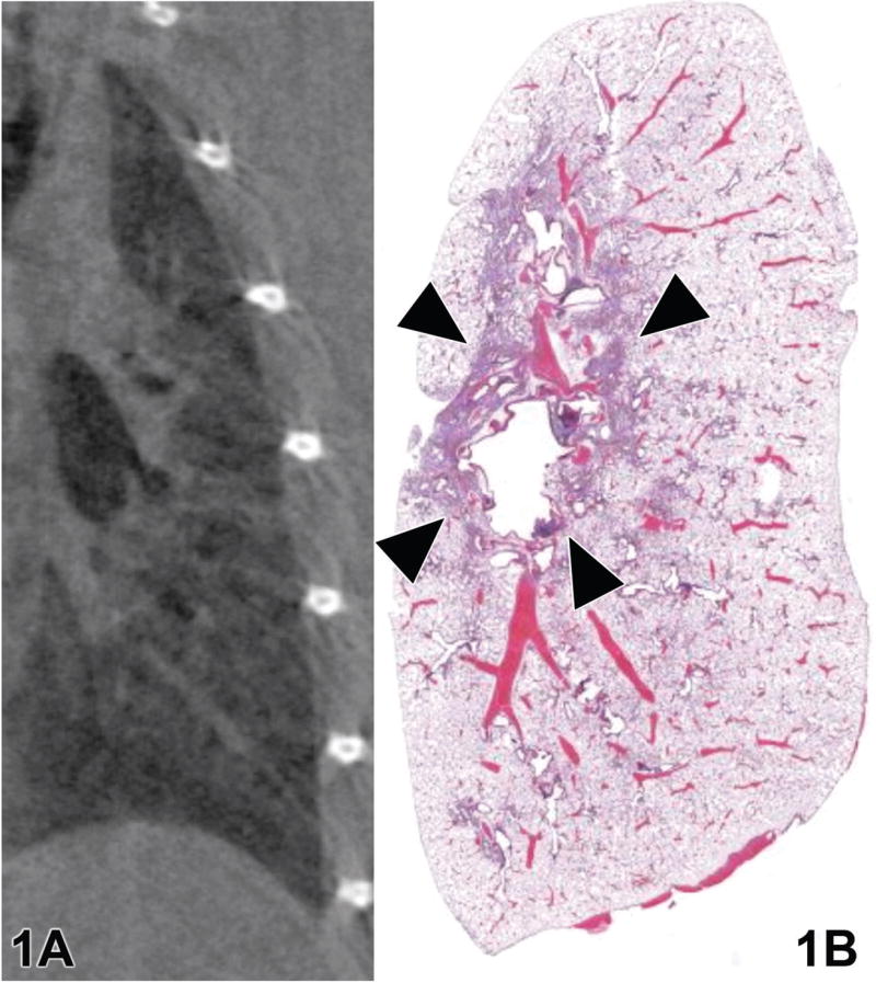
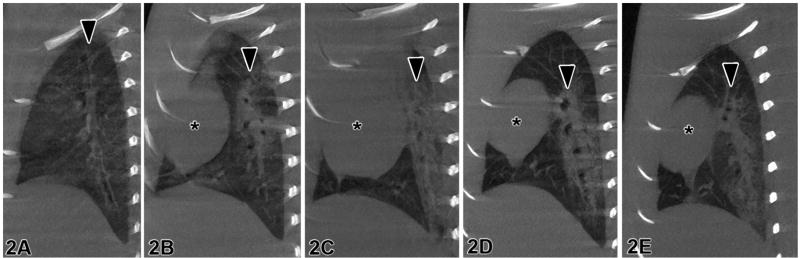
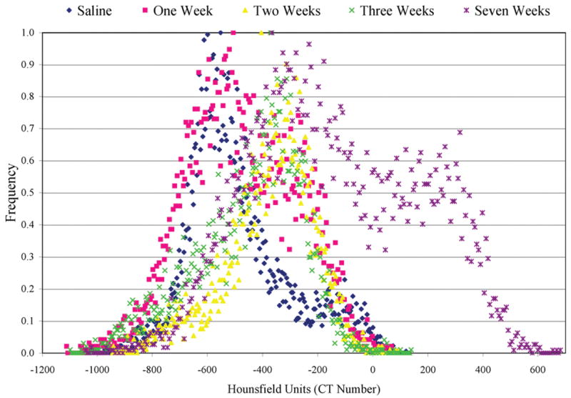
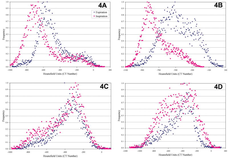
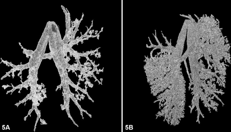
Microcomputed tomography (micro-CT) is ideal for quantifying pulmonary disease because of the inherent contrast between tissue and air that exists in the lungs. Both in vivo and in vitro studies can be performed using micro-CT. Live animal studies show function, while fixed specimen studies show structure. Through the use of image processing techniques, both acute and chronic lung diseases can be quantified. The information provided by micro-CT is complementary to histological evaluation, since CT is nondestructive. This paper discusses two examples, in vivo and in vitro, of how micro-CT can be used to assess pulmonary diseases in small animal models. With the use of micro-CT, we were able to quantify pulmonary fibrosis in the live rat and investigate the microstructure of the airway in fixed mouse lungs.
Figures
References
-
- Bushberg JT, Seibert JA, Leidholdt EM, Boone JM. Essential Physics of Medical Imaging. 2. Williams and Wilkins; Baltimore, MD: 1994.
-
- Cavanaugh D, Johnson E, Price RE, Kurie J, Travis EL, Cody DD. In vivo respiratory-gated micro-CT imaging in small-animal oncology models. Mol Imaging. 2004;3:55–62. - PubMed
-
- Chaturvedi A, Lee Z. Three-dimensional segmentation and skeletonization to build an airway tree data structure for small animals. Phys Med Biol. 2005;50:1405–19. - PubMed
-
- Ford NL, Thornton MM, Holdsworth DW. Fundamental image quality limits for microcomputed tomography in small animals. Med Phys. 2003;30:2869–77. - PubMed
