

Respiratory tract lesions in noninhalation studies
- PMID: 17325986
- PMCID: PMC3433271
- DOI: 10.1080/01926230601059969
Respiratory tract lesions in noninhalation studies
Abstract
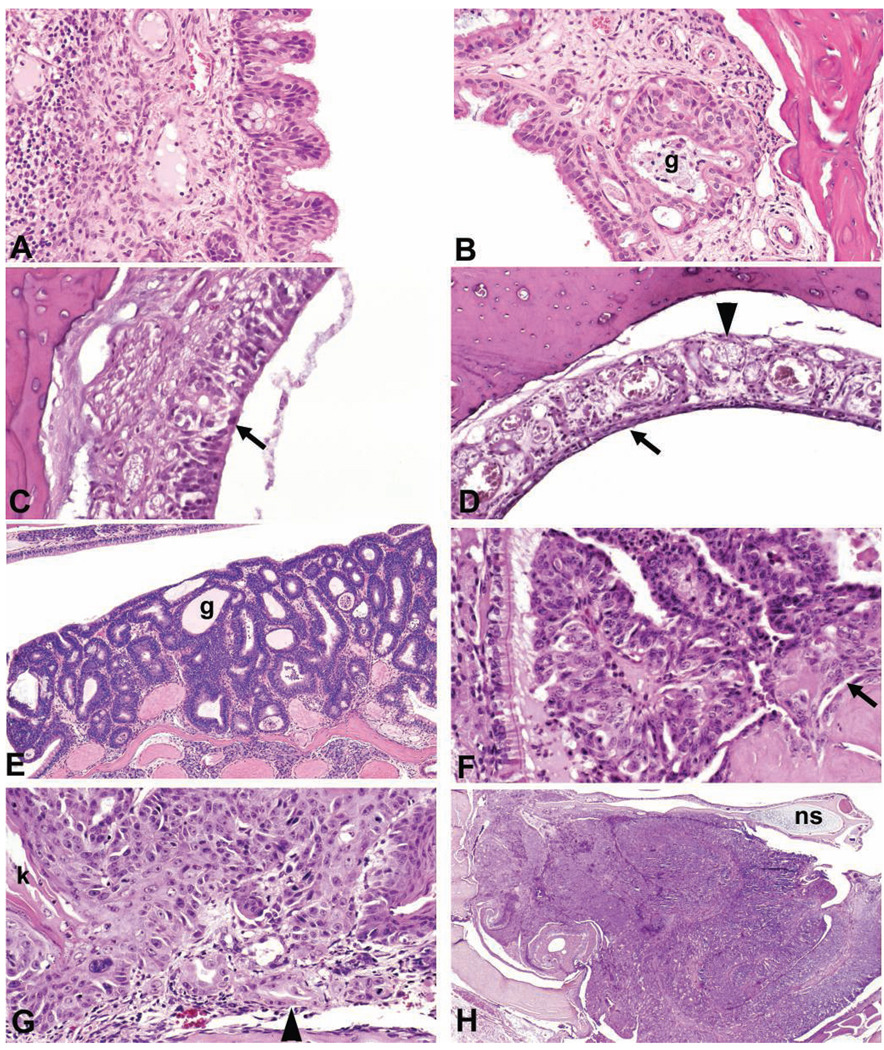
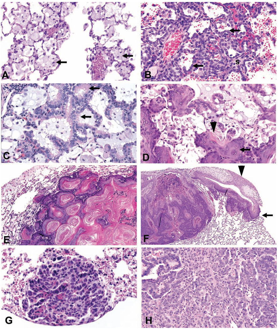
This paper reviews respiratory tract lesions observed in rodents administered various chemicals by noninhalation routes. Chemicals administered by inhalation caused lesions in the respiratory tract and were well described; however, when chemicals were administered by noninhalation routes the effort to evaluate tissues for lesions may have been less or not considered, especially in the upper respiratory tract, and some lesions may have gone undetected. Lesions described in this review mostly occurred in rodent chronic noninhalation studies conducted by the National Toxicology Program; however, some were noted in studies of shorter duration. The nasal cavity was vulnerable to damage when chemicals were administered by noninhalation routes. Changes included respiratory epithelial hyperplasia, degeneration and necrosis of olfactory epithelium, olfactory epithelial metaplasia, adenoma, adenocarcinoma, squamous cell carcinoma, and neuroblastoma. In the lung, compound-related lesions included alveolar histiocytosis, alveolar epithelial hyperplasia, bronchiolar metaplasia of the alveolar epithelium, squamous metaplasia, alveolar/bronchial adenoma and carcinoma, and squamous tumors. Pathogenesis of these lesions included regurgitation of volatiles, metabolites arriving from the blood stream, and additional metabolism by olfactory epithelium or Clara cells. The presence of respiratory tract lesions in noninhalation studies emphasizes the need for a thorough examination of the respiratory tract including nasal passages, regardless of the route of administration.
Figures
Similar articles
-
Toxicology and carcinogenesis studies of 1-bromopropane (CAS No. 106-94-5) in F344/N rats and B6C3F1 mice (inhalation studies).Natl Toxicol Program Tech Rep Ser. 2011 Aug;(564):1-190. Natl Toxicol Program Tech Rep Ser. 2011. PMID: 21921963
-
NTP Toxicology and Carcinogenesis Studies of Ozone (CAS No. 10028-15-6) and Ozone/NNK (CAS No. 10028-15-6/ 64091-91-4) in F344/N Rats and B6C3F1 Mice (Inhalation Studies).Natl Toxicol Program Tech Rep Ser. 1994 Oct;440:1-314. Natl Toxicol Program Tech Rep Ser. 1994. PMID: 12595923
-
NTP Toxicology and Carcinogenesis Studies of Allyl Glycidyl Ether (CAS No. 106-92-3) in Osborne-Mendel Rats and B6C3F1 Mice (Inhalation Studies).Natl Toxicol Program Tech Rep Ser. 1990 Jan;376:1-219. Natl Toxicol Program Tech Rep Ser. 1990. PMID: 12692645
-
Histologic changes in the respiratory tract induced by inhalation of xenobiotics: physiologic adaptation or toxicity?Toxicol Appl Pharmacol. 1989 Dec;101(3):521-42. doi: 10.1016/0041-008x(89)90200-7. Toxicol Appl Pharmacol. 1989. PMID: 2690398 Review.
-
Styrene respiratory tract toxicity and mouse lung tumors are mediated by CYP2F-generated metabolites.Regul Toxicol Pharmacol. 2002 Jun;35(3):308-19. doi: 10.1006/rtph.2002.1545. Regul Toxicol Pharmacol. 2002. PMID: 12202046 Review.
Cited by
-
Proceedings of the 2011 National Toxicology Program Satellite Symposium.Toxicol Pathol. 2012;40(2):321-44. doi: 10.1177/0192623311427713. Epub 2011 Nov 16. Toxicol Pathol. 2012. PMID: 22089839 Free PMC article.
-
Fourteen-week toxicity study of green tea extract in rats and mice.Toxicol Pathol. 2010 Dec;38(7):1070-84. doi: 10.1177/0192623310382437. Epub 2010 Sep 30. Toxicol Pathol. 2010. PMID: 20884815 Free PMC article.
-
The growing use of herbal medicines: issues relating to adverse reactions and challenges in monitoring safety.Front Pharmacol. 2014 Jan 10;4:177. doi: 10.3389/fphar.2013.00177. eCollection 2014 Jan 10. Front Pharmacol. 2014. PMID: 24454289 Free PMC article. Review.
-
Toxicity and carcinogenicity studies of Ginkgo biloba extract in rat and mouse: liver, thyroid, and nose are targets.Toxicol Pathol. 2014 Jul;42(5):830-43. doi: 10.1177/0192623313501235. Epub 2013 Aug 19. Toxicol Pathol. 2014. PMID: 23960164 Free PMC article.
-
Proceedings of the 2024 Division of Translational Toxicology Satellite Symposium.Toxicol Pathol. 2024 Dec;52(8):460-488. doi: 10.1177/01926233241298895. Epub 2024 Dec 11. Toxicol Pathol. 2024. PMID: 39660627 Free PMC article.
References
-
- Boorman GA, Brockmann M, Carlton WW, Davis JM, Dungworth DL, Hahn FF, Mohr U, Reichhelm HB, Turusov VS, Wagner BM. Classification of cystic keratinizing squamous lesions of the rat lung: report of a workshop. Toxicol Pathol. 1996;24:564–572. - PubMed
-
- Boorman GA, Eustis SL. Lung. In: Boorman GA, Eustis SL, Elwell MR, Montgonery CA Jr, MacKenzie WF, editors. Pathology of the Fischer Rat. San Diego, CA: Academic Press; 1990a. pp. 339–367.
-
- Boorman GA, Morgan KT, Uriah LC. Nose, larynx, and trachea. In: Boorman GA, Eustis SL, Elwell MR, Montgonery CA Jr, MacKenzie WF, editors. Pathology of the Fischer Rat. San Diego, CA: Academic Press; 1990b. pp. 315–337.
-
- Brix AE, Jokinen MP, Walker NJ, Sells DM, Nyska A. Characterization of bronchiolar metaplasia of the alveolar epithelium in female Sprague–Dawley rats exposed to 3,3′,4,4′,5-pentachlorobiphenyl (PCB126) Toxicol Pathol. 2004;32:333–337. - PubMed
Publication types
MeSH terms
Substances
Grants and funding
LinkOut - more resources
Full Text Sources
