. 2007 Feb 27:8:14.
doi: 10.1186/1471-2199-8-14.

Ets-2 and C/EBP-beta are important mediators of ovine trophoblast Kunitz domain protein-1 gene expression in trophoblast

Affiliations

Ets-2 and C/EBP-beta are important mediators of ovine trophoblast Kunitz domain protein-1 gene expression in trophoblast

Anindita Chakrabarty et al. BMC Mol Biol. .

Abstract

Background: The trophoblast Kunitz domain proteins (TKDPs) constitute a highly expressed, placenta-specific, multigene family restricted to ruminant ungulates and characterized by a C-terminal "Kunitz" domain, preceded by one or more unique N-terminal domains. TKDP-1 shares an almost identical expression pattern with interferon-tau, the "maternal recognition of pregnancy protein" in ruminants. Our goal here has been to determine whether the ovine (ov) Tkdp-1 and IFNT genes possess a similar transcriptional code.

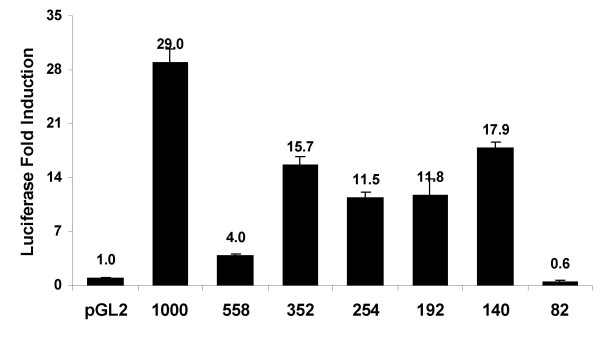
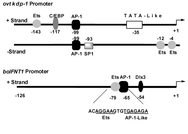
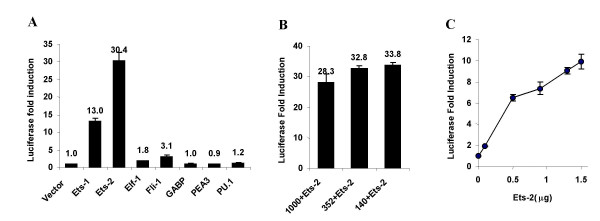
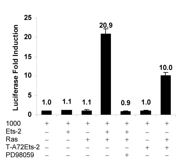
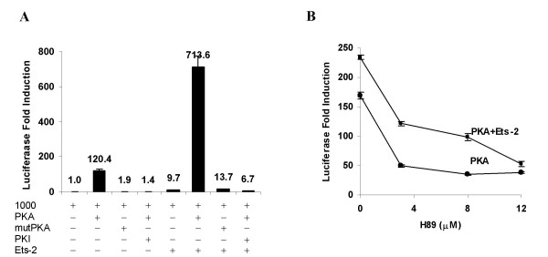
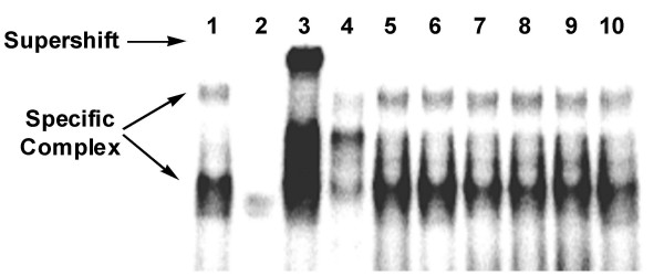
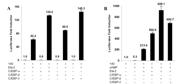
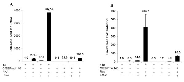
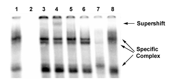
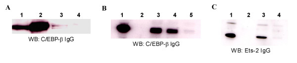
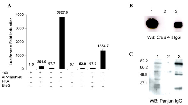
Results: The ovTkdp-1 promoter has been cloned and characterized. As with the IFNT promoter, the Tkdp-1 promoter is responsive to Ets-2, and promoter-driven reporter activity can be increased over 700-fold in response to over-expression of Ets-2 and a constitutively active form of protein Kinase A (PKA). Unexpectedly, the promoter element of Tkdp-1 responsible for this up-regulation, unlike that of the IFNT, does not bind Ets-2. However, mutation of a CCAAT/enhancer binding element within this control region not only reduced basal transcriptional activity, but prevented Ets-2 as well as cyclic adenosine 5'-monophosphate (cAMP)/PKA and Ras/mitogen-activated protein kinase (MAPK) responsiveness. In vitro binding experiments and in vivo protein-protein interaction assays implicated CCAAT/enhancer binding protein-beta (C/EBP-beta) as involved in up-regulating the Tkdp-1 promoter activity. A combination of Ets-2 and C/EBP-beta can up-regulate expression of the minimal Tkdp-1 promoter as much as 930-fold in presence of a cAMP analog. An AP-1-like element adjacent to the CCAAT enhancer, which binds Jun family members, is required for basal and cAMP/ C/EBP-beta-dependent activation of the gene, but not for Ets-2-dependent activity.

Conclusion: This paper demonstrates how Ets-2, a key transcription factor for trophoblast differentiation and function, can control expression of two genes (Tkdp-1 and IFNT) having similar spatial and temporal expression patterns via very different mechanisms.


Figures

Figure 1
Figure 1
Detection of ovTKDP-1, IFN-τ, Ets-2 and C/EBP-β mRNA in sheep trophectoderm. RT reactions were performed on sheep trophoblast RNA collected from different days of pregnancy. After binding to oligo dT primer, reverse transcription and PCR amplification with primers specific for each message were performed. A) Comparison of ovTKDP-1 and IFN-τ expression during the peri-implantation period of conceptus development. Lanes 1–3: positive control PCR with TKDP-1, IFN-τ and S25 plasmid templates, respectively; lanes 4–9: PCR with days 14, 15, 16, 17, 19 and 25 sheep trophoblast cDNA. B) Ets-2 mRNA expression during the peri-implantation period of conceptus development. Lane 1: DNA marker; lane 2: positive control PCR with Ets-2 expression plasmid; lanes 3–8: PCR with day 14, 15, 16, 17 and 25 cDNA. C) C/EBP-β mRNA expression;. Lane 1: DNA marker; lane 2: positive control PCR with C/EBPβ expression plasmid; lanes 3–8: PCR with day 14, 15, 16, 17 and 25 cDNA.
Figure 2
Figure 2
Expression of a series of Tkdp-1 promoter-Luc reporter constructs transfected into JEG-3 cells. Luc activities are expressed relative to that of the promoter-less pGL2-Basic Luc reporter construct, with data shown as means ± SEM (n = 3; P < 0.05: pGL2 vs. 1000, 352, 254, 192, 140).
Figure 3
Figure 3
Possible transcription factor-binding sites in the proximal ovTkdp-1 and boIFNT1 promoters. The sequence of the Ets-AP-1 composite enhancer in the boIFNT1 promoter is also shown [23]. The tsp is designated +1.
Figure 4
Figure 4
Transactivation of the Tkdp-1 promoter by Ets-2. A) Effects of different Ets family transcription factors on reporter expression from the 1000 bp Tkdp-1 promoter-Luc reporter construct in JEG-3 cells. Luc activities (means ± SEM; n = 3) are expressed relative to that of the basal activity of the 1000 bp construct. Following values differ significantly (p < 0.05): 1000 vs. 1000+Ets-1 and Ets-2; 1000+Ets-1 vs. 1000+Ets-2. B) Effect of Ets-2 on the transcriptional activities of three 5/-truncated Tkdp-1 promoter-Luc reporter constructs 1000, 352 and 140 in JEG-3 cells. Luc activities are expressed relative to that of the basal activity of each of the truncated constructs and data are shown as mean ± SEM (n = 3). For each truncated construct, the fold-induction in reporter activity after co-transfection with Ets-2 is significantly different from that without Ets-2 (P < 0.05). C) Effect of increasing concentrations of Ets-2 on the activity of the 1000 bp Tkdp-1 promoter-Luc reporter construct. Luc activities are expressed relative to that of the basal activity of the 1000 bp construct and data are shown as mean ± SEM (n = 3). Following values differ significantly (p < 0.05): 1000 vs. 0.5 μg, 0.9 μg, 1.3 μg and 1.5 μg Ets-2.
Figure 5
Figure 5
Effects of Ets-2 and activated Ras on the Tkdp-1 1000 bp promoter activity in NIH3T3 cells. Luc activities are expressed relative to that of the basal activity of the 1000 bp construct. Data are shown as means ± SEM (n = 3). The following values differ significantly (p < 0.05): 1000 vs. Ets-2+Ras and T-A72Ets-2+Ras; Ets-2+Ras vs. T-A72Ets-2+Ras.
Figure 6
Figure 6
Effects of Ets-2 and activated PKA on the Tkdp-1 1000 bp promoter activity in JEG-3 cells. Expression of the 1000 bp promoter-Luc construct A) following co-transfection with activated PKA, activated PKA+PKI, mutPKA, Ets-2, Ets-2+activated PKA, Ets-2+activated PKA+PKI, Ets-2+mutPKA and B) in cells exposed to the PKA inhibitor H89 in presence and absence of co-transfected Ets-2. Luc activities are expressed relative to that of the basal activity of the 1000 bp construct and data are shown as mean ± SEM (n = 3). The following values are significantly different at P < 0.05: A) 1000 vs. 1000+PKA, 1000+Ets-2, 1000+Ets-2+PKA; 1000+PKA vs. 1000+mutPKA, 1000+PKA+PKI; 1000+Ets-2+PKA vs. 1000+Ets-2+mutPKA, 1000+Ets-2+PKA+PKI and B) 1000+PKA vs. 1000+PKA+3 μMH89, 1000+PKA+8 μMH89; 1000+PKA+12 μM H89; 1000+Ets-2+PKA vs. 1000+Ets-2+PKA+3 μM H89, 1000+Ets-2+PKA+8 μM H89, 1000+Ets-2+PKA+12 μM H89
Figure 7
Figure 7
Competition EMSA with recombinant Ets-2 protein and a 32P-labeled ds Ets-2 consensus oligonucleotide. Lane 1: labeled oligonucleotide+Ets-2-GST-fusion protein; lane 2: labeled oligonucleotide+GST protein; lane 3: labeled oligonucleotide+Ets-2-GST-fusion protein+anti-Ets-2 IgG; lane 4: labeled oligonucleotide+Ets-2-GST-fusion protein+unlabeled Ets-2 consensus oligonucleotide (250-fold molar excess); lanes 5–10: labeled oligonucleotide+Ets-2-GST-fusion protein+unlabeled overlapping oligonucleotides from the Tkdp-1 promoter (-150 to +20) at 250-fold molar excess.
Figure 8
Figure 8
Effects of C/EBP-α, -β and -δ on the Tkdp-1 140 bp promoter activity in JEG-3 cells. Luc expression driven by the minimal promoter in cells co-transfected with each of the C/EBP isoforms A) in the absence and presence of over-expressed Ets-2 and B) in the presence of over-expressed Ets-2 and 250 μM 8-BrcAMP. Luc activities are expressed relative to that of the basal activity of the 140 bp promoter and data are shown as mean ± SEM (n = 3). The following values are significantly different at P < 0.05: A) 140 vs. Ets-2, C/EBP-α+Ets-2, C/EB-Pβ+Ets-2, C/EBP-δ+Ets-2 and B) 140 vs. cAMP+Ets-2, cAMP+Ets-2+C/EBP-α, cAMP+Ets-2+C/EBP-β, cAMP+Ets-2+C/EBP-δ; cAMP+Ets-2 vs. cAMP+Ets-2+C/EBP-β, cAMP+Ets-2+C/EBP-δ. cAMP+Ets-2+C/EBP-β is significantly different (P < 0.05) from both cAMP+Ets-2+C/EBP-α and cAMP+ Ets-2+C/EBP-δ.
Figure 9
Figure 9
Effects of the CCAAT/enhancer element (-117 to -109) mutation on the Tkdp-1 140 bp promoter activity in A) JEG-3 and B) NIH3T3 cells. A) Basal, activated PKA, Ets-2, Ets-2+activated PKA-dependent activation of the wild type and C/EBP-mutated minimal promoter in JEG-3 cells. B) Basal, activated Ras, Ets-2, Ets-2+activated Ras-dependent activation of the wild type and C/EBP-mutated promoter in NIH3T3 cells. Luc activities are expressed relative to that of the basal activity of the 140 bp promoter and data are shown as mean ± SEM (n = 3). Following values are significantly different at P < 0.05: A) 140+PKA vs. C/EBPmut140+PKA; 140+Ets-2 vs. C/EBPmut140+Ets-2; 140+Ets-2+PKA vs. C/EBPmut140+Ets-2+PKA and B) 140+Ets-2 vs. C/EBPmut140+Ets-2 and 140+Ets-2+Ras vs. C/EBPmut140+Ets-2+Ras.
Figure 10
Figure 10
EMSA with 32P-labeled C/EBP oligonucleotide (-117 to -109) and JEG-3 cell extracts (CE). Lane 1: labeled oligonucleotide+CE; lane 2: labeled oligonucleotide-CE; lane 3: labeled oligonucleotide+CE+C/EBP-α IgG; lane 4: labeled oligonucleotide+CE+C/EBP-β IgG; lane 5: labeled oligonucleotide+CE+C/EBP-δ IgG; lane 6: labeled oligonucleotide+CE+non-immune rabbit IgG; lanes 7–8: labeled oligonucleotide+CE+250-fold molar excess of unlabeled wild type (lane 7) and mutated (lane 8) C/EBP competitor oligonucleotides.
Figure 11
Figure 11
C/EBP-β and Ets-2 interaction demonstrated by A) co-immunoprecipitation, B) and C) pull-down assays with biotinylated C/EBP oligonucleotide (-117 to -109) in JEG-3 cell extracts (CE). A) Lane 1: JEG-3 CE (positive control for western blot); lane 2: immunoprecipitation (IP) with C/EBP-β IgG (positive control for IP); lane 3: IP with Ets-2 IgG; lane 4: IP with non-immune rabbit IgG (negative control for IP). Western blot (WB) was performed with C/EBP-β IgG. B) WB performed with C/EBP-β IgG as detection reagent following pull-down assays. Lane 1: JEG-3 CE (positive control for WB); lane 2: biotinylated C/EBP oligonucleotide; lane 3: JEG-3 CE+biotinylated C/EBP oligonucleotide; lane 4: JEG-3 CE+biotinylated C/EBP oligonucleotide+250-fold molar excess unlabeled mutated C/EBP oligonucleotide; lane 5: JEG-3 CE+biotinylated C/EBP oligonucleotide+250-fold molar excess unlabeled wild type C/EBP oligonucleotide. C) WB with Ets-2 IgG as the detection agent following pull-down assays. Lane 1: JEG-3 CE (positive control for WB); lane 2: biotinylated C/EBP oligonucleotide; lane 3: JEG-3 CE+biotinylated C/EBP oligonucleotide; lane 4: JEG-3 CE+biotinylated C/EBP oligonucleotide+250-fold molar excess unlabeled wild type C/EBP oligonucleotide.
Figure 12
Figure 12
Importance of the AP-1 element (-99 to -93) for the Tkdp-1.140 bp promoter activity in JEG-3 cells. A) Effects of the AP-1 element mutation on the basal, activated PKA, Ets-2 and Ets-2+activated PKA-dependent activation of the 140 bp Tkdp-1 promoter-Luc reporter construct in JEG-3 cells. Luc activities are expressed relative to that of the basal activity of the 140 bp promoter and data are shown as mean ± SEM (n = 3). Following values are significantly different at P < 0.05: 140+PKA vs. AP-1mut140+PKA and 140+Ets-2+PKA vs. AP-1mut140+Ets-2+PKA. B) and C) Pull-down assays with biotinylated AP-1 oligonucleotide (-99 to -93 in the tkdp-1 promoter) and JEG-3 cell extracts(CE). Following pull-down a ssays, WB analysis was performed with B) C/EBP-β IgG and C) pan-Jun IgG. Lane 1: JEG-3 CE (positive control for WB); lane 2: biotinylated AP-1 oligonucleotide; lane 3: JEG-3 CE+biotinylated AP-1 oligonucleotide. The molecular weight standards are shown on the left-hand side.

Similar articles

Cited by

References

    1. Chakrabarty A, Green JA, Roberts RM. Origin and evolution of the TKDP gene family. Gene. 2006;373:35–43. doi: 10.1016/j.gene.2005.12.033. - DOI - PubMed
    1. MacLean JA, II, Chakrabarty A, Xie S, Bixby JA, Roberts RM, Green JA. Family of Kunitz proteins from trophoblast: expression of the trophoblast Kunitz domain proteins (TKDP) in cattle and sheep. Mol Reprod Dev. 2003;65:30–40. doi: 10.1002/mrd.10262. - DOI - PubMed
    1. Kwong PD, McDonald NQ, Sigler PB, Hendrickson WA. Structure of beta 2-bungarotoxin: potassium channel binding by Kunitz modules and targeted phospholipase action. Structure. 1995;3:1109–1119. doi: 10.1016/S0969-2126(01)00246-5. - DOI - PubMed
    1. Harvey AL. Twenty Years of Dendrotoxins. Toxicon. 2001;39:15–26. doi: 10.1016/S0041-0101(00)00162-8. - DOI - PubMed
    1. Schweitz H, Heurteaux C, Bois P, Moinier D, Romey G, Lazdunski M. Calcicludine, a Venom Peptide of the Kunitz-type Protease Inhibitor Family, is a Potent blocker of high-threshold Ca+2 Channels with a High-affinity for L-type channels in cerebellar Granule Neurons. Proc Natl Acad Sci USA. 1994;91:878–882. doi: 10.1073/pnas.91.3.878. - DOI - PMC - PubMed

Publication types

MeSH terms