. 2007 Feb 27:8:20.
doi: 10.1186/1471-2474-8-20.

Whole body MR imaging in ankylosing spondylitis: a descriptive pilot study in patients with suspected early and active confirmed ankylosing spondylitis

Affiliations

Whole body MR imaging in ankylosing spondylitis: a descriptive pilot study in patients with suspected early and active confirmed ankylosing spondylitis

Ulrich Weber et al. BMC Musculoskelet Disord. .

Abstract

Background: Ankylosing spondylitis is a chronic inflammatory rheumatic disorder which usually begins in early adulthood. The diagnosis is often delayed by many years. MR imaging has become the preferred imaging method for detection of early inflammation of the axial skeleton in ankylosing spondylitis. The goal of this study was to assess the frequency and distribution of abnormalities on whole body MR imaging in patients with suspected early ankylosing spondylitis and with active confirmed ankylosing spondylitis.

Methods: Ten patients with suspected early ankylosing spondylitis and ten patients with confirmed ankylosing spondylitis were enrolled. On an 18-channel MR system, coronal and sagittal T1 weighted and STIR sequences were acquired covering the entire spine, sacrum, anterior chest wall, shoulder girdle, and pelvis. The total examination time was 30 minutes.

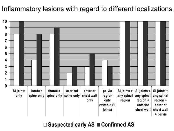
Results: In both groups inflammatory lesions of the lower thoracic spine were frequent (number of patients with suspected early/confirmed ankylosing spondylitis: 7/9). In confirmed ankylosing spondylitis the upper thoracic spine (3/6) and the lumbar spine (4/8) were more commonly involved. The inferior iliac quadrant of the sacroiliac joints was frequently altered in both groups (8/8). The superior iliac (2/5), inferior sacral (6/10) and superior sacral (3/6) quadrants were more frequently affected in confirmed ankylosing spondylitis. Abnormalities of the manubriosternal joint (2/4), the sternoclavicular joints (1/2) and hip joint effusion (4/3) were also seen.

Conclusion: In both suspected early ankylosing spondylitis and confirmed ankylosing spondylitis, whole body MR examinations frequently demonstrate inflammatory lesions outside the sacroiliac joints. These lesions are similarly distributed but occur less frequently in suspected early compared to confirmed ankylosing spondylitis. Due to the small sample size in this pilot study these results need to be confirmed in larger studies with this emerging technique.


Figures

Figure 1
Figure 1
Inflammatory lesions separated for each vertebral unit. Numbers are patients with a positive finding. The white bars represent the patients with suspected early AS; the black bars represent the patients with confirmed AS.
Figure 2
Figure 2
28-year old male patient with suspected early AS (duration of inflammatory back pain 7 months, BASDAI 3.7, BASDAI 2 5, HLA B27 positive). Coronal (left) and sagittal (right) STIR images show the most commonly seen signal abnormalities in suspected early AS. Inflammatory changes (open arrows) are seen in the lower aspects of the SI joints and subtle abnormalities in the anterior corner of the endplates in the thoracic spine (arrows).
Figure 3
Figure 3
30-year old male patient with confirmed AS (duration of inflammatory back pain 7 years, BASDAI 4.8, HLA B27 positive). Sagittal STIR images show inflammatory lesions in the thoracic and lumbar spine (arrows). Inflammatory lesions of the spinous process are shown at L4 (curved arrow)
Figure 4
Figure 4
47-year old male patient with confirmed AS (duration of inflammatory back pain 13 years, BASDAI 7.0, HLA B27 positive). Fatty replacement of subchondral bone marrow is shown in the lower sacral quadrant (arrows) on the coronal T1-weighted image.
Figure 5
Figure 5
34-year old male patient with confirmed AS (duration of inflammatory back pain 13 years, BASDAI 4.9, HLA B27 positive). Inflammatory lesions of the anterior chest wall are shown in the manubriosternal joint (curved arrows) on coronal (left) and sagittal (right) STIR images. Inflammatory changes are seen in the lower thoracic spine and L1 (arrows).
Figure 6
Figure 6
30-year old male patient with confirmed AS (duration of inflammatory back pain 7 years, BASDAI 4.8, HLA B27 positive). Two coronal STIR images demonstrate inflammatory lesions in the thoracic spine (solid arrows), in the sacroiliac joint (sacral side) (open arrows), and in the left hip. Extensive bone marrow changes in the acetabulum (curved arrow) and an effusion of the hip joint are shown (arrowheads).
Figure 7
Figure 7
MRI abnormalities suggestive of inflammatory lesions with regard to different localizations. Numbers are patients with a positive finding. The white bars represent the patients with suspected early AS; the black bars represent the patients with confirmed AS. The wording "only" refers to inflammatory lesions when analyzing the selected region only irrespective of any additional inflammatory lesions seen in other localizations.

References

    1. Dagfinrud H, Mengshoel AM, Hagen KB, Loge JH, Kvien TK. Health status of patients with ankylosing spondylitis: a comparison with the general population. Ann Rheum Dis. 2004;63:1605–1610. doi: 10.1136/ard.2003.019224. - DOI - PMC - PubMed
    1. Boonen A, van der Heijde D, Landewé R, Guillemin F, Rutten-van Mölken M, Dougados M, Mielants H, de Vlam K, van der Tempel H, Boesen S, Spoorenberg A, Schouten H, van der Linden Sj. Direct costs of ankylosing spondylitis and its determinants: an analysis among three European countries. Ann Rheum Dis. 2003;62:732–740. doi: 10.1136/ard.62.8.732. - DOI - PMC - PubMed
    1. Boonen A, van der Heijde D, Landewé R, Spoorenberg A, Schouten H, Rutten-van Mölken M, Guillemin F, Dougados M, Mielants H, de Vlam K, van der Tempel H, van der Linden Sj. Work status and productivity costs due to ankylosing spondylitis: comparison of three European countries. Ann Rheum Dis. 2002;61:429–437. doi: 10.1136/ard.61.5.429. - DOI - PMC - PubMed
    1. Boonen A, Chorus A, Miedema H, van der Heijde D, Landewé R, Schouten H, van der Tempel H, van der Linden S. Withdrawal from labour force due to work disability in patients with ankylosing spondylitis. Ann Rheum Dis. 2001;60:1033–1039. doi: 10.1136/ard.60.11.1033. - DOI - PMC - PubMed
    1. Braun J, Davis J, Dougados M, Sieper J, van der Linden S, van der Heijde D. First update of the international ASAS consensus statement for the use of anti-TNF agents in patients with ankylosing spondylitis. Ann Rheum Dis. 2006;65:316–320. doi: 10.1136/ard.2005.040758. - DOI - PMC - PubMed

Publication types

MeSH terms