

Case Reports
doi: 10.3201/eid1212.060750.
Spongiform encephalopathy in a miniature zebu
Affiliations
- PMID: 17326950
- PMCID: PMC3291368
- DOI: 10.3201/eid1212.060750
Item in Clipboard
Case Reports
Spongiform encephalopathy in a miniature zebu
Emerg Infect Dis.
2006 Dec.
Abstract
The first case of spongiform encephalopathy in a zebu (Bos indicus) was identified in a zoo in Switzerland. Although histopathologic and immunohistochemical analyses of the central nervous system indicated a diagnosis of bovine spongiform encephalopathy (BSE), molecular typing showed some features different from those of BSE in cattle (B. taurus).
Figures
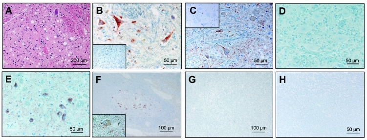
Histopathologic and immunohistochemical analyses. A) Spongiform lesions; B) partially proteinase K–resistant prion protein (PrPsc) deposits detected by immunohistochemistry (monoclonal antibodies [MAb] F99/97.6.1 diluted 1:500) in the nucleus of the solitary tract (STN) in the zebu under investigation. C–E) Comparative immunohistochemistry with MAb P4 (1:800) in the olivary nuclei of the zebu (C), a bovine spongiform encephalopathy (BSE)-positive cow (D), and a scrapie-positive sheep (E). Insets show control tissue slides of BSE-negative cattle. F–H) Immunohistochemistry for PrPsc in lymphoid tissue of the zebu (H, mediastinal lymph node), and a BSE-negative cow (G, mandibular lymph node) with MAb L42 (R-biopharm, 1:800). A retropharyngeal lymph node of a scrapie-affected sheep (F) and a brainstem tissue slide of the zebu (F, inset) served as positive controls. Pretreatment of the tissue slides comprised a proteinase K–digestion step (5 μg/mL, 15 min, 37°C).
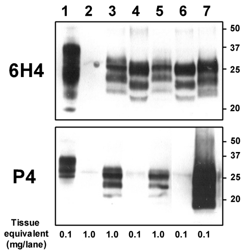
Molecular analyses of the zebu under investigation. Western immunoblot with monoclonal antibodies (MAbs) 6H4 (upper panel) and P4 (lower panel) after limited proteinase K digestion (100 μg/mL, 40 min, 48°C) of 10% brainstem (lanes 3 and 4) and thalamus (lanes 5, 6, and 7) tissue homogenates of the zebu (lanes 3 and 5), a cow with bovine spongiform encephalopathy (lanes 4 and 6), and a sheep with scrapie (lane 7). An undigested cattle brainstem tissue homogenate (lane 1) and a cerebrum tissue homogenate of a spongiform-encephalopathy-negative zebu (lane 2) were included as controls. All samples were processed equally as described by Stack et al. (7), and the membranes were exposed in parallel on the same photographic film. Molecular mass standards in kilodaltons are indicated on the right; tissue mass equivalents, at the bottom.
References
-
- Wyatt JM, Pearson GR, Smerdon TN, Gruffydd-Jones TJ, Wells GA. Spongiform encephalopathy in a cat [letter]. Vet Rec. 1990;126:513. - PubMed
-
- Eliot M, Adjou KT, Coulpier M, Fontaine JJ, Hamel R, Lilin T, et al. BSE agent signatures in a goat [letter] [Erratum in Vet Rec. 2005;156:620]. Vet Rec. 2005;156:523–4. - PubMed
Publication types
MeSH terms
Substances
LinkOut - more resources
Full Text Sources