Review
. 2007:79:515-42.
doi: 10.1016/S0091-679X(06)79020-3.

Electron microscopy analysis of viral morphogenesis

Affiliations
Review

Electron microscopy analysis of viral morphogenesis

Annegret Pelchen-Matthews et al. Methods Cell Biol. 2007.

Abstract

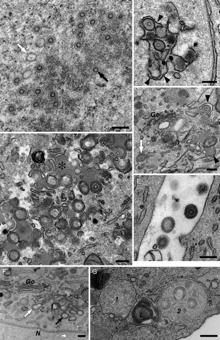
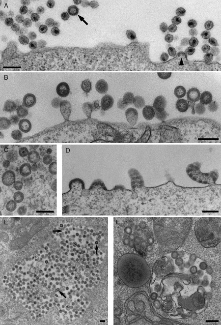
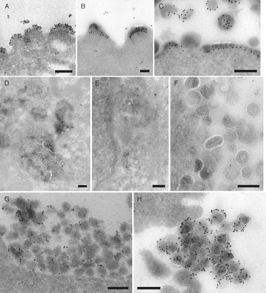
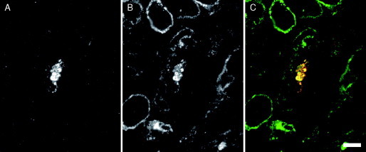
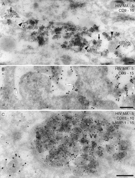
This chapter reviews the use of standard transmission electron microscopy (TEM) of plastic-embedded material, as well as protocols for the immunolabeling of cryosections, in the analysis of viral interactions with cells. It focuses particularly on the assembly of two types of enveloped viruses: (1) the beta herpesvirus—human cytomegalovirus (HCMV), and (2) the primate lentiviruses—the simian and human immunodeficiency viruses (SIV and HIV). The chapter discusses the ways EM is used to identify morphological features of the various stages in the assembly of virus particles, to distinguish immature and mature particles, or to analyze steps involved in the acquisition of lipid membranes by enveloped viruses. In addition, it demonstrates the way immunolabeling allows the quantification of viral components, even in individual virus particles, and comparisons between particles at different locations in the cell or at different stages in viral assembly. Together with the newly developed methods for electron tomography and correlative immunofluorescence studies and EM, huge potential exists to unravel more details about virus assembly in the near future.


Figures

Fig. 1
Fig. 1
Stages of HCMV assembly. HCMV assembly was documented in human embryonic lung or foreskin fibroblast cells fixed in 2% formaldehyde/1.5% glutaraldehyde and postfixed with reduced OsO4 and tannic acid. (A–E) Human embryonic lung cells. (A) The nucleus contains empty icosahedral capsids (e.g., white arrow) or nucleocapsids enclosing a darker ring composed of the viral DNA (black arrow). (B) The capsids bud into the lumen of the nuclear envelope, acquiring a first membrane from the inner nuclear envelope (black arrowheads). This membrane is lost during fusion with the outer membrane of the nuclear envelope, so that naked capsids are released into the cytoplasm. (C) A cytoplasmic “virus factory” composed of electron‐dense accumulations of viral tegument proteins (asterisk) and various membrane vesicles. HCMV capsid particles acquire a layer of tegument and then proceed to wrap into the membrane vesicles. (D) Detail of the envelopment of tegumented capsids at cytoplasmic membranes near the Golgi complex (Go). The membrane vesicles are deformed into characteristic crescents (black arrowheads). This leads to the production of mature enveloped HCMV particles within cytoplasmic vacuoles (white arrow). (E) Fusion of these vesicles with the plasma membrane releases HCMV particles into the extracellular space. (F and G) Human foreskin fibroblasts. (F) Three types of enveloping particles in the cytoplasm. The white and black arrows indicate envelopment of an empty capsid and a DNA‐filled nucleocapsid, respectively, while the asterisk marks an enveloped tegument aggregate or “dense body.” Note also the empty capsid (white arrowhead) in the nucleus (N), which lacks the tegument layer. (G) Two multivesicular late endosomes (1, 2) and a lysosome containing multilamellar membranes (L). Endosome 2 contains several assembled HCMV virions. Images were taken from cells infected for 72 h (A and B), 96 h (E, F, and G), or 120 h (C and D). Scale bars = 200 nm.
Fig. 2
Fig. 2
Assembly of SIV viruses and VLPs. (A) CEMx174 T2 cells acutely infected with a SIVmac251‐derived virus produce many virions at the cell surface. A budding virus can be recognized by the electron‐dense Gag layer accumulating under the membrane (arrowhead). The Gag layer is also visible in the immature virus particle (arrow). (B) VLPs with the morphology of immature viruses and budding figures accumulate at the surface of COS cells that have been transfected with the SIV Gag protein. (C and D) COS cells expressing a chimeric SIV Gag‐GFP show more irregular VLPs (C) or cell surface‐budding figures (D). The particles lack the thin electron‐dense line next to the VLP lumen, and the electron‐dense Gag protein layer is frequently interrupted due to steric interference by the GFP moiety. (E and F) Intracellular vacuoles containing viruses or VLPs can occasionally be found in CEMx174 cells infected with SIV NC‐MAC (LaBranche et al., 1995) (E) or COS cells expressing SIV Gag (F), respectively. Arrows in (E) indicate virus particles with immature morphology. Scale bars = 200 nm.
Fig. 3
Fig. 3
Analysis of SIV assembly by immunolabeling of cryosections. (A, B, and C) SIV Gag‐GFP VLPs budding from the surface of transfected COS cells. The buds were immunolabeled with mouse monoclonal antibodies against SIV MA (mAb KK59 in A) or CA (mAb KK64 in B, antibodies provided by Dr. K. Kent through the NIBSC Centralised Facility for AIDS Reagents, Potters Bar, UK) and a rabbit anti‐mouse IgG‐bridging antibody (Dako UK Limited, Ely, UK), or with a rabbit polyclonal antiserum recognizing GFP (C; Living Colors™ peptide antibody, Clontech Laboratories, Inc., Mountain View, CA, USA). All antibodies were detected with 10‐nm PAG. (D, E, and F) COS cells expressing SIV Gag and assembling VLPs in intracellular vacuoles resembling multivesicular bodies were immunolabeled with mouse monoclonal antibodies against CD63 (Fraile‐Ramos et al., 2001) or against LBPA (Kobayashi et al., 1998, provided by Dr. J. Gruenberg, University of Geneva, Switzerland) and 10‐nm PAG. The LBPA antibody labels intracellular (E), but not cell surface (F) VLPs. (G and H) Virus particles at the surface of CEMx174 cells chronically infected with SIVmac239 expressing an Env glycoprotein with a long cytoplasmic domain (G) or SIVmac239/251T with a cytoplasmically truncated Env protein (H) were stained with an antibody against SIV Env SU (Edinger et al., 2000, provided by Dr. R. W. Doms, University of Pennsylvania, Philadelphia) and 10‐nm PAG. The immunolabeling suggests that these two virus strains incorporate different amounts of the Env protein. PAG reagents were purchased from The Cell Microscopy Center, University Medical Center, Utrecht, The Netherlands. Scale bars = 200 nm.
Fig. 4
Fig. 4
Screening semithin cryosections by immunofluorescence. Cryosections (0.5‐μm thick) from macrophages infected for 13 days with HIV‐1BaL were immunolabeled with a rabbit antiserum against HIV‐1 MA (UP595, provided by Dr. M. Malim, King's College London) and a monoclonal antibody against CD9 (mouse monoclonal anti‐CD9, Serotec, Kidlington, Oxford, UK), and the bound antibodies were detected with goat anti‐rabbit Alexa Fluor‐594 and goat anti‐mouse Alexa Fluor‐488 secondary antibodies (Invitrogen Molecular Probes, Paisley, UK). (A) Red fluorescence. MA staining reveals an accumulation of HIV viruses in one of the cells. (B) Green fluorescence. CD9 staining. (C) Merged image shows colocalization in yellow. Scale bar = 10 μm.
Fig. 5
Fig. 5
Double‐ and triple‐staining immunolabeling with PAG. (A and B) Ultrathin cryosections of 7‐day HIV‐1BaL infected (A) or uninfected macrophages (B) were stained for CD9 (mouse monoclonal anti‐CD9, rabbit anti‐mouse bridging antibody and 15‐nm PAG) followed by the rabbit antiserum against HIV‐1 MA and 5‐nm PAG. (A) An intracellular vacuole containing many virus particles is stained strongly for CD9, both on the virus particles (black arrows) and on smaller intralumenal vesicles (black arrowheads). (B) CD9 labeling at the cell surface of uninfected macrophages. A small number of 5‐nm PAG particles are seen close to 15‐nm PAG (white arrows), indicating a minor amount of cross‐reaction in this double‐labeling protocol. (C) Triple staining with a mouse monoclonal anti‐HIV MA antibody (4C9, Ferns et al., 1987, provided by Drs. R. B. Ferns and R. S. Tedder through the NIBSC Centralised Facility for AIDS Reagents) and 5‐nm PAG, the mouse monoclonal anti‐CD63 and 10‐nm PAG and a rabbit anti‐LAMP‐1 (provided by Dr. S. Carlsson, Umea University, Sweden) and 15‐nm PAG. The large vacuole is packed with virus particles that are also labeled for CD63, but there is little labeling for LAMP‐1. By contrast, a lysosome (L) nearby is strongly labeled for LAMP‐1 on its limiting membrane. Scale bars = 200 nm.

Similar articles

Cited by

References

    1. Briggs J.A., Grunewald K., Glass B., Forster F., Krausslich H.G., Fuller S.D. The mechanism of HIV‐1 core assembly: Insights from three‐dimensional reconstructions of authentic virions. Structure. 2006;14:15–20. - PubMed
    1. Briggs J.A., Simon M.N., Gross I., Krausslich H.G., Fuller S.D., Vogt V.M., Johnson M.C. The stoichiometry of Gag protein in HIV‐1. Nat. Struct. Mol. Biol. 2004;11:672–675. - PubMed
    1. Britt W.J., Boppana S. Human cytomegalovirus virion proteins. Hum. Immunol. 2004;65:395–402. - PubMed
    1. Britt W.J., Mach M. Human cytomegalovirus glycoproteins. Intervirology. 1996;39:401–412. - PubMed
    1. Canto‐Nogues C., Hockley D., Grief C., Ranjbar S., Bootman J., Almond N., Herrera I. Ultrastructural localization of the RNA of immunodeficiency viruses using electron microscopy in situ hybridization and in vitro infected lymphocytes. Micron. 2001;32:579–589. - PubMed

Publication types

LinkOut - more resources