. 2007 Apr 20;282(16):11849-57.
doi: 10.1074/jbc.M609283200. Epub 2007 Feb 27.

Endothelial cell protein C receptor acts as a cellular receptor for factor VIIa on endothelium

Affiliations

Endothelial cell protein C receptor acts as a cellular receptor for factor VIIa on endothelium

Samit Ghosh et al. J Biol Chem. .

Abstract

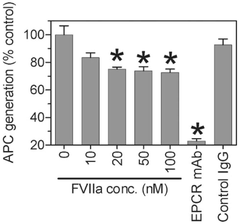
Although factor VII/factor VIIa (FVII/FVIIa) is known to interact with many non-vascular cells, activated monocytes, and endothelial cells via its binding to tissue factor (TF), the interaction of FVII/FVIIa with unperturbed endothelium and the role of this interaction in clearing FVII/FVIIa from the circulation are unknown. To investigate this, in the present study we examined the binding of radiolabeled FVIIa to endothelial cells and its subsequent internalization. (125)I-FVIIa bound to non-stimulated human umbilical vein endothelial cells (HUVEC) in time- and dose-dependent manner. The binding is specific and independent of TF and negatively charged phospholipids. Protein C and monoclonal antibodies to endothelial cell protein C receptor (EPCR) blocked effectively (125)I-FVIIa binding to HUVEC. FVIIa binding to EPCR is confirmed by demonstrating a marked increase in (125)I-FVIIa binding to CHO cells that had been stably transfected with EPCR compared with the wild-type. Binding analysis revealed that FVII, FVIIa, protein C, and activated protein C (APC) bound to EPCR with similar affinity. FVIIa binding to EPCR failed to accelerate FVIIa activation of factor X or protease-activated receptors. FVIIa binding to EPCR was shown to facilitate FVIIa endocytosis. Pharmacological concentrations of FVIIa were found to impair partly the EPCR-dependent protein C activation and APC-mediated cell signaling. Overall, the present data provide convincing evidence that EPCR serves as a cellular binding site for FVII/FVIIa. Further studies are needed to evaluate the pathophysiological consequences and relevance of FVIIa binding to EPCR.


Figures

FIGURE 1
FIGURE 1. FVIIa binding and internalization in HUVEC at 4 and 37 °C
Monolayers of confluent non-stimulated HUVEC (in a 24-well plate) were incubated with 125I-FVIIa (10 nM) for varying time periods (0 –180 min) at 4 °C (○) and at 37 °C (●). At the end of time period, the amount of 125I-FVIIa associated with the cell surface (A) and internalized (B) was determined as described under “Experimental Procedures.” Data are expressed as mean ± S.E. (n = 3).
FIGURE 2
FIGURE 2. FVIIa binding specificity to HUVEC
A, unperturbed HUVEC were incubated with 125I-FVIIa (10 nM) for 3 h at 4 °C in the presence or the absence of unlabeled FVIIa (1 μM), anti-human TF IgG (25 μg/ml), or annexin V (200 nM), and 125I-FVIIa bound to the cell surface was determined (mean ± S.E.; n = 4 –5). B, HUVEC were stimulated with 20 ng/ml of each of IL-1β and TNF-α for 6 h at 37 °C. The stimulated HUVEC were then incubated with 125I-FVIIa (10 nM) with or without unlabeled FVIIa, anti-human TF IgG, or annexin V for 3 h at 4 °C as in panel A, and the amount 125I-FVIIa bound to the cell surface was determined. *, denotes the value significantly (p < 0.05) differs from the control value, i.e. FVIIa bound to HUVEC cells in absence of competitors. C, HUVEC were incubated with wild type 125I-FVIIa (10 nM), 125I-des-FVIIa (10 nM), or 125I-asialo-FVIIa (10 nM) at 4 °C for 3 h, and the FVIIa binding to the cell surface was determined. Values were shown as the percent relative to the binding of wild type FVIIa (n = 3, mean ± S.E.). D, HUVEC were incubated with various concentrations of 125I-FVIIa (0 –100 nM) in the presence or the absence of 2 μM of unlabeled FVIIa, and the specific FVIIa binding to HUVEC was calculated by subtracting the amount of 125I-FVIIa associated with the cells in the presence of unlabeled FVIIa (nonspecific binding) from the amount of 125I-FVIIa associated in the absence of unlabeled FVIIa (total binding). The data shown in the figure represent mean ± S.E. (n = 2–5).
FIGURE 3
FIGURE 3. FVIIa binding site on HUVEC is not a common binding site for vitamin K-dependent clotting proteins
125I-FVIIa (10 nM) was added to HUVEC in the presence or the absence of a 100-fold molar excess (1 μM) of vitamin K-dependent clotting proteins, factor VIIa, prothrombin, FIX, FX, and protein C alone (A) or in combination (B). At the end of 3 h of incubation at 4 °C, 125I-FVIIa bound to the cell surface was determined (mean ± S.E., n = 3–5). *, the value significantly (p < 0.05) differs from the control value, i.e. FVIIa bound to HUVEC in the absence of competitor. #, the value differs significantly from all other values shown in the graph.
FIGURE 4
FIGURE 4. FVIIa binds to endothelial cell protein C receptor (EPCR)
A, HUVEC were incubated with varying concentration of anti-EPCR mAb (1–10 μg/ml) for 30 min at 4 °C, followed by incubation with 125I-FVIIa (10 nM) for 3 h at 4 °C. The surface binding of 125I-FVIIa was then determined (n = 3, mean ± S.E.). B, wild-type CHO and EPCR-transfected CHO (CHO/EPCR+) cells were incubated with 125I-FVIIa (10 nM) in the presence or absence of various competitors. At the end of 3 h of incubation at 4 °C, the surface binding of 125I-FVIIa was determined. Data are expressed as mean ± S.E. (n = 3), *, value(s) significantly (p < 0.05) differs from the control value, i.e. FVIIa bound to CHO/EPCR+ cells in the absence of competitor/inhibitor.
FIGURE 5
FIGURE 5. Specific binding kinetics of FVIIa, FVII, protein C and APC to EPCR
HUVEC were incubated with varying concentrations (0 –150 nM) of 125I-FVIIa (A), 125I-FVII (B), 125I-protein C (C), or 125I-APC (D) in the presence or the absence of anti-EPCR mAb (10 μg/ml) at 4 °C for 3 h. E, HUVEC were incubated with varying concentrations (1–10 nM) of 125I-EPCR mAb ± 50-fold molar excess of unlabeled EPCR mAb. The surface binding of the raidoligand was then determined and the EPCR specific binding was calculated for the each ligand by subtracting the binding values obtained in the presence of EPCR antibody from that obtained in the absence of the antibody. Data are represented in the figure as mean ± S.E. (n = 3–5).
FIGURE 6
FIGURE 6. EPCR-dependent FVIIa binding and internalization
Wild type CHO (○), CHO/EPCR+ (▲), and CHO/EPCR+ cells pretreated with anti EPCR mAb (10 μg/ml) (■) were incubated with 125I-FVIIa (10 nM) for varying time periods (0 –120 min) at 37 °C, and the 125I-FVIIa surface binding (A) and internalization (B) were determined. Data are represented as mean ± S.E. (n = 3). C, HUVEC were incubated with a control vehicle or anti-EPCR mAb (10 μg/ml) in buffer B for 30 min at room temperature. The cells were then chilled on ice and incubated with 10 nM 125I-FVIIa in the absence or presence of unlabeled FVIIa, PC or APC (250 nM) for 2 h at 4 °C to allow 125I-FVIIa binding to the cells. Unbound radioactivity was removed by washing the cells thrice with buffer B, the cells were transferred to 37 °C, and at the end of 15 min of incubation 125I-FVIIa internalized was measured. 125I-FVIIa internalized at 4 °C, immediately prior to their incubation at 37 °C, was subtracted from those obtained at 15 min at 37 °C. The values are expressed as mean ± S.E. (n = 3). *, the value differs significantly (p < 0.05) from the control value. D, HUVEC were incubated with a control vehicle (○) or anti-EPCR mAb (10 μg/ml) (■) in buffer B for 30 min at room temperature and then 125I-FVIIa (10 nM) was added to the wells and allowed to incubate at 37 °C. At varying time periods, small aliquots were removed and subjected to trichloroacetic acid precipitation to determine FVIIa degradation (n = 3, mean ± S.E.).
FIGURE 7
FIGURE 7. FVIIa binding to EPCR does not influence FVIIa activation of FX
Non-stimulated (A) and stimulated (B) HUVEC were incubated with FVIIa (10 nM), FX (175 nM) in the presence or the absence of anti-EPCR mAb (10 μg/ml), control IgG (10 μg/ml), and anti-TF IgG (25 μg/ml). FXa generation was then determined as described under “Experimental Procedures” (n = 3– 4, mean ± S.E.).
FIGURE 8
FIGURE 8. Effect of FVIIa and activated protein C on PAR1 and PAR2 activation in cells expressing EPCR
A, CHO/EPCR+ cells were transfected transiently with AP-PAR1 or AP-PAR2 reporter constructs. The cells expressing AP-PAR1 and AP-PAR2 were treated with FVIIa, APC, thrombin, or trypsin (10 nM each). At the end of 60 min, an aliquot was removed from overlying conditioned medium and the activity of alkaline phosphatase, which was released into the medium following the activation of AP-PAR1 or AP-PAR2, was measured (n = 3, mean ± S.E.). In one experiment, hirudin 2 units/ml, was included with APC and thrombin treatments. B, CHO/EPCR+ cells transfected transiently with AP-PAR1 were exposed to APC (10 nM), FVIIa (100 nM), or APC (10 nM) + FVIIa (100 nM). At the end of 30 min, an aliquot was removed from overlying conditioned medium, and the activity of alkaline phosphatase released into the medium was measured (n = 3, mean ± S.E.). *, value differs significantly (p < 0.05) from the value obtained in the absence of FVIIa.
FIGURE 9
FIGURE 9. Effect of FVIIa on protein C activation
CHO/EPCR+ cells were incubated with protein C (80 nM) and thrombin (2 nM) in the presence or the absence of varying concentrations of FVIIa (0 –100 nM), anti EPCR mAb (10 μg/ml), and control IgG (10 μg/ml) for 30 min at 37 °C. The reaction was stopped by adding hirudin (2 units/ml) and the activated protein C (APC) generated was measured using the substrate Spectrozyme PCa (2 mM). Data are presented as mean ± S.E. (n = 3) of the percent control of APC generated in absence of FVIIa. *, value differs significantly (p < 0.05) from the control value obtained in the absence of FVIIa or anti-EPCR antibody.

References

    1. Fair DS. Blood. 1983;62:784–791. - PubMed
    1. Loeliger EA, van der Esch B, ter Haar Romney-Wachter CC, Booij HL. Thromb Diath Haemorrh. 1960;4:196–200. - PubMed
    1. Seligsohn U, Kasper CK, Osterud B, Rapaport SI. Blood. 1979;53:828–837. - PubMed
    1. Erhardtsen E. Semin Thromb Hemost. 2000;26:385–391. - PubMed
    1. Hasselback R, Hjort PF. J Appl Physiol. 1960;15:945–948. - PubMed

Publication types