. 2007 Jun 15;109(12):5270-5.
doi: 10.1182/blood-2006-12-064188. Epub 2007 Feb 27.

Fatal hemorrhage in mice lacking gamma-glutamyl carboxylase

Affiliations

Fatal hemorrhage in mice lacking gamma-glutamyl carboxylase

Aihua Zhu et al. Blood. .

Abstract

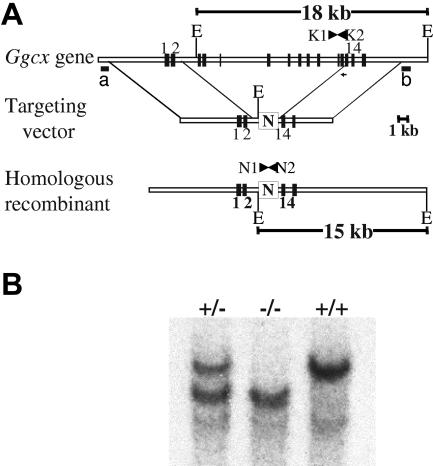
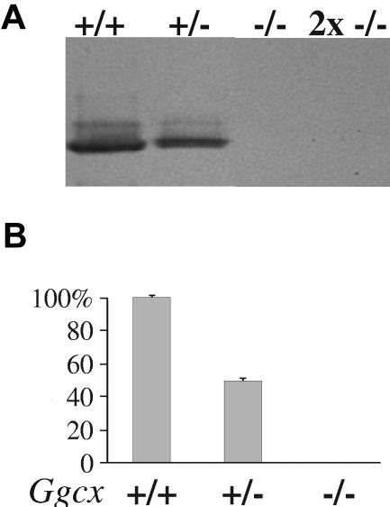
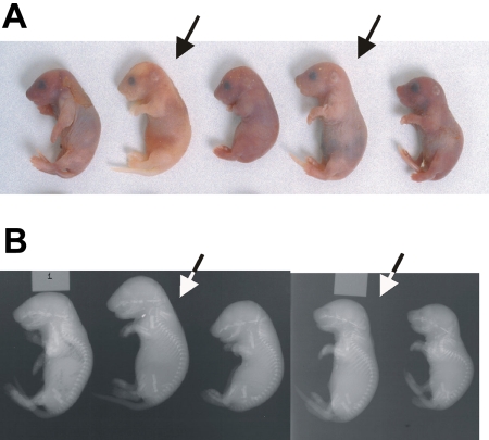
The carboxylation of glutamic acid residues to gamma-carboxyglutamic acid (Gla) by the vitamin K-dependent gamma-glutamyl carboxylase (gamma-carboxylase) is an essential posttranslational modification required for the biological activity of a number of proteins, including proteins involved in blood coagulation and its regulation. Heterozygous mice carrying a null mutation at the gamma-carboxylase (Ggcx) gene exhibit normal development and survival with no evidence of hemorrhage and normal functional activity of the vitamin K-dependent clotting factors IX, X, and prothrombin. Analysis of a Ggcx(+/-) intercross revealed a partial developmental block with only 50% of expected Ggcx(-/-) offspring surviving to term, with the latter animals dying uniformly at birth of massive intra-abdominal hemorrhage. This phenotype closely resembles the partial midembryonic loss and postnatal hemorrhage previously reported for both prothrombin- and factor V (F5)-deficient mice. These data exclude the existence of a redundant carboxylase pathway and suggest that functionally critical substrates for gamma-carboxylation, at least in the developing embryo and neonate, are primarily restricted to components of the blood coagulation cascade.


Figures

Figure 1
Figure 1
Targeting of the murine γ-carboxylase gene by homologous recombination. (A) structure of the endogenous γ-carboxylase gene. The targeting vector contains a neomycin resistance cassette (N), driven by the phosphoglycerokinase promoter, that replaces murine exons 3 to 13 flanked by 5.8 kb of 5′ and 7 kb of 3′ homologous DNA arms. The predicted product of successful homologous recombination is shown at the bottom. The location of hybridization probes a and b, used to detect successful targeting, are indicated. (B) Southern blot analyses demonstrating the expected genomic fragments from the 3′ end of the locus. Genomic DNA was prepared from tail biopsies of Ggcx+/+, Ggcx+/−, and Ggcx−/− mice and was analyzed by restriction digestion with EcoRV and hybridization with probe b. Restriction of Ggcx+/+ genomic DNA yields a fragment of 18 kb after hybridization with probe b. The targeted Ggcx+/− DNA yields 2 distinct bands of 15 kb and 18 kb, indicating that correct targeting of the Ggcx gene has occurred. Animals harboring copies of the targeted allele at both chromosomal loci show the expected hybridization band only at 15 kb, indicating that both copies of the Ggcx gene have been successfully deleted. The presence of the PCR primers (N1, N2, K1, and K2) used for subsequent genotyping are indicated in panel A.
Figure 2
Figure 2
Analysis of γ-carboxylase expression. (A) Western blot analysis of mouse fetal liver microsomes probed with an antibody raised against recombinant bovine γ-carboxylase. All lanes were loaded with 5 ng of microsomal protein except for the 2x −/− lane, which contains 10 ng of microsomal protein. (B) Incorporation of 14CO2 into FLEEL by solubilized γ-carboxylase from the livers of Ggcx+/+ mice was considered 100% γ-carboxylase activity, and the incorporation of 14CO2 by solubilized carboxylase from Ggcx+/− and Ggcx−/− mice were expressed as a percentage of the wild-type. Mean values; error bars indicate ± SD.
Figure 3
Figure 3
Phenotype of Ggcx−/− mice. (A) Newborn progeny from a Ggcx+/− intercross. The homozygous null animals (arrows) exhibit intra-abdominal hemorrhage. (B) X-rays from the same animals shown in panel A. Arrows again indicate the homozygous null mice.

References

    1. Furie B, Bouchard BA, Furie BC. Vitamin K-dependent biosynthesis of γ-carboxyglutamic acid. Blood. 1999;93:1798–1808. - PubMed
    1. Stafford DW. The vitamin K cycle. J Thromb Haemost. 2005;3:1873–1878. - PubMed
    1. Romero EE, Velazquez-Estades LJ, Deo R, Schapiro B, Roth DA. Cloning of rat vitamin K-dependent γ-glutamyl carboxylase and developmentally regulated gene expression in post-implantation embryos. Exp Cell Res. 1998;243:334–346. - PubMed
    1. Hanumanthaiah R, Thankavel B, Day K, Gregory M, Jagadeeswaran P. Developmental expression of vitamin K-dependent gamma-carboxylase activity in zebrafish embryos: effect of warfarin. Blood Cells Mol Dis. 2001;27:992–999. - PubMed
    1. Kulman JD, Harris JE, Nakazawa N, et al. Vitamin K-dependent proteins in Ciona intestinalis, a basal chordate lacking a blood coagulation cascade. Proc Natl Acad Sci U S A. 2006;103:15794–15799. - PMC - PubMed

Publication types