

The developmental regulation of CD81 in the rat retina
- PMID: 17327823
- PMCID: PMC2610370
The developmental regulation of CD81 in the rat retina
Abstract
Purpose: The tetraspanin CD81 is expressed in Müller glial cells and retinal pigment epithelium (RPE). CD81 and other members of the tetraspanin family link extracellular interactions of cells into intracellular cascades. This study examined the developmental expression of CD81 and protein-protein interactions linking CD81 to intracellular proteins.
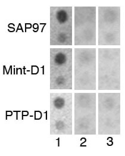
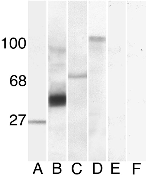
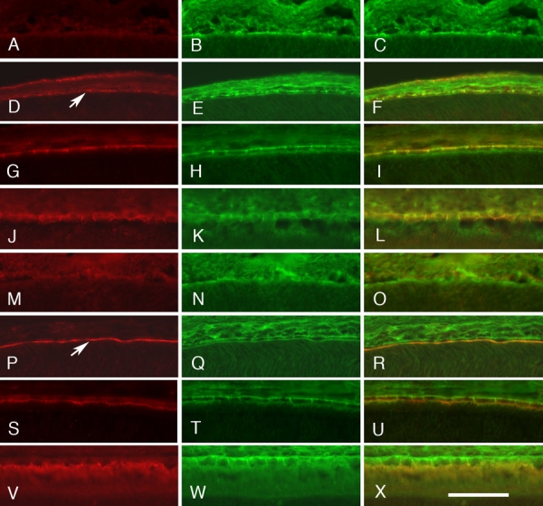
Methods: We used synthetic peptides of the C-terminal intracellular domains of CD81 to probe fusion proteins of PDZ domains blotted to nitrocellulose membranes, then confirmed the relationships between the PDZ proteins using immunoprecipitation methods. Colocalization of the associated proteins was analyzed across development, using double-label immunohistochemical methods in the retina of Sprague-Dawley rats.
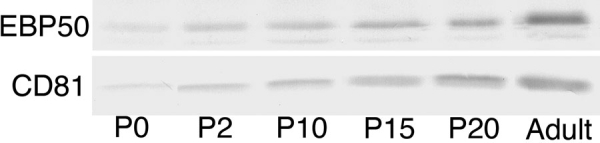
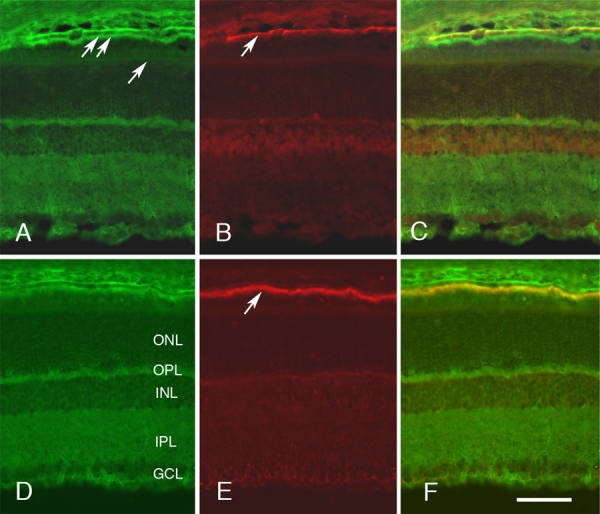
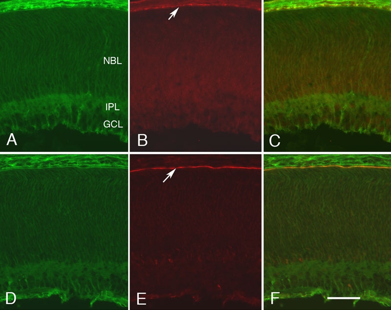
Results: The C-terminal intracellular sequences of CD81 bound to three putative PDZ domains that potentially represented domains on Sap97 and EBP50. In immunoprecipitation experiments using RPE cells, CD81 coprecipitated with both proteins, EBP50 and Sap97. Like CD81, EBP50 and Sap97 are expressed at low levels immediately after birth and upregulated during the first two postnatal weeks, reaching almost adult levels at postnatal day 20. In the RPE layer, synapse-associated protein 97 (Sap97) and CD81 were associated with the basolateral surface of the cells; ezrin-radixin-moesin-binding phosphoprotein 50 (EBP50) localizing with CD81 was found on microvilli at the inner surface of RPE cells.
Conclusions: These results support the hypothesis that CD81 is associated with the final stages of RPE cell maturation, establishing key molecular interactions linking the cell membrane proteins into macromolecular complexes containing PDZ protein scaffolds.
Figures
Similar articles
-
Polarity and developmental regulation of two PDZ proteins in the retinal pigment epithelium.Invest Ophthalmol Vis Sci. 2001 Dec;42(13):3274-82. Invest Ophthalmol Vis Sci. 2001. PMID: 11726633
-
Cellular retinaldehyde-binding protein interacts with ERM-binding phosphoprotein 50 in retinal pigment epithelium.Invest Ophthalmol Vis Sci. 2004 Feb;45(2):393-401. doi: 10.1167/iovs.03-0989. Invest Ophthalmol Vis Sci. 2004. PMID: 14744877
-
Identification of EBP50: A PDZ-containing phosphoprotein that associates with members of the ezrin-radixin-moesin family.J Cell Biol. 1997 Oct 6;139(1):169-79. doi: 10.1083/jcb.139.1.169. J Cell Biol. 1997. PMID: 9314537 Free PMC article.
-
ERM-Merlin and EBP50 protein families in plasma membrane organization and function.Annu Rev Cell Dev Biol. 2000;16:113-43. doi: 10.1146/annurev.cellbio.16.1.113. Annu Rev Cell Dev Biol. 2000. PMID: 11031232 Review.
-
Role of the PDZ-scaffold protein NHERF1/EBP50 in cancer biology: from signaling regulation to clinical relevance.Oncogene. 2017 Jun 1;36(22):3067-3079. doi: 10.1038/onc.2016.462. Epub 2017 Jan 9. Oncogene. 2017. PMID: 28068322 Review.
Cited by
-
Tspan2: a tetraspanin protein involved in oligodendrogenesis and cancer metastasis.Biochem Soc Trans. 2017 Apr 15;45(2):465-475. doi: 10.1042/BST20160022. Biochem Soc Trans. 2017. PMID: 28408487 Free PMC article. Review.
-
CD81 inhibits the proliferation of astrocytes by inducing G(0)/G (1) arrest in vitro.J Huazhong Univ Sci Technolog Med Sci. 2010 Apr;30(2):201-5. doi: 10.1007/s11596-010-0214-1. Epub 2010 Apr 21. J Huazhong Univ Sci Technolog Med Sci. 2010. PMID: 20407874
-
Tetraspanin CD81 is required for the alpha v beta5-integrin-dependent particle-binding step of RPE phagocytosis.J Cell Sci. 2007 Sep 1;120(Pt 17):3053-63. doi: 10.1242/jcs.006361. Epub 2007 Aug 7. J Cell Sci. 2007. PMID: 17684062 Free PMC article.
-
Tetraspanins in mammalian reproduction: spermatozoa, oocytes and embryos.Med Microbiol Immunol. 2020 Aug;209(4):407-425. doi: 10.1007/s00430-020-00676-0. Epub 2020 May 18. Med Microbiol Immunol. 2020. PMID: 32424440 Review.
-
Laminin-binding integrins and their tetraspanin partners as potential antimetastatic targets.Expert Rev Mol Med. 2010 Jan 18;12:e3. doi: 10.1017/S1462399409001355. Expert Rev Mol Med. 2010. PMID: 20078909 Free PMC article. Review.
References
-
- Geisert EE, Jr, Abel HJ, Fan L, Geisert GR. Retinal pigment epithelium of the rat express CD81, the target of the anti-proliferative antibody (TAPA). Invest Ophthalmol Vis Sci. 2002;43:274–80. - PubMed
-
- Clarke K, Geisert EE., Jr The target of the antiproliferative antibody (TAPA) in the normal and injured rat retina. Mol Vis. 1998;4:3. http://www.molvis.org/molvis/v4/a3/ - PubMed
-
- Young RW. Cell differentiation in the retina of the mouse. Anat Rec. 1985;212:199–205. - PubMed
-
- Stroeva OG, Panova IG. Retinal pigment epithelium: pattern of proliferative activity and its regulation by intraocular pressure in postnatal rats. J Embryol Exp Morphol. 1983;75:271–91. - PubMed
Publication types
MeSH terms
Substances
Grants and funding
LinkOut - more resources
Full Text Sources
Other Literature Sources
Medical
Research Materials