. 2006 Dec 28;39(6):145-53.
doi: 10.1267/ahc.06023. Epub 2006 Dec 6.

Interstitial cells of cajal are involved in neurotransmission in the gastrointestinal tract

Affiliations

Interstitial cells of cajal are involved in neurotransmission in the gastrointestinal tract

Satoshi Iino et al. Acta Histochem Cytochem. .

Abstract

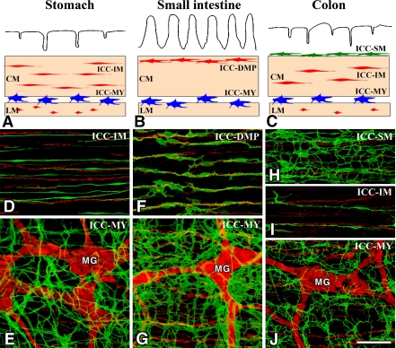
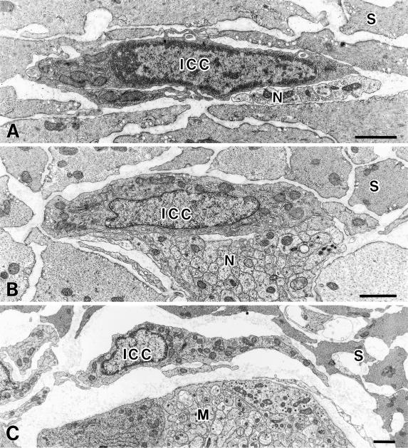
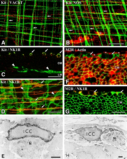
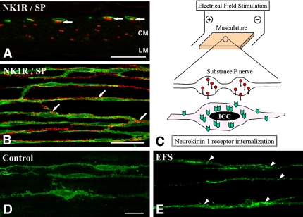
Interstitial cells of Cajal (ICC) are important cells which coordinate gastrointestinal motility. ICC express Kit receptor tyrosine kinase, and Kit immunohistochemistry reveals ICC morphology and distribution in the gastrointestinal musculature. ICC show a highly branched morphology and form unique networks. Myenteric ICC (ICC-MY) are located at the layer of the myenteric plexus and serve as electrical pacemakers. Intramuscular ICC (ICC-IM) and ICC in the deep muscular plexus (ICC-DMP) are distributed within the muscular layers, and are densely innervated by excitatory and inhibitory enteric motor neurons and in close contact with nerve terminals. Recent studies combined with morphological and functional techniques directly revealed that ICC-IM and ICC-DMP are mediators of enteric motor neuro-transmission. These types of ICC express several receptors for neurotransmitters such as acetylcholine and substance P and show responses to excitatory nerve stimulations. ICC also express receptive mechanisms for nitric oxide, which is an inhibitory neurotransmitter in the gastrointestinal tract. They can respond to nitrergic nerve stimulation by cyclic GMP production. Kit mutant mice lack ICC-IM and show attenuated postsynaptic responses after intrinsic nerve stimulation. These findings indicate the importance for ICC in neurotransmission in the gastrointestinal tract.


Figures

Fig. 1
Fig. 1
Overview of the types of ICC in the gastrointestinal tract. A–C: Schematic representations of ICC in the stomach (A), small intestine (B) and colon (C). ICC-MY (blue) are located between circular (CM) and longitudinal (LM) muscle layers. ICC-IM (red) and ICC-DMP (red) are located within circular and longitudinal muscles. ICC-SM (green) are located at the submucosal surface of the circular muscle layer in the colon. D–J: ICC and nerve networks in the murine stomach (D, E), small intestine (F, G) and colon (H–J). ICC and nerves in whole mount preparations are demonstrated by immunohistochemistry for Kit (ICC marker, green) and PGP9.5 (pan-neuronal marker, red), respectively. ICC-MY with a multipolar shape are associated with myenteric ganglia (Auerbach’s ganglia) (MG). ICC-IM in the stomach and colon have a bipolar shape and are associated with intramuscular nerve fibers. ICC-DMP in the small intestine are associated with deep muscular plexus. ICC-SM are multipolar cells having many thin processes. Bar=100 µm.
Fig. 2
Fig. 2
Ultrastructural characteristics of ICC. A: ICC-IM (ICC) in the rat stomach circular muscle layer have an electron-dense cytoplasm, many mitochondria and endoplasmic reticulum. Nerve terminals (N) are in close contact with ICC-IM. S shows smooth muscle cells in the circular layer. B: ICC-DMP (ICC) in the rat jejunum have caveolae, many mitochondria and well developed Golgi apparatus and endoplasmic reticulum. Nerve fiber bundles (N) are in close contact with ICC-DMP. S shows smooth muscle cell in the circular layer. C: ICC-MY (ICC) in the rat jejunum have caveolae, many mitochondria and well developed Golgi apparatus and endoplasmic reticulum. Myenteric ganglion (M) is located near ICC-MY. S shows smooth muscle cells in the circular layer. Bars=1 µm.
Fig. 3
Fig. 3
ICC are associated with nerve fibers and express receptors. A, B: ICC-IM (Kit immunoreactivity, green) in the murine fundus are associated with cholinergic nerve fibers containing VAChT (A, red) and nitrergic nerve fibers containing NOS (B, red). ICC-IM are aligned horizontally in the circular muscle layer and vertically in the longitudinal muscle layer (arrows). N shows NOS-containing nerve cell bodies in the myenteric ganglia. Whole mount preparations were imaged by confocal microscope. C–E: Neurokinin 1 receptor (NK1R) distribution in the murine small intestine. ICC-DMP (arrows in C and D) labeled with Kit antibody (green) specifically express NK1R (red) in the circular muscle layer (CM), whereas ICC-MY (arrowheads) are immunonegative for NK1R between circular and longitudinal (LM) muscle layers. Immunoelectron microscopy (E) reveals ICC-DMP (ICC) expressing NK1R and associated with nerve fibers (N) in the DMP. F–H: Muscarinic M2 receptors (M2R) distribution in the guinea pig small intestine. M2R (green, arrows) positive cells are observed between the inner circular (asterisks) and outer circular muscles (CM) shown by actin immunoreactivity (F, red). Smooth muscle cells in the outer circular muscle also showed M2R. ICC-DMP (arrows in G) contain both M2R (green) and NK1R (red) immunoreactivity in the musculature. Immunoelectron microscopy (H) shows that M2R-immunopositive cells (ICC) are typical ICC-DMP and associated with DMP (N). Bars=50 µm (A, B, D), 20 µm (C), 10 µm (F, G), 1 µm (E, H).
Fig. 4
Fig. 4
Neurokinin 1 receptor (NK1R) internalization of ICC-DMP in the murine small intestine. A, B: ICC-DMP expressing NK1R (green) are in close contact with substance P (red) containing nerve terminals (arrows). CM and LM indicate circular muscle and longitudinal muscle layers, respectively. C: Experimental scheme of electrical field stimulation (EFS). Musculature between the parallel platinum electrodes is stimulated by EFS (10 Hz, 0.5 ms duration, 15V) for 1 min. Stimulated nerve terminals release neurotransmitters such as substance P. ICC-DMP near the nerve terminals receive substance P and substance P-NK1R complexes are internalized into the cell bodies. D, E: NK1R (green) distribution in ICC-DMP before and after nerve stimulation. Before stimulation (D), NK1R are distributed around the peripheries of ICC-DMP. After EFS (E), NK1R are concentrated into granular structures (arrowheads) in ICC-DMP. Bars=20 µm (A, D, E), 50 µm (B).

References

    1. Beckett E. A., Horiguchi K., Khoyi M., Sanders K. M., Ward S. M. Loss of enteric motor neurotransmission in the gastric fundus of Sl/Sld mice. J. Physiol. 2002;543:871–887. - PMC - PubMed
    1. Beckett E. A., Takeda Y., Yanase H., Sanders K. M., Ward S. M. Synaptic specializations exist between enteric motor nerves and interstitial cells of Cajal in the murine stomach. J. Comp. Neurol. 2005;493:193–206. - PubMed
    1. Burns A. J., Lomax A. E., Torihashi S., Sanders K. M., Ward S. M. Interstitial cells of Cajal mediate inhibitory neurotransmission in the stomach. Proc. Natl. Acad. Sci. U S A. 1996;93:12008–12013. - PMC - PubMed
    1. Burns A. J., Herbert T. M., Ward S. M., Sanders K. M. Interstitial cells of Cajal in the guinea-pig gastrointestinal tract as revealed by c-Kit immunohistochemistry. Cell Tissue Res. 1997;290:11–20. - PubMed
    1. Burnstock G., Lavin S. Interstitial cells of Cajal and purinergic signalling. Auton. Neurosci. 2002;97:68–72. - PubMed