. 2007 Feb 28:8:15.
doi: 10.1186/1471-2199-8-15.

The coding/non-coding overlapping architecture of the gene encoding the Drosophila pseudouridine synthase

Affiliations

The coding/non-coding overlapping architecture of the gene encoding the Drosophila pseudouridine synthase

Sara Riccardo et al. BMC Mol Biol. .

Abstract

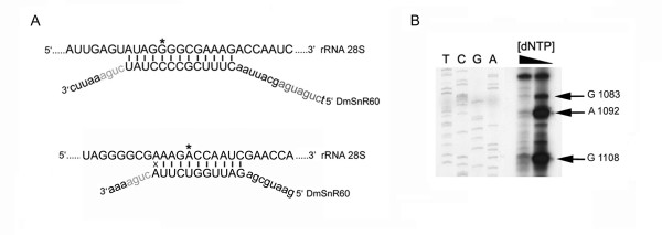
Background: In eukaryotic cells, each molecule of H/ACA small nucleolar RNA (snoRNA) assembles with four evolutionarily conserved core proteins to compose a specific ribonucleoprotein particle. One of the four core components has pseudouridine synthase activity and catalyzes the conversion of a selected uridine to pseudouridine. Members of the pseudouridine synthase family are highly conserved. In addition to catalyzing pseudouridylation of target RNAs, they carry out a variety of essential functions related to ribosome biogenesis and, in mammals, to telomere maintenance. To investigate further the molecular mechanisms underlying the expression of pseudouridine synthase genes, we analyzed the transcriptional activity of the Drosophila member of this family in great detail.

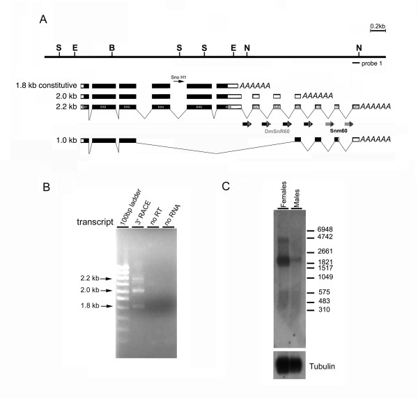
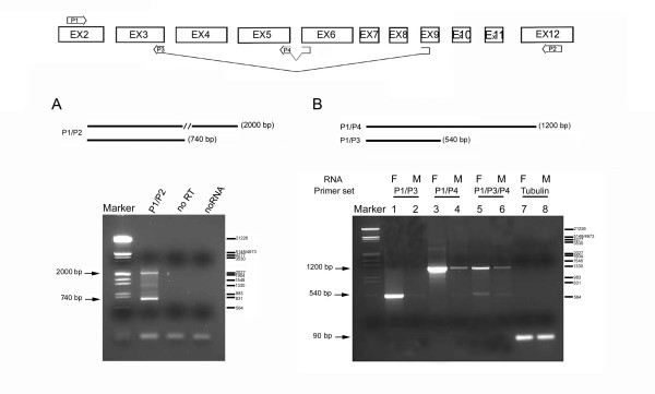
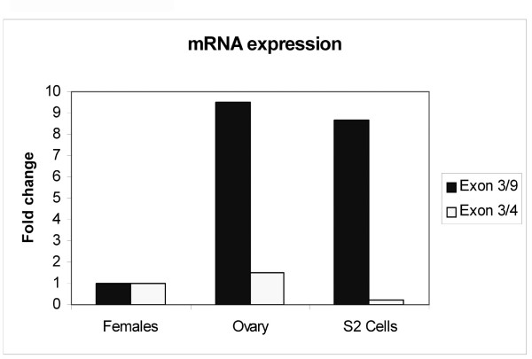
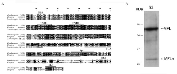
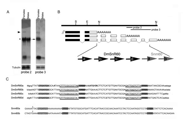
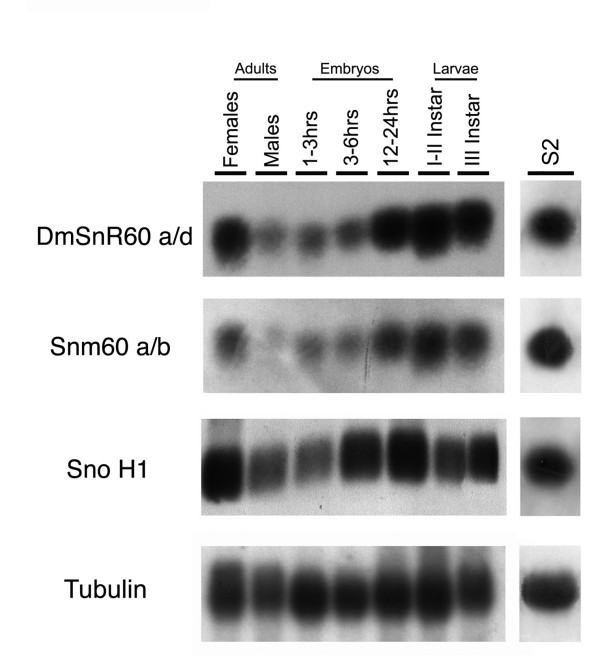
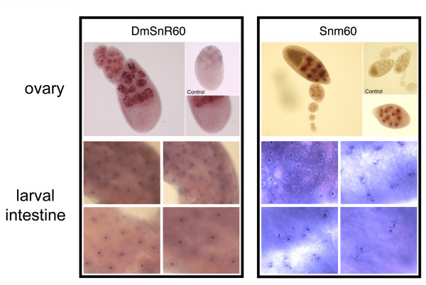
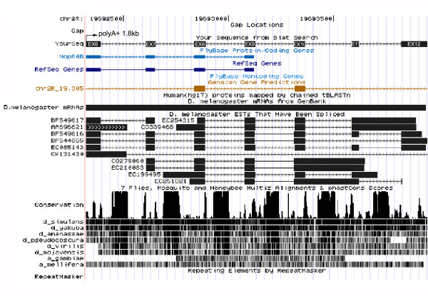
Results: The Drosophila gene for pseudouridine synthase, minifly/Nop60b (mfl), encodes two novel mRNAs ending at a downstream poly(A) site. One species is characterized only by an extended 3'-untranslated region (3'UTR), while a minor mRNA encodes a variant protein that represents the first example of an alternative subform described for any member of the family to date. The rare spliced variant is detected mainly in females and is predicted to have distinct functional properties. We also report that a cluster comprising four isoforms of a C/D box snoRNA and two highly related copies of a small ncRNA gene of unknown function is intron-encoded at the gene-variable 3'UTRs. Because this arrangement, the alternative 3' ends allow mfl not only to produce two distinct protein subforms, but also to release different ncRNAs. Intriguingly, accumulation of all these intron-encoded RNAs was found to be sex-biased and quantitatively modulated throughout development and, within the ovaries, the ncRNAs of unknown function were found not ubiquitously expressed.

Conclusion: Our results expand the repertoire of coding/non-coding transcripts derived from the gene encoding Drosophila pseudouridine synthase. This gene exhibits a complex and interlaced organization, and its genetic information may be expressed as different protein subforms and/or ncRNAs that may potentially contribute to its biological functions.


Figures

Figure 1
Figure 1
Molecular structure of mfl mRNAs. (A) Restriction map of the genomic region encompassing the minifly gene (S, SalI; E, EcoRI; B, BamHI; N: NotI) [GenBank: AF 097634]. Below, organization of the previously-described 1.8 [GenBank: AF017230] and 2.0 kb [GenBank: AF089837] mfl mRNAs [7], compared with that of the newly identified 2.2 [GenBank: DQ857345] and 1.0 kb mRNA species [GenBank: DQ857346]; note that subsequent releases of the Drosophila genome sequence have revealed that a small intron splits the formerly-designated exon 8 [7] into two moieties, currently indicated as exon 8 and 9. Exonic regions spanned by mfl ORFs are depicted in black. The positions of the intron-encoded H/ACA snoRNA H1 gene [7] and of the DmSnR60 and snm60 isoforms are also shown. (B) The products of the 3'RACE reactions were separated on 2% agarose gels and visualised by ethidium bromide staining. Lane 1, GeneRuler 100 bp DNA ladder (MBI Fermentas). Lane 2, products obtained after amplification with a forward primer derived from exon 5, in combination with the oligo-adaptor reverse primer. Specific fragments of about 800, 650 and 400 bp were obtained; each fragment represents the specific 3' end of a different mfl mRNA, the length of which (in kb) is indicated on the left. Lanes 2 and 3 show negative controls in which no reverse transcriptase or no input RNA were added to the reaction. (C) Northern blot analysis of poly(A)+ RNA extracted from male and female adult flies with a genomic probe derived from exon 12 (probe1: see the genomic map for position). This probe specifically detects the novel 2.2 kb mRNA, most abundant in females, and a large transcript of about 4.4 kb of which the structure remains to be defined. The amount of RNA loaded on each lane was checked by hybridization with a probe derived from αTub84B. The RNA marker I (Roche) was utilised (on the right).
Figure 2
Figure 2
mfl encodes a novel alternatively-spliced mRNA. At the top, a schematic drawing of the splicing patterns generating mfl mRNAs; the position and structure of the primers utilised in RT-PCR assays are indicated. HindIII-digested λ DNA was used as marker; base pairs are indicated on the right. (A) RT-PCR performed with poly(A)+RNA isolated from adult females and an exon 2–12 primer pair (P1/P2). The 2000 bp fragment is specific for the 2.2 kb mRNA, while the 740 bp fragment derives from the 1.0 kb alternatively-spliced subform. (B) RT-PCR was performed with poly(A)+RNA isolated from male (M) and female (F) adult flies. Female or male samples were amplified using the same forward primer (P1, derived from exon 2) in combination with a reverse primer spanning the alternative 3–9 exon junction (P3; lanes 1–2) or the canonical 5–6 exon junction (P4; lanes 3–4); in lanes 5–6, the three primers were added to the same reaction. The 1200 bp fragment represents all three transcripts generated by the canonical splicing pattern, while the 540 bp fragment derives specifically from the 1.0 kb mRNA. In lanes 7–8, a 90 bp fragment representating the αTub84B transcript was amplified as internal control of the quantity of RNA.
Figure 3
Figure 3
Relative abundance of alternatively- and canonically-spliced mfl mRNA subforms. The abundances of the alternatively- and canonically- spliced mfl mRNAs were measured by quantitative real-time RT-PCR in poly(A)+ RNA from adult Drosophila females, manually-dissected ovaries and cultured S2 cells. Three different RNA extractions were examined for each sample, and each reaction was performed in triplicate. Data were normalized to αTub84B expression and are presented relative to the female sample; they represent three independent experiments.
Figure 4
Figure 4
Amino acid sequence and expression of the novel MFLα protein subform. (A) Alignment of MFL (top), dyskerin (middle) and MFLα (bottom) amino acid sequences. White letters highlight identical amino acids on a black background, and different but conserved amino acids on a grey background; block letters on a white background indicate different and non-conserved residues. Lines above or below the sequences indicate putative functional domains. NLS: nuclear localisation signal; TruB I, Trub II: regions having homology with members of the bacterial tRNA pseudouridine synthase family; tyr: tyrosine domain containing a putative uracil binding pocket; PUA: a conserved RNA binding domain observed in archaeal, bacterial and eukaryotic RNA modifying proteins. (B) Western blot analysis of extracts from S2 cells. Affinity-purified rabbit polyclonal antibodies recognizing both MFL and MFLα were utilised (see Methods). A major band corresponding to the MFL subform (56 kDa) and a minor band corresponding to the MFLα subform (29 kDa) were detected. Positions of the protein ladder (Precision Plus Prestained Protein Standard Dual Colour; Biorad Laboratories) are shown on the left.
Figure 5
Figure 5
An intronic cluster of ncRNA genes maps at the variable mfl 3' UTRs. (A) Northern blot analysis of total RNA from male and female adult flies with genomic probes derived from the mfl 3' region (see B for genomic position). Probes 2 and 3 both detect a large transcript of about 4.4 kb (marked by the triangle) and small RNAs of about 100 nt. Note that probe 2, derived from intron 9, strongly cross-hybridizes with rRNA (asterisk on the left) because of bipartite DmSnR60 complementarity to 28S sequences. Since Drosophila 28S rRNA is processed into two fragments that migrate in a similar manner to the 18S rRNA (2.0 kb), cross-reaction labels a band with a mobility similar to that of the mfl 2.2 kb mRNA (indicated by the arrow), which is specifically recognized by probe 3. (B) Genomic organization of the cluster of small ncRNA genes intron-encoded at the mfl 3'UTRs; the four copies of the DmSnR60 C/D box snoRNA gene (a, b, c, d) and the two copies of the snm60 (a, b) exhibit a one gene-per intron organization. (C) Nucleotide sequences of DmSnR60 and snm60 isoforms. Within DmSnR60 sequences (capital letters; flanking sequences are indicated as lower case letters), dark-shaded regions indicate the D and D' boxes, while the 5'-terminal C box is grey-shaded. The D and D' antisenses able to target the Drosophila 28S rRNA are underlined. Positions of nucleotide polymorphisms among the DmSnR60 isoforms are indicated in italics. At the bottom, nucleotide sequences of the snm60 a and b isoforms [GenBank: DQ142641; DQ142642]. The vertical arrows mark the position of the 5' end of the products mapped by primer-extension (see Methods). The putative D and D' boxes are dark-shaded, the 5'-terminal C box is grey-shaded and the internal segment of perfect identity shared by the two snm60 subforms is in italics.
Figure 6
Figure 6
Functional roles of DmSnR60 molecules. (A) Base-pairing interactions between DmSnR60 and Drosophila 28S rRNA sequences. The upper strand represents the rRNA sequence; the G1083 and the A1092 residues selected for methylation by D' and D antisense elements are marked by the asterisk. (B) Reverse transcription at low dNTP concentration of the 28S Drosophila rRNA region, including the G1083 and the A1092 methylation sites; on the left, lanes T, C, G and A are dideoxy sequence reactions performed using the primer utilised for reverse transcription and a plasmid carrying the Drosophila 28S rRNA gene. One additional 2'-O-methylation, at position Gm1108 of 28S rRNA, is detected in this experiment. This methylation has not yet been described in other organisms and may be specific to Drosophila rRNA.
Figure 7
Figure 7
Developmental expression of mfl intron-encoded ncRNAs. Total RNA extracted at various developmental stages of the Drosophila life cycle was hybridized to probes specific to DmSnR60, snm60 and snoH1. On the right, Northern blot analysis of total RNA extracted from the Drosophila S2 cell line. The amount of RNA loaded on each lane was checked by hybridization with a probe derived from Drosophila αTub84B.
Figure 8
Figure 8
Cellular location of DmSnR60 and snm60 molecules. Top-left panel: whole mount in situ hybridization of DmSnR60 specific probes to adult ovaries. DmSnR60 molecules are detected at all stages of oogenesis, in the nurse and the follicle cells (see enlargement on bottom-right). Analysis of the large polyploid nurse-cell nuclei reveals that the DmSnR60 molecules are located in the peripheral nucleolar structures. Hybridization of the sense probe was performed as control (top-right). Bottom-left panel: in situ hybridization of DmSnR60 probes to larval intestines. Top-right panel: whole mount in situ hybridization of snm60 specific probes to adult ovaries. snm60 molecules accumulate specifically in the nurse cell nuclei from stage 7 of oogenesis, and are not detected in the follicle cells (see also the enlargement at bottom-right). Hybridization of the sense probe was performed as control (top-right). Bottom-right panel: in situ hybridazion of snm60 probes to larval intestines.
Figure 9
Figure 9
Screen shot of the UCSC Genome Browser () conservation tracks of the mfl 3' genomic region. The region examined, spanning exons 6–12 (black boxes), is shown at the top. The conservation track has two parts: a plot of conservation scores, and beneath it, a display showing where each of the other genomes aligns to the reference sequence (darker shading indicates higher BLASTZ scores; white indicates no alignment). Peaks of cross-species conservation are observed at each 3 'UTR intron, but only sequences of the DmSnR60 isoforms are highly conserved among all the annotated genomes of the Drosophila species.

References

    1. Bachellerie JP, Cavaillé J, Hüttenhofer A. The expanding snoRNA world. Biochimie. 2002;84:775–790. doi: 10.1016/S0300-9084(02)01402-5. - DOI - PubMed
    1. Kiss T. Small nucleolar RNAs: an abundant group of noncoding RNAs with diverse cellular functions. Cell. 2002;19:145–8. doi: 10.1016/S0092-8674(02)00718-3. - DOI - PubMed
    1. Baker DL, Youssef OA, Chastkofsky MI, Dy DA, Terns RM, Terns MP. RNA-guided RNA modification: functional organization of the archaeal H/ACA RNP. Genes Dev. 2005;19:1238–48. doi: 10.1101/gad.1309605. - DOI - PMC - PubMed
    1. Cadwell C, Yoon HJ, Zebarjadian Y, Carbon J. The yeast nucleolar protein Cbf5p is involved in rRNA biosynthesis and interacts with the RNA polymerase I transcription factor RRN3. Mol Cell Biol. 1997;17:6175–6183. - PMC - PubMed
    1. Barth S, Hury A, Liang XH, Michaeli S. Elucidating the role of H/ACA-like RNAs in trans-splicing and rRNA processing via RNA interference silencing of the Trypanosoma brucei CBF5 pseudouridine synthase. J Biol Chem. 2005;280:34558–68. doi: 10.1074/jbc.M503465200. - DOI - PubMed

Publication types