. 2007 Apr 20;355(4):993-9.
doi: 10.1016/j.bbrc.2007.02.067. Epub 2007 Feb 22.

Induction of apoptosis by tumor suppressor FHIT via death receptor signaling pathway in human lung cancer cells

Affiliations

Induction of apoptosis by tumor suppressor FHIT via death receptor signaling pathway in human lung cancer cells

Wu-Guo Deng et al. Biochem Biophys Res Commun. .

Abstract

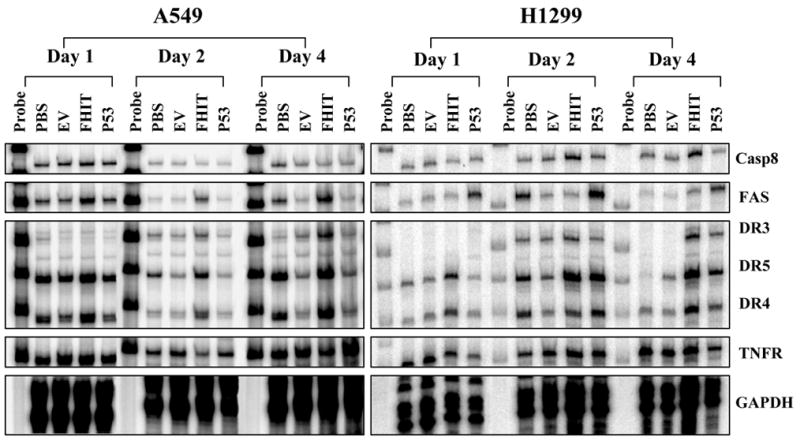
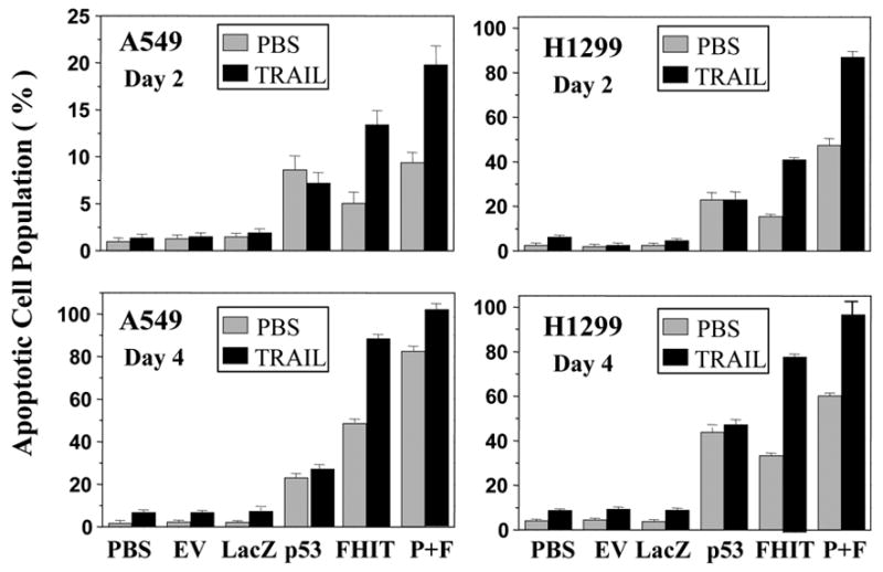
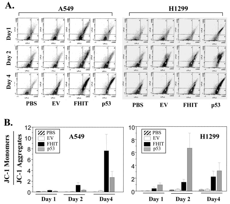
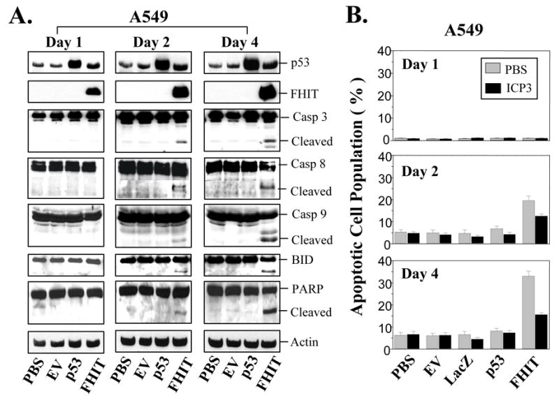
FHIT is a novel tumor suppressor gene located at human chromosome 3p14.2. Restoration of wild-type FHIT in 3p14.2-deficient human lung cancer cells inhibits cell growth and induces apoptosis. In this study, we analyzed potential upstream/downstream molecular targets of the FHIT protein and found that FHIT specifically targeted and regulated death receptor (DR) genes in human non-small-cell lung cancer (NSCLC) cells. Exogenous expression of FHIT by a recombinant adenoviral vector (Ad)-mediated gene transfer upregulated expression of DR genes. Treatment with a recombinant TRAIL protein, a DR-specific ligand, in Ad-FHIT-transduced NSCLC cells considerably enhanced FHIT-induced apoptosis, further demonstrating the involvement of DRs in FHIT-induced apoptosis. Moreover, we also found that FHIT targeted downstream of the DR-mediated signaling pathway. FHIT overexpression disrupted mitochondrial membrane integrity and activated multiple pro-apoptotic proteins in NSCLC cell. These results suggest that FHIT induces apoptosis through a sequential activation of DR-mediated pro-apoptotic signaling pathways in human NSCLC cells.


Figures

Fig. 1
Fig. 1
FHIT specifically targets and upregulates apoptosis-related DRs. The regulation of gene expression by FHIT was analyzed using a multiprobe RPA with specific probes for human DR genes and their effectors, caspase 8 (Casp-8), FASL, FAS, DCR1, DR3, DR4, DR5, TRAIL, TNFR, TRADD, RIP, and L32. The NSCLC cell lines A549 and H1299 were transduced with Ad-FHIT. PBS and an empty vector (EV) were used as negative controls, and Ad-p53 was used as a positive control. On days 1, 2, and 4 after transduction, cells were harvested, and the total RNAs were isolated and RPA was performed. Representative RPA images indicated the expression of the selected genes. Three separate experiments were performed.
Fig. 2
Fig. 2
Treatment of TRAIL proteins enhances FHIT-mediated apoptosis. The NSCLC cell lines A549 and H1299 were transduced with Ad-FHIT and treated with a recombinant TRAIL protein (10 ng/ml). On days 2 and 4 after transduction, cells were harvested, and apoptosis was examined using TUNEL-based FACS analysis. PBS, an empty vector (EV), and LacZ were used as negative controls, and Ad-p53 was used as a positive control. Apoptosis is shown as the relative percentage of apoptotic cells. Three separate experiments were performed in duplicate and each bar denotes mean ± SEM of three experiments. P + F means p53 + FHIT.
Fig. 3
Fig. 3
FHIT overexpression disrupts the integrity of the mitochondrial membrane. The NSCLC cell lines A549 and H1299 were transduced with Ad-FHIT. PBS and an empty vector (EV) were used as negative controls, and Ad-p53 was used as a positive control. On days 1, 2, and 4 after transduction, cells were harvested, and the mitochondrial membrane potential was assessed using JC-1 staining by flow cytometry. Panel (A) shows representative images of JC-1 staining. The mitochondrial membrane potential (B) in cells is shown as the relative value of JC-1 monomers and JC-1 aggregates. Each bar denotes mean ± SEM of three experiments.
Fig. 4
Fig. 4
FHIT overexpression activates caspase-dependent signaling pathway. (A), FHIT-mediated activation of caspases. The NSCLC cell line A549 was transduced with Ad-FHIT. On days 1, 2, and 4 after transduction, the activation of caspases was assessed using Western blot analysis with specific antibodies. The activation of caspase 3, caspase 8, caspase 9 and PARP is indicated by detected cleavage of these proteins. (B), Inhibition of ICP3 on FHIT-mediated apoptosis. A549 cells were transduced with Ad-FHIT and treated with or without 10 μM ICP3. After 1, 2, or 4 days, apoptosis was examined using TUNEL-based FACS analysis. Apoptosis is shown as the relative percentage of apoptotic cells. Three separate experiments were performed in duplicate. Each bar denotes mean ± SEM of three experiments.

Similar articles

Cited by

References

    1. Peto R, Lopez AD, Boreham J, Thun M, Heath C, Jr, Doll R. Mortality from smoking worldwide. Br Med Bull. 1996;52:12–21. - PubMed
    1. Zabarovsky ER, Lerman MI, Minna JD. Tumor suppressor genes on chromosome 3p involved in the pathogenesis of lung and other cancers. Oncogene. 2002;21:6915–6935. - PubMed
    1. Sozzi G, Pastorino U, Moiraghi L, Tagliabue E, Pezzella F, Ghirelli C, Tornielli S, Sard L, Huebner K, Pierotti MA, Croce CM, Pilotti S. Loss of FHIT function in lung cancer and preinvasive bronchial lesions. Adv Cancer Res. 1998;74:141–166. - PubMed
    1. Ji L, Fang B, Yen N, Fong K, Minna JD, Roth JA. Induction of apoptosis and inhibition of tumorigenicity and tumor growth by adenovirus vector-mediated fragile histidine triad (FHIT) gene overexpression. Cancer Res. 1999;59:3333–3339. - PubMed
    1. Pekarsky Y, Palamarchuk A, Huebner K, Croce CM. FHIT as tumor suppressor: mechanisms and therapeutic opportunities. Cancer Biol Ther. 2002;1:232–236. - PubMed

Publication types

MeSH terms