

Directed mutagenesis identifies amino acid residues involved in elongation factor Tu binding to yeast Phe-tRNAPhe
- PMID: 17328911
- PMCID: PMC2246379
- DOI: 10.1016/j.jmb.2007.01.075
Directed mutagenesis identifies amino acid residues involved in elongation factor Tu binding to yeast Phe-tRNAPhe
Abstract
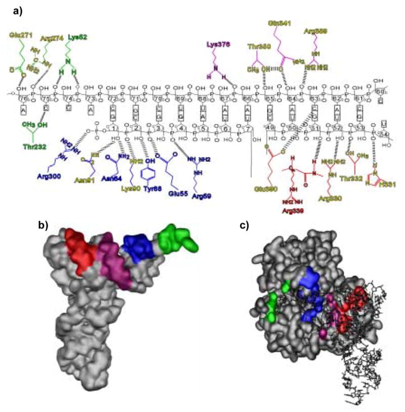
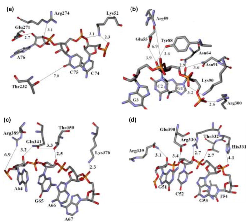
The co-crystal structure of Thermus aquaticus elongation factor Tu.guanosine 5'- [beta,gamma-imido]triphosphate (EF-Tu.GDPNP) bound to yeast Phe-tRNA(Phe) reveals that EF-Tu interacts with the tRNA body primarily through contacts with the phosphodiester backbone. Twenty amino acids in the tRNA binding cleft of Thermus Thermophilus EF-Tu were each mutated to structurally conservative alternatives and the affinities of the mutant proteins to yeast Phe-tRNA(Phe) determined. Eleven of the 20 mutations reduced the binding affinity from fourfold to >100-fold, while the remaining ten had no effect. The thermodynamically important residues were spread over the entire tRNA binding interface, but were concentrated in the region which contacts the tRNA T-stem. Most of the data could be reconciled by considering the crystal structures of both free EF-Tu.GTP and the ternary complex and allowing for small (1.0 A) movements in the amino acid side-chains. Thus, despite the non-physiological crystallization conditions and crystal lattice interactions, the crystal structures reflect the biochemically relevant interaction in solution.
Figures
Similar articles
-
The crystal structure of Cys-tRNACys-EF-Tu-GDPNP reveals general and specific features in the ternary complex and in tRNA.Structure. 1999 Feb 15;7(2):143-56. doi: 10.1016/s0969-2126(99)80021-5. Structure. 1999. PMID: 10368282
-
The ternary complex of aminoacylated tRNA and EF-Tu-GTP. Recognition of a bond and a fold.Biochimie. 1996;78(11-12):921-33. doi: 10.1016/s0300-9084(97)86714-4. Biochimie. 1996. PMID: 9150869 Review.
-
Purification and crystallization of the ternary complex of elongation factor Tu:GTP and Phe-tRNA(Phe).FEBS Lett. 1994 Dec 19;356(2-3):165-8. doi: 10.1016/0014-5793(94)01254-7. FEBS Lett. 1994. PMID: 7805830
-
Identification of thermodynamically relevant interactions between EF-Tu and backbone elements of tRNA.J Mol Biol. 2001 May 18;308(5):895-905. doi: 10.1006/jmbi.2001.4612. J Mol Biol. 2001. PMID: 11352580
-
The affinity of elongation factor Tu for an aminoacyl-tRNA is modulated by the esterified amino acid.Biochemistry. 2004 May 25;43(20):6159-66. doi: 10.1021/bi036290o. Biochemistry. 2004. PMID: 15147200
Cited by
-
Histidine 66 in Escherichia coli elongation factor tu selectively stabilizes aminoacyl-tRNAs.J Biol Chem. 2012 Jan 6;287(2):1229-34. doi: 10.1074/jbc.M111.294850. Epub 2011 Nov 21. J Biol Chem. 2012. PMID: 22105070 Free PMC article.
-
Elongation Factor Tu Prevents Misediting of Gly-tRNA(Gly) Caused by the Design Behind the Chiral Proofreading Site of D-Aminoacyl-tRNA Deacylase.PLoS Biol. 2016 May 25;14(5):e1002465. doi: 10.1371/journal.pbio.1002465. eCollection 2016 May. PLoS Biol. 2016. PMID: 27224426 Free PMC article.
-
Lack of discrimination against non-proteinogenic amino acid norvaline by elongation factor Tu from Escherichia coli.Croat Chem Acta. 2013;86(1):73-82. doi: 10.5562/cca2173. Croat Chem Acta. 2013. PMID: 23750044 Free PMC article.
-
Fine-Tuning of Alanyl-tRNA Synthetase Quality Control Alleviates Global Dysregulation of the Proteome.Genes (Basel). 2020 Oct 18;11(10):1222. doi: 10.3390/genes11101222. Genes (Basel). 2020. PMID: 33081015 Free PMC article.
-
Elongation factor Ts directly facilitates the formation and disassembly of the Escherichia coli elongation factor Tu·GTP·aminoacyl-tRNA ternary complex.J Biol Chem. 2013 May 10;288(19):13917-28. doi: 10.1074/jbc.M113.460014. Epub 2013 Mar 28. J Biol Chem. 2013. PMID: 23539628 Free PMC article.
References
-
- Louie A, Ribeiro NS, Reid BR, Jurnak F. Relative affinities of all Escherichia coli aminoacyl-tRNAs for elongation factor Tu-GTP. J Biol Chem. 1984;259:5010–6. - PubMed
-
- Ott G, Schiesswohl M, Kiesewetter S, Forster C, Arnold L, Erdmann VA, Sprinzl M. Ternary complexes of Escherichia coli aminoacyl-tRNAs with the elongation factor Tu and GTP: thermodynamic and structural studies. Biochim Biophys Acta. 1990;1050:222–5. - PubMed
-
- Abrahamson JK, Laue TM, Miller DL, Johnson AE. Direct determination of the association constant between elongation factor Tu X GTP and aminoacyl-tRNA using fluorescence. Biochemistry. 1985;24:692–700. - PubMed
-
- Janiak F, Dell VA, Abrahamson JK, Watson BS, Miller DL, Johnson AE. Fluorescence characterization of the interaction of various transfer RNA species with elongation factor Tu.GTP: evidence for a new functional role for elongation factor Tu in protein biosynthesis. Biochemistry. 1990;29:4268–77. - PubMed
-
- LaRiviere FJ, Wolfson AD, Uhlenbeck OC. Uniform binding of aminoacyl-tRNAs to elongation factor Tu by thermodynamic compensation. Science. 2001;294:165–8. - PubMed
MeSH terms
Substances
Grants and funding
LinkOut - more resources
Full Text Sources
