

A non-invasive in vitro technique for the three-dimensional quantification of microdamage in trabecular bone
- PMID: 17329178
- PMCID: PMC3312747
- DOI: 10.1016/j.bone.2006.10.031
A non-invasive in vitro technique for the three-dimensional quantification of microdamage in trabecular bone
Abstract
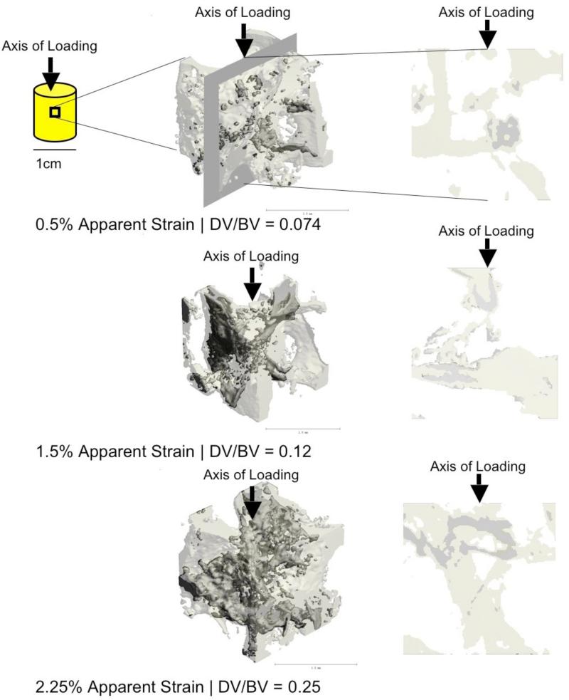
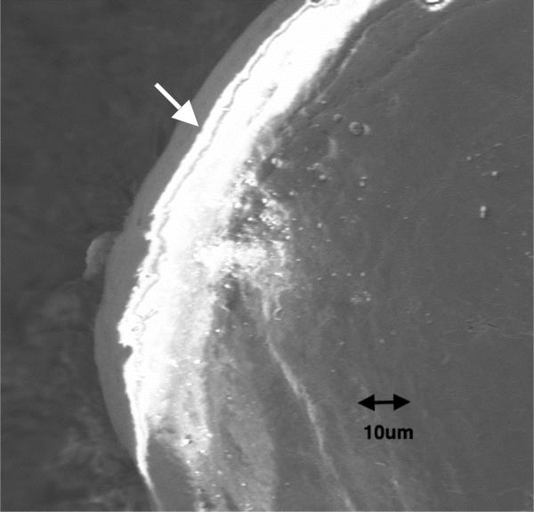
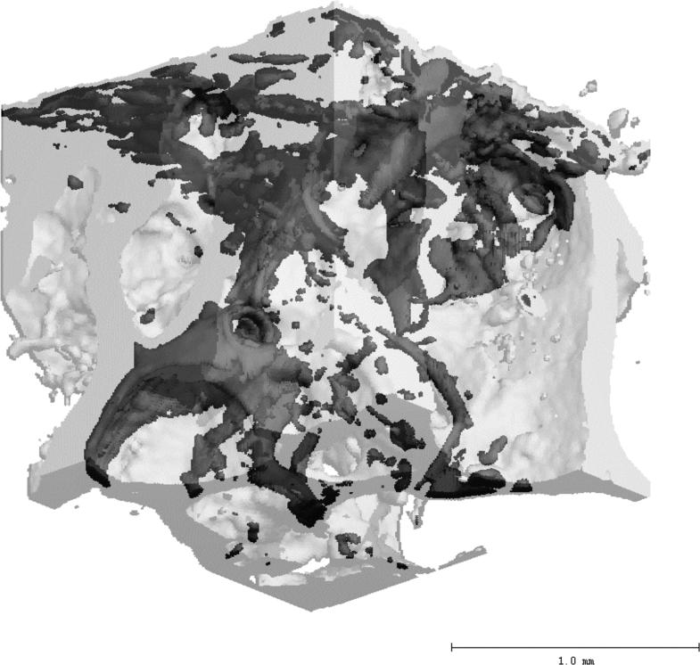
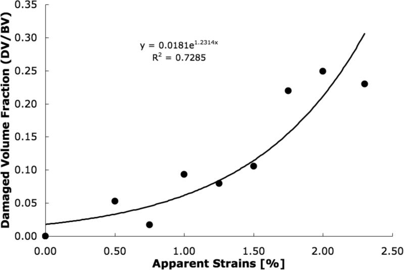
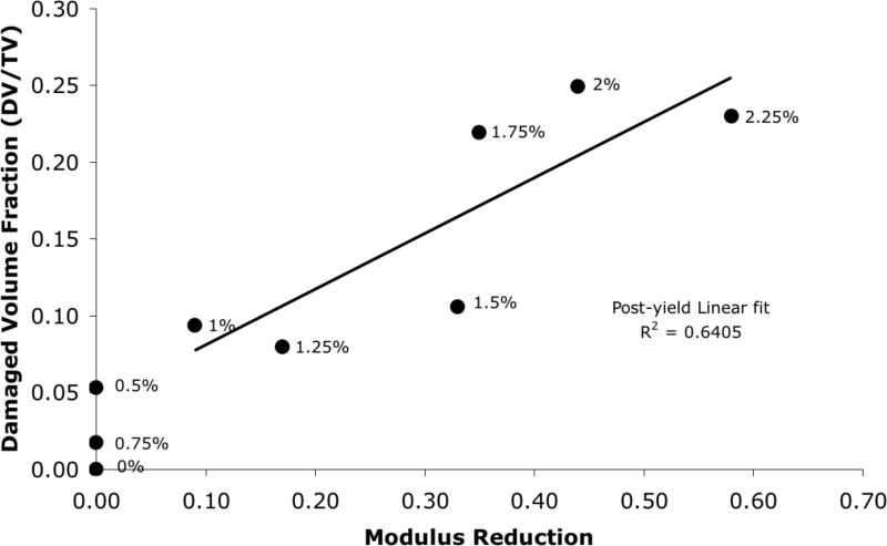
An accurate analysis and quantification of microdamage is critical to understand how microdamage affects the mechanics and biology of bone fragility. In this study we demonstrate the development and validation of a novel in vitro micro-computed tomography (microCT) method that employs lead-uranyl acetate as a radio-opaque contrast agent for automated quantification of microdamage in trabecular bone. Human trabecular bone cores were extracted from the femoral neck, scanned via microCT, loaded in unconfined compression to a range of apparent strains (0.5% to 2.25%), stained in lead-uranyl acetate, and subsequently re-scanned via microCT. An investigation of the regions containing microdamage using the backscatter mode of a scanning electron microscope (BSEM) showed that the lead-uranyl sulfide complex was an effective contrast agent for microdamage in bone. Damaged volume fraction (DV/BV), as determined by microCT, increased exponentially with respect to applied strains and proportionately to mechanically determined modulus reduction (p<0.001). Furthermore, the formation of microdamage was observed to occur before any apparent stiffness loss, suggesting that the localized tissue yielding occurs prior to the structural yielding of trabecular bone. This non-invasive in vitro technique for the detection of microdamage using microCT may serve as a valuable complement to existing morphometric analyses of bone.
Figures

Similar articles
-
Assessment of subchondral bone microdamage quantification using contrast-enhanced imaging techniques.J Anat. 2024 Jul;245(1):58-69. doi: 10.1111/joa.14035. Epub 2024 Mar 13. J Anat. 2024. PMID: 38481117 Free PMC article.
-
Non-enzymatic glycation alters microdamage formation in human cancellous bone.Bone. 2010 Jan;46(1):148-54. doi: 10.1016/j.bone.2009.09.003. Epub 2009 Sep 9. Bone. 2010. PMID: 19747573 Free PMC article.
-
Detection of trabecular bone microdamage by micro-computed tomography.J Biomech. 2007;40(15):3397-403. doi: 10.1016/j.jbiomech.2007.05.009. Epub 2007 Jun 22. J Biomech. 2007. PMID: 17588588 Free PMC article.
-
Multiscale imaging of bone microdamage.Connect Tissue Res. 2015 Apr;56(2):87-98. doi: 10.3109/03008207.2015.1008133. Epub 2015 Feb 9. Connect Tissue Res. 2015. PMID: 25664772 Free PMC article. Review.
-
Detecting microdamage in bone.J Anat. 2003 Aug;203(2):161-72. doi: 10.1046/j.1469-7580.2003.00211.x. J Anat. 2003. PMID: 12924817 Free PMC article. Review.
Cited by
-
X-ray-computed tomography contrast agents.Chem Rev. 2013 Mar 13;113(3):1641-66. doi: 10.1021/cr200358s. Epub 2012 Dec 5. Chem Rev. 2013. PMID: 23210836 Free PMC article. Review. No abstract available.
-
Eigenstrain Toughening in Presence of Elastic Heterogeneity with Application to Bone.Int J Solids Struct. 2018 Jul;144-145:137-144. doi: 10.1016/j.ijsolstr.2018.04.019. Epub 2018 Apr 26. Int J Solids Struct. 2018. PMID: 31105330 Free PMC article.
-
Distributions of Microdamage Are Altered Between Trabecular Rods and Plates in Cancellous Bone From Men With Type 2 Diabetes Mellitus.J Bone Miner Res. 2022 Apr;37(4):740-752. doi: 10.1002/jbmr.4509. Epub 2022 Feb 15. J Bone Miner Res. 2022. PMID: 35064941 Free PMC article.
-
Microdamage caused by fatigue loading in human cancellous bone: relationship to reductions in bone biomechanical performance.PLoS One. 2013 Dec 30;8(12):e83662. doi: 10.1371/journal.pone.0083662. eCollection 2013. PLoS One. 2013. PMID: 24386247 Free PMC article.
-
Nanoscale examination of microdamage in sheep cortical bone using synchrotron radiation transmission x-ray microscopy.PLoS One. 2013;8(3):e57942. doi: 10.1371/journal.pone.0057942. Epub 2013 Mar 5. PLoS One. 2013. PMID: 23472121 Free PMC article.
References
-
- Krajcinovic D. Continuum damage mechanics. Appl Mech Rev. 1984;37:1–5.
-
- Mori S, Burr DB. Increased intracortical remodeling following fatigue damage. Bone. 1996;14(2):103–9. - PubMed
-
- Zioupos P, Currey JD, Sedman AJ. An examination of the micromechanics of failure of bone and antler by acoustic emission tests and Laser Scanning Confocal Microscopy. Med Eng Phys. 1994;16(3):203–12. - PubMed
-
- Burr DB, Turner CH, Naick P, Forwood MR, Ambrosius W, Hasan MS, Pridaparti R. Does microdamage accumulation affect the mechanical properties of bone? J Biomech. 1998;21(1998):337–45. - PubMed
-
- Morgan EF, Yeh OC, Keaveny TM. Damage in trabecular bone at small strains. Eur J Morphol. 2005 Feb-Apr;42(1-2):13–21. - PubMed
Publication types
MeSH terms
Grants and funding
LinkOut - more resources
Full Text Sources
Medical
