

Progression of radiographic damage in patients with ankylosing spondylitis: defining the central role of syndesmophytes
- PMID: 17329306
- PMCID: PMC1955120
- DOI: 10.1136/ard.2006.066415
Progression of radiographic damage in patients with ankylosing spondylitis: defining the central role of syndesmophytes
Abstract
Background: Structural changes such as erosions, syndesmophytes and ankylosis are characteristic of ankylosing spondylitis (AS). These can be quantified by the modified Stokes Anklylosing Spondylitis Spinal Score (mSASSS). It is unknown which radiographic feature is most relevant for the assessment of change and the prediction of future damage in AS.
Objectives: To analyse radiographic progression in AS by using different assessments to define the most important changes.
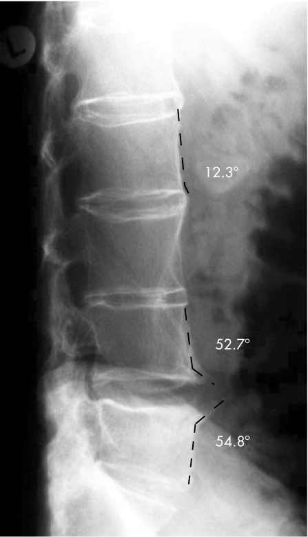
Methods: Spinal radiographs of 116 patients with AS were scored by the mSASSS at baseline (BL) and after 2 years. Radiographic progression was assessed by differentiating (1) any change; (2) progression to syndesmophytes/ankylosis (definite change); and (3) changes exceeding the smallest detectable change (SDC) as predefined. A growth angle of 45 degrees was used to differentiate syndesmophytes from spondylophytes.
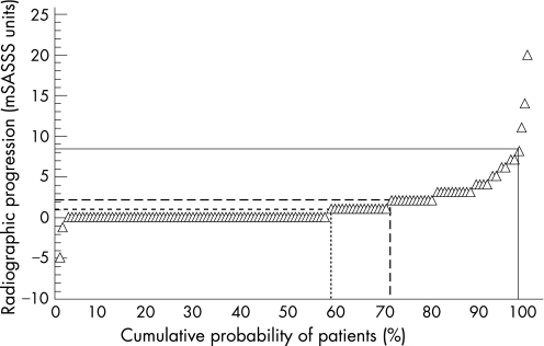
Results: Some radiographic progression after 2 years was detected in 42% of patients, novel syndesmophytes in 31% of patients, and, using the SDC (calculated at 2 mSASSS units) as cut-off, progression was seen in 28% of patients. Thus, in 74% of the patients changes were because of syndesmophytes and/or ankylosis. Using the predefined cut-off, only 12% of all syndesmophytes were spondylophytes. Patients with such changes were of older age. Definite radiographic progression was found in 44% of the patients with syndesmophytes/ankylosis at BL (n = 57) versus 19% (p = 0.03) of the patients without such changes (n = 59).
Conclusions: Syndesmophytes and ankylosis are the most relevant structural changes in AS, and also in the mSASSS. Development of just one syndesmophyte within 2 years indicates progression of structural changes in AS; this is relevant for clinical practice. Syndesmophytes are the best predictors of radiographic progression.
Conflict of interest statement
Competing interests: None declared.
References
-
- Braun J, Bollow M, Remlinger G, Eggens U, Rudwaleit M, Distler A.et al Prevalence of spondylarthropathies in HLA‐B27 positive and negative blood donors. Arthritis Rheum 19984158–67. - PubMed
-
- Braun J, Sieper J. The sacroiliac joint in the spondyloarthropathies. Curr Opin Rheumatol 19968275–287. - PubMed
-
- Braun J, van der Heijde D. Imaging and scoring in ankylosing spondylitis. Best Pract Res Clin Rheumatol 200216573–604. - PubMed
-
- Braun J B M, Sieper J. Radiology and pathology of the spondyloarthropathies. Rheum Dis Clin North Am 199824697–735. - PubMed
MeSH terms
LinkOut - more resources
Full Text Sources
Medical
Research Materials
